. 2020 Mar;39(13):2819-2834.
doi: 10.1038/s41388-020-1187-6. Epub 2020 Feb 6.

Mitotic slippage is determined by p31comet and the weakening of the spindle-assembly checkpoint

Affiliations

Mitotic slippage is determined by p31comet and the weakening of the spindle-assembly checkpoint

Tsun Ming Lok et al. Oncogene. 2020 Mar.

Abstract

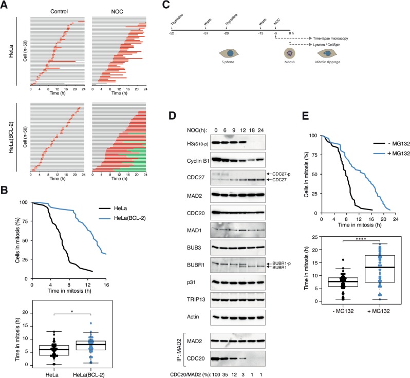
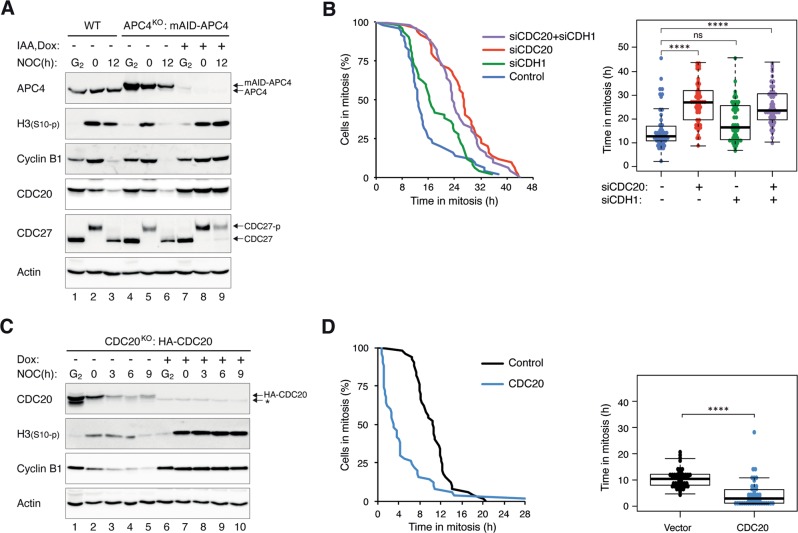
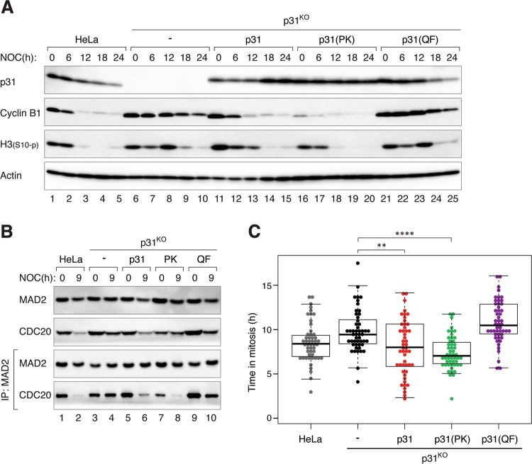
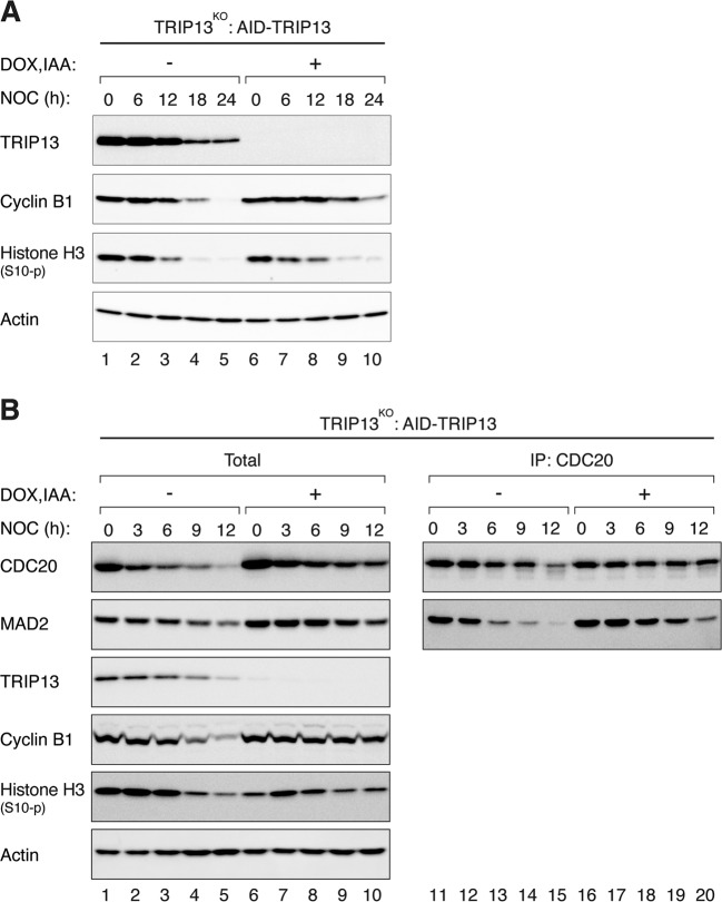
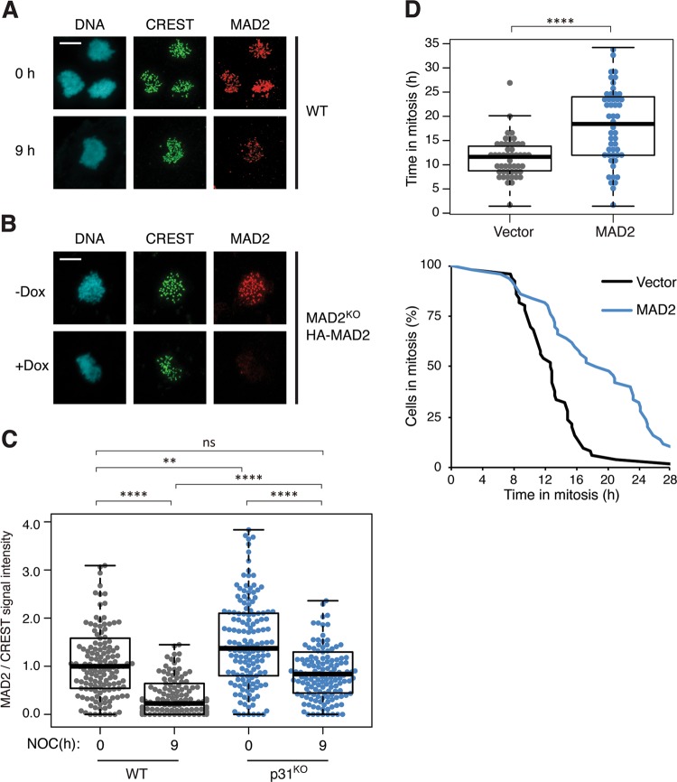
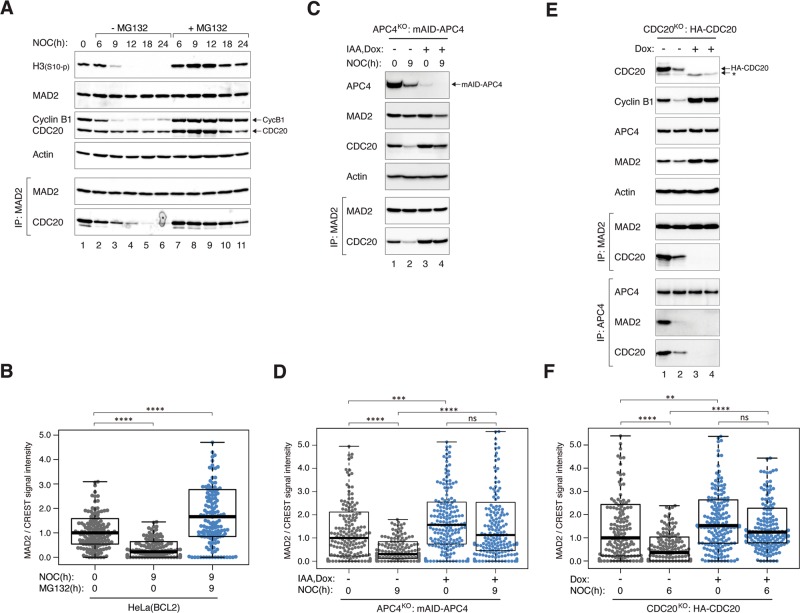
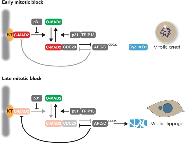
Mitotic slippage involves cells exiting mitosis without proper chromosome segregation. Although degradation of cyclin B1 during prolonged mitotic arrest is believed to trigger mitotic slippage, its upstream regulation remains obscure. Whether mitotic slippage is caused by APC/CCDC20 activity that is able to escape spindle-assembly checkpoint (SAC)-mediated inhibition, or is actively promoted by a change in SAC activity remains an outstanding issue. We found that a major culprit for mitotic slippage involves reduction of MAD2 at the kinetochores, resulting in a progressive weakening of SAC during mitotic arrest. A further level of control of the timing of mitotic slippage is through p31comet-mediated suppression of MAD2 activation. The loss of kinetochore MAD2 was dependent on APC/CCDC20, indicating a feedback control of APC/C to SAC during prolonged mitotic arrest. The gradual weakening of SAC during mitotic arrest enables APC/CCDC20 to degrade cyclin B1, cumulating in the cell exiting mitosis by mitotic slippage.


Conflict of interest statement

The authors declare that they have no conflict of interest.

Figures

Fig. 1
Fig. 1. Inhibition of apoptosis promotes proteasome-dependent mitotic slippage.
a Inhibition of apoptosis promotes mitotic slippage. HeLa or HeLa expressing BCL-2 were incubated with NOC. Individual cells were then tracked using live-cell imaging for 24 h. Key: interphase (gray); mitosis (red); interphase after mitotic slippage (green); truncated bars (cell death). b Inhibition of apoptosis extends the duration of mitotic arrest. The duration of mitotic arrest of cells from a is plotted by using Kaplan–Meier estimator. Box-and-whisker plots show the elapsed time between mitotic entry and mitotic cell death/exit. Note that cells that were still in mitosis at the end of the imaging were also included in the statistical analysis. c Schematic diagram of the synchronization procedure to obtain cells arrested in mitosis. HeLa cells expressing BCL-2 were synchronized at G1/S with a double-thymidine procedure, and released into the cell cycle before treatment with NOC. The cells were then monitored using live-cell imaging to analyze cell fate at single cells level. Alternatively, mitotic cells were isolated by mechanical shake off, continued to be incubated with NOC, and harvested at different time points for biochemical analyses. d Mitotic markers, SAC components, and MCC during mitotic slippage. Cells were synchronized and treated with NOC as described in c. Lysates were prepared from cells harvested at the indicated time points and subjected to immunoprecipitation (IP) using antiserum against MAD2. The immunoprecipitates and total lysates were then analyzed using immunoblotting. The positions of the phosphorylated and unphosphorylated forms of BUBR1 and CDC27 are indicated. Uniform loading of lysates was confirmed by immunoblotting for actin. The band intensities of CDC20 in the MAD2 immunoprecipitates were quantified, normalized with MAD2, and expressed as % max. e Mitotic slippage is delayed after inhibition of the proteasome. HeLa cells expressing both BCL-2 and histone H2B-GFP were synchronized and treated with NOC as described in c. The cells were incubated with either buffer or MG132. Individual cells were then tracked using live-cell imaging. The duration of mitotic arrest and the elapsed time between mitotic entry and mitotic cell death/slippage are shown. The live-cell imaging profile of individual cells is shown in Fig. S1C.
Fig. 2
Fig. 2. Mitotic slippage requires APC/CCDC20.
a Depletion of APC4 prevents mitotic slippage. HeLa (WT) or APC4KO expressing mAID-APC4 cells were synchronized as described in Fig. 1c with minor modifications: IAA and Dox were added (at 15 h after second thymidine release) to turn off mAID-APC4. The cells were harvested either at G2 (22 h after second thymidine release) or at different time points in mitosis. Lysates were prepared and the indicated proteins were detected with immunoblotting. b Downregulation of CDC20 delays mitotic slippage. HeLa cells were transfected with siRNA against CDC20 and/or CDH1. The cells were treated with NOC as described in Fig. 1c (transfection was performed at first thymidine release) before analyzed using live-cell imaging. The duration of mitotic arrest and the elapsed time between mitotic entry and mitotic cell death/slippage are shown. The immunoblotting or CDC20 and CDH1 and the live-cell imaging profile of individual cells are shown in Fig. S2A. c Conditional depletion of CDC20 delays mitotic slippage. CDC20KO cells expressing HA-CDC20 were synchronized as described in Fig. 1c with minor modifications: Dox was added to turn off the expression of HA-CDC20 (at 14 h after first thymidine release). The cells were harvested either at G2 (22 h after second thymidine release) or at different time points in mitosis. Lysates were prepared and the indicated proteins were detected with immunoblotting. The asterisk indicates the position of a cross-reactive band by the CDC20 antibodies (see Fig. 7e). d Overexpression of CDC20 promotes mitotic slippage. Cells were transfected with either vector or a plasmid expressing FLAG-CDC20. A plasmid expressing ECFP was co-transfected. After 40 h, the cells were treated with NOC and analyzed using live-cell imaging. The duration of mitotic arrest and the elapsed time between mitotic entry and mitotic cell death/slippage are shown. The live-cell imaging profile of individual cells and CDC20 expression are shown in Fig. S2C.
Fig. 3
Fig. 3. p31comet controls the timing of mitotic slippage by regulating MCC.
a Mitotic slippage is delayed after knockdown of p31comet. HeLa cells expressing BCL-2 were transfected with either control or siRNA against p31comet (sip31). Knockdown of p31comet was confirmed with immunoblotting (Fig. S3A). The cells were synchronized and treated with NOC as described in Fig. 1c. Individual cells were then tracked using live-cell imaging. The duration of mitotic arrest and the elapsed time between mitotic entry and mitotic cell death/slippage are shown. The live-cell imaging profile of individual cells is shown in Fig. S3B. b Mitotic slippage is delayed in p31comet-deficient cells. HeLa (WT) and p31comet-deficient cells (p31KO) (both stably expressing BCL-2) were incubated with NOC before analyzed using live-cell imaging. The duration of mitotic arrest and the elapsed time between mitotic entry and mitotic cell death/slippage are shown. The live-cell imaging profile of individual cells is shown in Fig. S4A. c MCC inactivation is delayed in the absent of p31comet. WT and p31KO (both stably expressing BCL-2) were synchronized and treated with NOC as described in Fig. 1c. The cells were harvested at the indicated time points. Lysates were prepared and subjected to immunoprecipitation (IP) using antiserum against MAD2 followed by immunoblotting analysis. d Knockout of p31comet delays MPS1i-mediated mitotic slippage. WT and p31KO (both stably expressing BCL-2) were synchronized and treated with NOC as described in Fig. 1c. At t = 0 h, the cells were treated with the MPS1 inhibitor AZ 3146. Lysates were prepared at the indicated time and analyzed with immunoblotting.
Fig. 4
Fig. 4. The promotion of mitotic slippage by p31comet does not require binding to TRIP13.
a Mitotic slippage in p31KO cells is restored by a non-TRIP13-binding mutant. HeLa, p31KO, p31KO expressing HA-tagged p31, p31(PK), or p31(QF) were synchronized and treated with NOC as described in Fig. 1c. Caspase inhibitors were added in this experiment to inhibit cell death. The cells were harvested at the indicated time points. Lysates were prepared and analyzed with immunoblotting. b Reduction of MCC during mitotic arrest is restored by a non-TRIP13-binding p31comet. Cells were treated with NOC as described in a and harvested at the indicated time points. Lysates were prepared and subjected to immunoprecipitation (IP) using antiserum against MAD2 followed by immunoblotting analysis. c Mitotic slippage in p31KO cells is restored by a non-TRIP13-binding mutant. Cells were treated with NOC as described in a and analyzed using live-cell imaging. Box-and-whisker plots show the elapsed time between mitotic entry and mitotic cell death/exit (n = 50).
Fig. 5
Fig. 5. TRIP13 plays only a minor role in mitotic slippage.
a TRIP13 only exerts a minor effect on mitotic slippage. TRIP13KO cells expressing AID-TRIP13 were synchronized and treated with NOC as described in Fig. 1c. IAA and Dox were added at 6 h after addition of the second thymidine block. Lysates were prepared at the indicated time and analyzed with immunoblotting. b Reduction of MCC during mitotic arrest is only marginally affected by the absence of TRIP13. Cells were treated with NOC as described in a and harvested at the indicated time points. Lysates were prepared and subjected to immunoprecipitation (IP) using antiserum against CDC20 followed by immunoblotting analysis.
Fig. 6
Fig. 6. Weakening of SAC during mitotic arrest involves reduction of MAD2 at the kinetochores.
a Reduction of kinetochore MAD2 during mitotic arrest. HeLa cells expressing BCL-2 were synchronized and treated with NOC as described in Fig. 1c. At t = 0 and 9 h after NOC treatment, mitotic cells were isolated, fixed, and analyzed using immunostaining for MAD2 and CREST. DNA was stained with Hoechst 33342. Examples of confocal microscopy images are shown. Scale bar: 10 µm. b Specificity of the MAD2 antibodies. MAD2KO cells expressing HA-MAD2 were cultured in the absence or presence of Dox to turn on or off the HA-MAD2, respectively. The cells were first blocked in G2 using RO3306, before released for 1 h and incubated with NOC and MG132 for 2 h. Mitotic cells were isolated, fixed, and analyzed using immunostaining for MAD2 and CREST. DNA was stained with Hoechst 33342. Examples of confocal microscopy images are shown. Scale bar: 10 µm. c Kinetochore MAD2 is present at a higher level in p31comet-deficient cells than normal cells. The abundance of kinetochore MAD2 was analyzed in WT and p31KO cells (both expressing FLAG-BCL-2) as described in a. The signal intensity of MAD2 at kinetochores was normalized with that of CREST (n = 150). Outliers that are higher than 1.5 times of the upper quartile and less than 1.5 times of the lower quartile are removed. d Overexpression of MAD2 delays mitotic slippage. HeLa cells expressing both BCL-2 and histone H2B-GFP were transiently transfected with either control vector or one expressing HA-MAD2 (a ECFP-expressing plasmid was co-transfected as a marker). Individual transfected cells were then tracked using live-cell imaging. The duration of mitotic arrest and the elapsed time between mitotic entry and mitotic cell death/slippage are shown (n = 50).
Fig. 7
Fig. 7. Weakening of SAC during mitotic arrest is dependent on APC/C and proteasome.
a MG132 delays MCC inactivation and mitotic slippage. HeLa cells were synchronized and treated with NOC as described in Fig. 1c. The cells were incubated with either buffer or MG132. Lysates were prepared from cells harvested at the indicated time points and subjected to immunoprecipitation and immunoblotting. b Reduction of kinetochore MAD2 during mitotic arrest is proteasome-dependent. HeLa cells expressing BCL-2 were synchronized and treated with NOC either in the absence or presence of MG132 as described in Fig. 1c. At t = 0 and 9 h, mitotic cells were isolated, fixed, and analyzed by immunostaining. The signal intensity of MAD2 at kinetochores was normalized with that of CREST (n = 125). c APC/C activity is required for MCC inactivation. APC4KO expressing mAID-APC4 cells were synchronized and treated with NOC as described in Fig. 1c with minor modifications: IAA and Dox were introduced (at 15 h after second thymidine release) to turn off mAID-APC4. Lysates were prepared at different time points and analyzed with immunoprecipitation and immunoblotting. d Reduction of kinetochore MAD2 during mitotic arrest is independent on APC/C. APC4KO expressing mAID-APC4 cells were synchronized and treated with NOC as in c. At t = 0 and 9 h, mitotic cells were isolated, fixed, and analyzed using immunostaining. The signal intensity of MAD2 at kinetochores was normalized with that of CREST (n = 200). e Conditional depletion of CDC20. CDC20KO cells expressing HA-CDC20 were synchronized as described in Fig. 1c with minor modifications: Dox was added to turn off the expression of HA-CDC20 (at 14 h after first thymidine release). Lysates were prepared from cells harvested at the indicated time points and subjected to immunoprecipitation (IP) using antiserum against MAD2 or APC4. The immunoprecipitates and total lysates were then analyzed using immunoblotting. The asterisk indicates the position of a cross-reactive band by the CDC20 antibodies (the band was absent in the MAD2 immunoprecipitates). f Reduction of kinetochore MAD2 during mitotic arrest is independent on CDC20. CDC20KO cells expressing HA-CDC20 were synchronized were synchronized and treated with NOC as described in Fig. 1c with minor modifications: Dox were introduced (at 15 h after second thymidine release) to turn off HA-CDC20. At t = 0 and 6 h, mitotic cells were isolated, fixed, and analyzed using immunostaining. The signal intensity of MAD2 at kinetochores was normalized with that of CREST (n = 180).
Fig. 8
Fig. 8. The proposed role of the weakening SAC in mitotic slippage.
SAC is activated during early mitotic arrest: C-MAD2 at unattached kinetochores (KT) promotes the conversion of O- to C-MAD2, resulting in the formation of MCC, APC/CCDC20 inhibition, and cyclin B1 stabilization. This study provides evidence that the timing of mitotic slippage is controlled by the level of p31comet. This is achieved by the blocking MAD2 activation by p31comet, instead of the conversion of C- to O-MAD2 by p31comet–TRIP13. We also provide evidence that during late mitotic arrest, the level of MAD2 is reduced at unattached kinetochores. The progressive weakening of SAC during mitotic arrest enables APC/CCDC20 to gradually degrade cyclin B1, cumulating in mitotic slippage.

References

    1. Pesin JA, Orr-Weaver TL. Regulation of APC/C activators in mitosis and meiosis. Annu Rev Cell Dev Biol. 2008;24:475–99. doi: 10.1146/annurev.cellbio.041408.115949. - DOI - PMC - PubMed
    1. Musacchio A. The molecular biology of spindle assembly checkpoint signaling dynamics. Curr Biol. 2015;25:R1002–18. doi: 10.1016/j.cub.2015.08.051. - DOI - PubMed
    1. Yu H. Structural activation of Mad2 in the mitotic spindle checkpoint: the two-state Mad2 model versus the Mad2 template model. J Cell Biol. 2006;173:153–7. doi: 10.1083/jcb.200601172. - DOI - PMC - PubMed
    1. Ma HT, Poon RY. TRIP13 regulates both the activation and inactivation of the spindle-assembly checkpoint. Cell Rep. 2016;14:1086–99. doi: 10.1016/j.celrep.2016.01.001. - DOI - PubMed
    1. Eytan E, Wang K, Miniowitz-Shemtov S, Sitry-Shevah D, Kaisari S, Yen TJ, et al. Disassembly of mitotic checkpoint complexes by the joint action of the AAA-ATPase TRIP13 and p31(comet) Proc Natl Acad Sci USA. 2014;111:12019–24. doi: 10.1073/pnas.1412901111. - DOI - PMC - PubMed

Publication types

MeSH terms