

Inter-tumor heterogeneity of PD-L1 expression in non-small cell lung cancer
- PMID: 32030214
- PMCID: PMC6988081
- DOI: 10.21037/jtd.2019.12.24
Inter-tumor heterogeneity of PD-L1 expression in non-small cell lung cancer
Abstract
Background: The Dako PD-L1 immunohistochemistry (IHC) 22C3 pharmDx and the Dako 28-8 IHC pharmDx assays were approved by the US Food and Drug Administration, as a companion diagnostic test for pembrolizumab (Keytruda, Merk, Kenilworth, NJ, USA) and a complementary diagnostic test for nivolumab (Opdivo, Bristol Meyer Squibb, New York, NY, USA) in non-small cell lung cancer (NSCLC), respectively. Increased PD-L1 expression levels can be associated with greater therapeutic efficacy of pembrolizumab relative to other anti-PD-1 agents. However, in treatment decision making, little is known about which tissue (primary or metastatic lesion) should be stained by 22C3 antibody. We investigated the relationship between PD-L1 expression in primary tumors and paired metastatic lymph nodes using the 22C3 assay, and evaluated the concordance between the 22C3 and 28-8 assays.
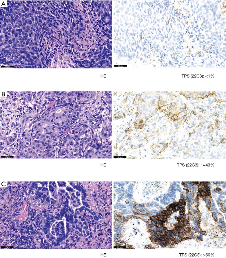
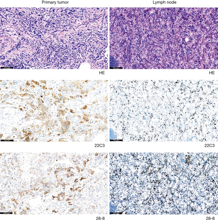
Methods: PD-L1 expression was evaluated in cells from primary tumors and paired metastatic lymph nodes using the 22C3 and 28-8 IHC assays. Total 35 patients with primary tumor and paired metastatic lymph node were enrolled into this study, and all samples were surgically resected, formalin-fixed, and paraffin-embedded NSCLC tissues. Tumor cells exhibiting complete or partial membrane staining, were considered as PD-L1 positive. On the basis of tumor proportion score (TPS), all samples were classified as no expression (TPS: <1%), low expression (TPS: 1-49%), or high expression (TPS: ≥50%).
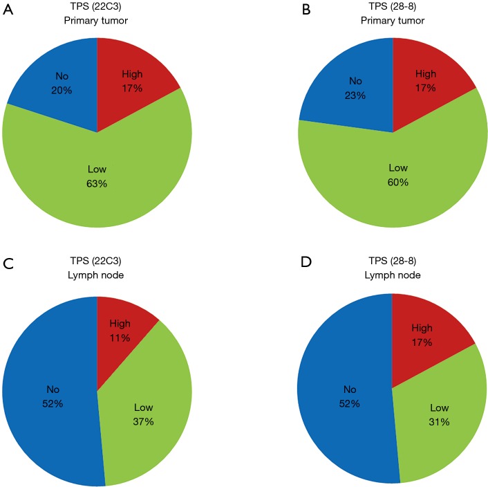
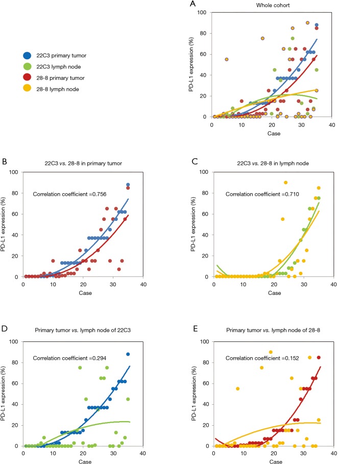
Results: TPS distribution was markedly different between primary tumors and paired metastatic lymph nodes. In 22C3 IHC assay, TPS similar to that of metastatic lymph nodes was demonstrated in 10 primary tumors, and concordance rate between them was 28.6%. Concurrently, in 28-8 IHC assay, 11 primary tumors had TPS similar to that of metastatic lymph nodes, with a concordance rate of 31.4%.
Conclusions: TPS concordance rates (for both 22C3 and 28-8 antibodies) between primary tumors and paired lymph nodes were low. Inter-tumor heterogeneity of PD-L1 expression is an important issue for clinical oncologists during treatment planning.
Keywords: Lung cancer; heterogeneity; immunotherapy; programmed death-ligand 1 (PD-L1); tumor proportion score (TPS).
2019 Journal of Thoracic Disease. All rights reserved.
Conflict of interest statement
Conflicts of Interest: The authors have no conflicts of interests to declare.
Figures
Comment in
-
Inter-tumor heterogeneity of PD-L1 status: is it important in clinical decision making?J Thorac Dis. 2020 May;12(5):1770-1775. doi: 10.21037/jtd-20-1661. J Thorac Dis. 2020. PMID: 32642082 Free PMC article. No abstract available.
-
Programmed death-ligand 1 expression discrepancy between primary tumor and metastatic lymph nodes in non-small cell lung cancer.J Thorac Dis. 2020 Aug;12(8):3918-3920. doi: 10.21037/jtd.2020.04.45. J Thorac Dis. 2020. PMID: 32944298 Free PMC article. No abstract available.
References
LinkOut - more resources
Full Text Sources
Other Literature Sources
Research Materials