. 2020 Jan 29:8:e8294.
doi: 10.7717/peerj.8294. eCollection 2020.

The immunostimulatory activity of polysaccharides from Glycyrrhiza uralensis

Affiliations

The immunostimulatory activity of polysaccharides from Glycyrrhiza uralensis

Adila Aipire et al. PeerJ. .

Abstract

Background: The enhancement of immunity is very important for immunocompromised patients such as cancer patients with radiotherapy or chemotherapy. Glycyrrhiza uralensis has been used as food and medicine for a long history. G. uralensis polysaccharides (GUPS) were prepared and its immunostimulatory effects were investigated.

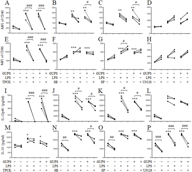
Methods: Human monocyte-derived dendritic cells (DCs) and murine bone marrow-derived DCs were treated with different concentrations of GUPS. The DCs maturation and cytokine production were analyzed by flow cytometry and ELISA, respectively. Inhibitors and Western blot were used to study the mechanism of GUPS. The immunostimulatory effects of GUPS were further evaluated by naïve mouse model and immunosuppressive mouse model induced by cyclophosphamide.

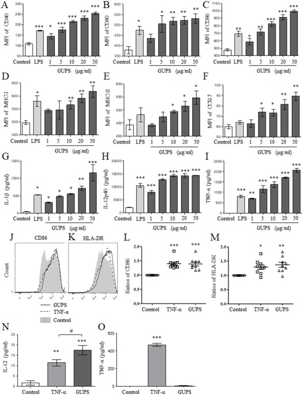
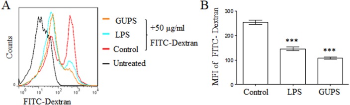
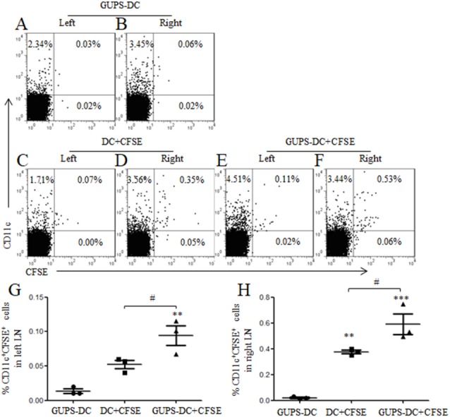
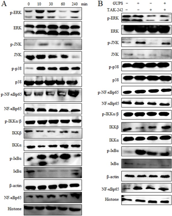
Results: GUPS significantly promoted the maturation and cytokine secretion of human monocyte-derived DCs and murine bone marrow-derived DCs through TLR4 and down-stream p38, JNK and NF-κB signaling pathways. Interestingly, the migration of GUPS treated-DCs to lymph node was increased. In the mouse model, GUPS increased IL-12 production in sera but not for TNF-α. Moreover, GUPS ameliorated the side effect of cyclophosphamide and improved the immunity of immunosuppressive mice induced by cyclophosphamide. These results suggested that GUPS might be used for cancer therapy to ameliorate the side effect of chemotherapy and enhance the immunity.

Keywords: Cytokine production; Dendritic cell; Glycyrrhiza uralensis polysaccharides; Immunity; Immunosuppressive mouse model; Maturation; Migration; Signaling pathway.


Conflict of interest statement

The authors declare there are no competing interests.

Figures

Figure 1
Figure 1. The phenotype and cytokine secretion of DCs.
(A–F) BM-DCs were treated with different concentrations (1, 5, 10, 20 and 50 µg/ml) of GUPS for 12 h. The expression of surface molecules on DCs was detected by flow cytometry. The mean fluorescence intensity (MFI) (mean ± SEM) of these molecules is shown. (G–I) The cytokine production in supernatant of BM-DCs was detected by ELISA. Data are from 3 independent experiments. (J–L) Mo-DCs were treated with 20 µg/ml of GUPS for 18 h. The expression of CD86 and HLA-DR on DCs was detected by flow cytometry. The MFI (mean ± SEM) of CD86 and HLA-DR is shown. (M–N) The cytokine production in supernatant of Mo-DCs was detected by ELISA. Data were analyzed by ANOVA. p < 0.05; ∗∗ p < 0.01; ∗∗∗ p < 0.001 compared to untreated DC. # p < 0.05 compared to TNF- α treated DC.
Figure 2
Figure 2. GUPS suppressed the phagocytosis of DCs.
DCs were treated with GUPS or LPS for 12 h, then co-cultured with FITC-Dextran for 1 h. (A) The fluorescence of FITC in DCs was analyzed by flow cytometry. (B) MFI of FITC was shown. Data were analyzed by ANOVA. ∗∗∗ p < 0.001 compared to Control.
Figure 3
Figure 3. DC migration in vivo.
DCs were treated with or without 20 µg/ml of GUPS and stained with CFSE. Cells were subcutaneously injected to the right flank. GUPS-DCs without CFSE staining were used as control. After 24 h, lymphocytes in both right and left inguinal LN were isolated. (A–F) CFSE + DCs were analyzed by flow cytometry. (G–H) Percentages of CD11c +CFSE + cells in left and right LN were shown, respectively. Data are from 2 independent experiments and the representative data are shown and analyzed by ANOVA. ∗∗ p < 0.01; ∗∗∗ p < 0.001 compared to GUPS-DCs.
Figure 4
Figure 4. The effect of TLR4 and TLR2 blockade on DC maturation and cytokine production.
DCs were pretreated with or without 1 µM TAK-242 or 100 ng/ml TLR2 mAb for 1 h, and then treated with GUPS (20 µg/ml) or LPS for 12 h. (A–F) The expression of CD40 and CD86 were detected by flow cytometry. (G–J) MFI of CD40 and CD86 was shown. (K–N) The levels of IL-12 and TNF- α in supernatant were tested by ELISA. Data are from 3 independent experiments. ∗∗ p < 0.01; ∗∗∗ p < 0.001 compared to untreated DCs (ANOVA). ### p < 0.001 compared to TAK-242 treated DCs (paired t-test).
Figure 5
Figure 5. The activation of MAPK and NF- κ B signaling pathways.
(A) Nuclear and cytoplasmic proteins were isolated at the indicated time points from DCs treated with 20 µg/ml of GUPS. The levels of protein and their phosphorylation in cytoplasm or nuclei were detected by Western blot. (B) DCs were pretreated with TAK-242 for 1 h, then treated with 20 µg/ml of GUPS for 10 min. Nuclear and cytoplasmic proteins were prepared and detected by Western blot.
Figure 6
Figure 6. The effect of MAPK and NF-κ B blockade on DC maturation and cytokine production.
DCs were pretreated with or without 10 µg/ml TPCK, 10 µM SB, 25 µM SP or 10 µM U0126 for 1 h, and then treated with GUPS (20 µg/ml) or LPS for 12 h. (A–H) The expression of CD40 and CD86 were detected by flow cytometry. MFI of CD40 and CD86 was shown. (I–P) The concentrations of IL-12p40 and IL-10 in supernatant were measured by ELISA. p < 0.05; ∗∗ p < 0.01; ∗∗∗ p < 0.001 compared to untreated DCs (ANOVA). # p < 0.05; ## p < 0.01; ### p < 0.001 compared to inhibitor treated DCs (paired t-test).
Figure 7
Figure 7. The effect of GUPS on naïve mice. Naïve BALB/c mice were injected with GUPS or LPS intraperitoneally.
(A–B) The production of IL-12 and TNF-α in sera after 3 and 6 h. (C–F) The organs of mice after 3 days of injection. p < 0.05; ∗∗ p < 0.01; ∗∗∗ p < 0.001 compared to control group (ANOVA).
Figure 8
Figure 8. The effect of GUPS on immunosuppressive mouse model.
Immunosuppressive mouse model was induced by CTX and treated with GUPS or λ-CGN. (A) The body weight of mice. (B–F) The proportions of immune cells in spleens of mice. On day 11, spleens were isolated to analyze the proportions of immune cells by flow cytometry. (G–J) The proportions and activation of CD4+ and CD8+ T cells in spleens of mice. ∗∗ p < 0.05; ∗∗ p < 0.01; ∗∗∗ p < 0.001 compared to control group (ANOVA). # p < 0.05; ## p < 0.01 compared to model group (ANOVA).

References

    1. Aipire A, Li J, Yuan P, He J, Hu Y, Liu L, Feng X, Li Y, Zhang F, Yang J, Li J. Glycyrrhiza uralensis water extract enhances dendritic cell maturation and antitumor efficacy of HPV dendritic cell-based vaccine. Scientific Reports. 2017;7:43796. doi: 10.1038/srep43796. - DOI - PMC - PubMed
    1. Bhattacharyya S, Liu H, Zhang Z, Jam M, Dudeja PK, Michel G, Linhardt RJ, Tobacman JK. Carrageenan-induced innate immune response is modified by enzymes that hydrolyze distinct galactosidic bonds. Journal of Nutritional Biochemistry. 2010;21:906–913. doi: 10.1016/j.jnutbio.2009.07.002. - DOI - PMC - PubMed
    1. Carreno BM, Becker-Hapak M, Huang A, Chan M, Alyasiry A, Lie WR, Aft RL, Cornelius LA, Trinkaus KM, Linette GP. IL-12p70-producing patient DC vaccine elicits Tc1-polarized immunity. The Journal of Clinical Investigation. 2013;123:3383–3394. doi: 10.1172/JCI68395. - DOI - PMC - PubMed
    1. Chen J, Li WC, Gu XL. Optimized extraction, preliminary characterization, and in vitro antioxidant activity of polysaccharides from glycyrrhiza uralensis fisch. Medical Science Monitor. 2017;23:1783–1791. doi: 10.12659/MSM.900471. - DOI - PMC - PubMed
    1. Chen X, Nie W, Fan S, Zhang J, Wang Y, Lu J, Jin L. A polysaccharide from Sargassum fusiforme protects against immunosuppression in cyclophosphamide-treated mice. Carbohydrate Polymers. 2012;90(12):1114–1119. doi: 10.1016/j.carbpol.2012.06.052. - DOI - PubMed