Review
. 2020 Jul;160(3):233-247.
doi: 10.1111/imm.13176. Epub 2020 Feb 27.

V(D)J recombination, somatic hypermutation and class switch recombination of immunoglobulins: mechanism and regulation

Affiliations
Review

V(D)J recombination, somatic hypermutation and class switch recombination of immunoglobulins: mechanism and regulation

Xiying Chi et al. Immunology. 2020 Jul.

Abstract

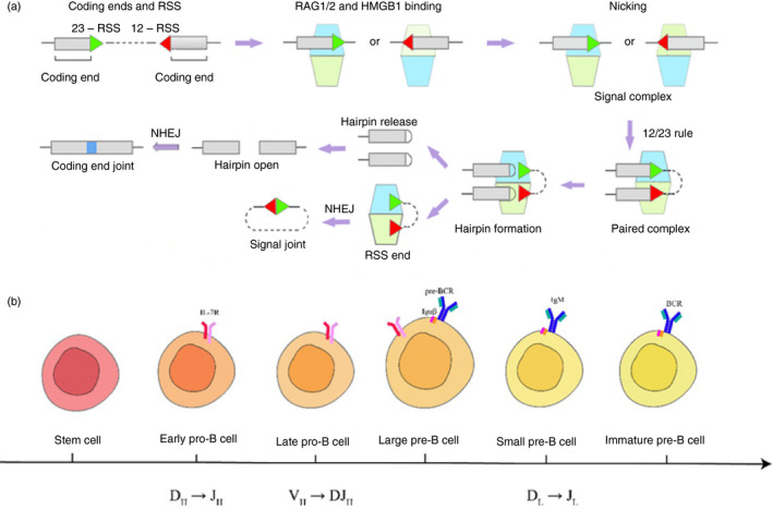
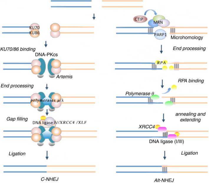
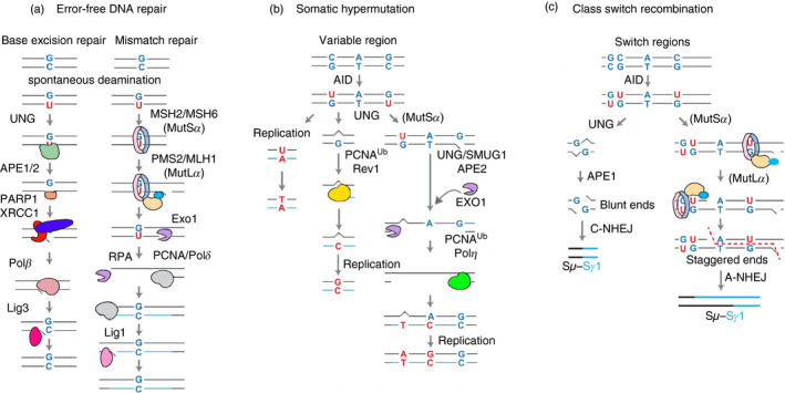
Immunoglobulins emerging from B lymphocytes and capable of recognizing almost all kinds of antigens owing to the extreme diversity of their antigen-binding portions, known as variable (V) regions, play an important role in immune responses. The exons encoding the V regions are known as V (variable), D (diversity), or J (joining) genes. V, D, J segments exist as multiple copy arrays on the chromosome. The recombination of the V(D)J gene is the key mechanism to produce antibody diversity. The recombinational process, including randomly choosing a pair of V, D, J segments, introducing double-strand breaks adjacent to each segment, deleting (or inverting in some cases) the intervening DNA and ligating the segments together, is defined as V(D)J recombination, which contributes to surprising immunoglobulin diversity in vertebrate immune systems. To enhance both the ability of immunoglobulins to recognize and bind to foreign antigens and the effector capacities of the expressed antibodies, naive B cells will undergo class switching recombination (CSR) and somatic hypermutation (SHM). However, the genetics mechanisms of V(D)J recombination, CSR and SHM are not clear. In this review, we summarize the major progress in mechanism studies of immunoglobulin V(D)J gene recombination and CSR as well as SHM, and their regulatory mechanisms.

Keywords: B cell; V(D)J recombination; class switch recombination; mechanism; somatic hypermutation.


Figures

Figure 1
Figure 1
Overview of V(D)J recombination. (a) The RAG1–RAG2 complex is shown as light blue and light green trapezoids. Briefly, RAG binds a single recombination signal sequence (RSS) in the presence of HMGB1 to form a 12 or 23 signal complex. Another RSS is captured to form a paired complex. The DNA is cleaved, generating hairpin coding ends and RSS ends with a 3′ hydroxyl (OH) group in the presence of Mg2+. The hairpin is released and opened and rejoined by non‐homologous end joining (NHEJ), resulting in imprecise coding joints that contain added nucleotides (blue bars). RSS ends are processed through the NHEJ pathway as well, creating a signal joint. (b) B‐cell ontogeny during V(D)J recombination. This figure shows the chronological order of B cells in different stages of development in the bone marrow. B cells progress from stem cells to pro‐B cells, pre‐B cells and immature pre‐B‐cell stages. During this differentiation, V(D)J rearrangement of heavy chains occurs in pro‐B‐cell periods, resulting in the expression of pre‐B‐cell receptors (pre‐BCR), which are composed of Igμ and surrogate light chains (composed of VpreB and λ‐5). After receiving the pre‐BCR signaling, the light chains rearrange in small pre‐B cells, resulting in the expression of a mature BCR (composed by rearranged heavy and light chains).
Figure 2
Figure 2
A model for canonical non‐homologous end joining (C‐NHEJ) and alternate NHEJ (Alt‐NHEJ). C‐NHEJ (left) and Alt‐NHEJ (right) are shown. For C‐NHEJ, Ku70/80 binds to the double strand breaks (DSB) first and protects DNA ends from digestion. The Ku:DNA complex recruits DNA‐dependent protein kinase catalytic subunit (DNA‐PKcs) in complex with Artemis, generating DNA‐PK. DNA‐PKcs undergoes autophosphorylation and activates Artemis, which then gains various nuclease activities, ensuring the DSB are compatible by resecting damaged DNA or non‐ligatable end groups. The gap created is filled by Polμ/λ recreated by Ku. DNA‐PK also facilitates recruitment of DNA ligase IV, XRCC4 and XLF to complete the ligation of DSB. For Alt‐NHEJ, PARP1 recognizes the broken DNA ends and recruits Mre11‐Rad50‐Nbs1 (MRN) and CtIP complex to initiate 5′‐3′ DNA resection, creating ssDNA overhangs. Resection exposes microhomology internal to break sites, facilitating spontaneous annealing of ssDNA. The binding of replication protein A (RPA) to the ssDNA overhangs removes secondary structure and prevents annealing of overhangs, which hinders Alt‐NHEJ. Polθ‐helicase acts as an ATP‐dependent annealing helicase that dissociates RPA to promote DNA annealing and stimulate Alt‐NHEJ. The paired ssDNA overhangs are subsequently extended by Po lθ‐polymerase. Finally, end rejoining is carried out by the DNA ligase I or III (Lig I or Lig III)/XRCC1 complex.
Figure 3
Figure 3
Overview of error‐free basic excision repair (BER) and mismatch repair (MMR), somatic hypermutation (SHM) and class switching recombination (CSR). (a) Error‐free repair pathways for BER (left) and MMR (right). Canonical BER is initiated by uracil‐DNA glycosylase, creating an abasic site that is recognized by APE1/2. APE1/2 nick the DNA 5′ of the abasic site. PARP1 and XRCC1 are activated and recruit proliferating cell nuclear antigen (PCNA) and polymerase β (Polβ). Polβ removes the remaining 5′ deoxyribose and insert a single nucleotide, followed by ligation with Lig3. In MMR, MutSα recognizes the U:G mispair and recruits MutLα. MutLα nicks the DNA 5′ of the mismatch via PMS2. Exo1 creates an ssDNA from the nick going past the mismatch site. PCNA subsequently recruits Polδ to fill over the gap, following ligation by Lig1. (b) During SHM, uracil can act as a template for replication leading to a C–T transition mutation. Alternatively, non‐canonical BER or MMR recruit low‐fidelity polymerases Polη through PCNA ubiquitination (PCNA‐Ub) leads to transition or transversion mutations. (c) For CSR, non‐canonical BER can lead to double strand breaks (DSB) when two uracils in opposite strands are closely spaced. MMR can process distantly spaced uracils, leading to staggered DSB. Blunt DSB are joined by canonical non‐homologous end joining, whereas staggered breaks are repaired by alternative non‐homologous end joining.
Figure 4
Figure 4
Chromosome looping and R‐loop. (a) This picture shows the structure of the chromosome looping, and each colorful circle representing the specific S‐C loci. Eμ and 3′Eα interact, causing Sμ and the downstream S regions located in a chromosomal loop. (b) After the Igε transcription promoter (located at the upstream of Igε acceptor S region) activated, Sε‐Cε loci are close to the 3′Eα segment and Eμ‐Sμ‐Cμ, inducing the B cell to switch to IgE. (c) Structures that recruit activation‐induced cytidine deaminase (AID): the switch sequence has a high G‐richness on the non‐template chain. During transcription, these G‐rich regions form secondary structures such as the R‐loop, G‐quadruplexes (G4) and G‐loop, which help recruit AIDs in the S region. (d) DDX1 promotes R‐loop formation: RNA polymerase II (Pol II) produces switch transcripts, whose intron contains the G4. After a splicing step, the intron lariat intermediate is debranched by RNA lariat (intron) debranching (DBR1) enzyme. Bound G4–AID complex, DDX1 targets to the S‐region DNA. At last G4 is resolved, and an R‐loop is formed in the S region.

References

    1. Bashford‐Rogers RJM, Smith KGC, Thomas DC. Antibody repertoire analysis in polygenic autoimmune diseases. Immunology 2018; 155:3–17. - PMC - PubMed
    1. Boyd SD, Joshi SA. High‐throughput DNA sequencing analysis of antibody repertoires. Microbiol Spectr 2014; 2:345–62. - PubMed
    1. Hwang JK, Alt FW, Yeap LS. Related mechanisms of antibody somatic hypermutation and class switch recombination. Microbiol Spectr 2015; 3:MDNA3‐0037‐2014. - PMC - PubMed
    1. Xu Z, Zan H, Pone EJ, Mai T, Casali P. Immunoglobulin class‐switch DNA recombination: induction, targeting and beyond. Nat Rev Immunol 2012; 12:517–31. - PMC - PubMed
    1. Carmona LM, Schatz DG. New insights into the evolutionary origins of the recombination‐activating gene proteins and V(D)J recombination. FEBS J 2017; 284:1590–605. - PMC - PubMed

Publication types

MeSH terms

LinkOut - more resources