. 2020 May;108(5):1186-1202.
doi: 10.1002/jbm.a.36893. Epub 2020 Feb 26.

Biodistribution and toxicity of epitope-functionalized dextran iron oxide nanoparticles in a pregnant murine model

Affiliations

Biodistribution and toxicity of epitope-functionalized dextran iron oxide nanoparticles in a pregnant murine model

Amir Bolandparvaz et al. J Biomed Mater Res A. 2020 May.

Abstract

In pursuit of a preventive therapeutic for maternal autoantibody-related (MAR) autism, we assessed the toxicity, biodistribution, and clearance of a MAR specific peptide-functionalized dextran iron oxide nanoparticle system in pregnant murine dams. We previously synthesized ~15 nm citrate-coated dextran iron oxide nanoparticles (DIONPs), surface-modified with polyethylene glycol and MAR peptides to produce systems for nanoparticle-based autoantibody reception and entrapments (SNAREs). First, we investigated their immunogenicity and MAR lactate dehydrogenase B antibody uptake in murine serum in vitro. To assess biodistribution and toxicity, as well as systemic effects, we performed in vivo clinical and post mortem pathological evaluations. We observed minimal production of inflammatory cytokines-interleukin 10 (IL-10) and IL-12 following in vitro exposure of macrophages to SNAREs. We established the maximum tolerated dose of SNAREs to be 150 mg/kg at which deposition of iron was evident in the liver and lungs by histology and magnetic resonance imaging but no concurrent evidence of liver toxicity or lung infarction was detected. Further, SNAREs exhibited slower clearance from the maternal blood in pregnant dams compared to DIONPs based on serum total iron concentration. These findings demonstrated that the SNAREs have a prolonged presence in the blood and are safe for use in pregnant mice as evidenced by no associated organ damage, failure, inflammation, and fetal mortality. Determination of the MTD dose sets the basis for future studies investigating the efficacy of our nanoparticle formulation in a MAR autism mouse model.

Keywords: MAR autism; clearance; distribution; histopathology; nanoformulation; peptide-functionalized.


Conflict of interest statement

CONFLICT OF INTEREST

All other authors have no conflict of interest to declare.

Figures
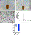
FIGURE 1
FIGURE 1
Physico-chemical characterization of iron oxide nanoparticles and SNAREs. Images of (a) DIONPs and (b) SNAREs at 1 mg/mL dispersed in water. (c) Transmission Electron Microscopy (TEM) of DIONPs. (d) Dynamic Light Scattering (DLS) of DIONPs. (e) The zeta potential of 1 mg/ml of DIONPs and SNAREs in water at physiological temperature
FIGURE 2
FIGURE 2
Investigating IL-10 and IL-12 release from bone marrow-derived macrophages in the presence of DIONPs and SNAREs. (a) ELISA on treatment supernatants and immature macrophages demonstrated significantly lower secretion of IL-10 compared to LPS-treated macrophages at 24 and 48 hr. (b) Supernatant from immature macrophages as well as DIONP and SNARE treated cultures showed lowered secretion of IL-12 compared to LPS-treated cells at same time points. The mean of every treatment was compared to LPS treatments and significance is denoted by (****) for p < .0001
FIGURE 3
FIGURE 3
Capture of MAR LDH B autoantibody from mouse serum. SNAREs significantly reduced LDH B antibody titer in mouse serum by capturing up to 87% of LDH B antibodies in vitro. Scrambled peptide-modified DIONPs and plain DIONPs served as negative controls. Data shown represent mean ± standard error (n = 3 biological replicates). The mean of every treatment was compared to the control and significance is denoted by (*) for p < .05 and (***) for p < .001
FIGURE 4
FIGURE 4
Maternal and Fetal toxicity of SNAREs and DIONPs in pregnant dams. (a) Weight gain of a subset of pergnant dams (n = 55) from GD 0 to GD 12 compared to unmated control (n = 7). Weight gain was used as a second confimatory parameter asides from observing copulation plug to establish timed pregnancies. The weight of pregnant dams were signficiantly different at GD 17 compared to the control. (b) Maternal weight gain determined by subtracting the weight of the dam at GD 0 and uteri on GD 17 from the weight of the dam at GD 17. (c) Frequency of fetal resorption calculated as number of resorption/number of fetuses of treated compared to control mice. Legend applies to graphs b,c
FIGURE 5
FIGURE 5
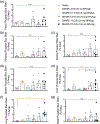
Serum biochemical composition of pregnant mice treated with SNAREs and DIONPs. Serum composition of total Bilirubin, Alanine Transaminase (ALT), Creatinine, Blood Urea Nitrogen (BUN), and total Iron of treated and control mice assessing liver and kidney functionalities. Blood was collected via cardiac puncture on GD 17 and serum separated via centrifugation in gel separator yellow-capped tubes. (a) Total bilirubin (range: 0–0.2 mg/dl). (b) Alanine transaminase (ALT) (range: 0–403 U/L). (c) Creatinine (range: 0–0.3 mg/dl). (d) Blood urea nitrogen (range: 15.2–34.7 mg/dl). (e) Total iron (range: 176–347 μg/dl). Legend applies to all graphs. Ranges shown grey dervied from nonpregnant BALB/c strain mice from UCD Pathology lab
FIGURE 6
FIGURE 6
Hematological analysis of SNARE and DIONP-treated mice. Blood count of white blood cells (WBC), red blood cells (RBC), and hemoglobin (Hgb) of treated and control mice. Blood was collected via cardiac Puncture on GD 17 and serum separated via centrifugation in gel separator yellow-capped tubes. (a) WBC, (b) RBC, (c) Hgb. Legend applies to all graphs. Reference range for nonpregnant C57BL/6j shown in grey (Santos et al., 2016)
FIGURE 7
FIGURE 7
Percent body weight of major maternal organs upon necropsy on gestational day 17. Percent body weight on GD 17 of uteri, liver, kidneys, spleen of treated and control mice assessing toxicity-related weight changes in major organs involved in clearance of iron oxide nanoparticles. (a) Uteri, (b) Liver, (c) Kidneys, (d) Spleen. Legend applies to all graphs
FIGURE 8
FIGURE 8
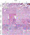
Representative histological images of major organs and their grades. In Grade 0, no iron aggregates are present in any of the organs shown. Hepatocytes are occasionally binucleated (inset), which is a typical feature of the murine liver. In the spleen, physiological iron accumulation is shown in the macrophage of the red pulp (inset). In Grade I, one iron aggregate was detected per ×20 power field. In the lung, these aggregates were present within pulmonary capillaries (inset). In the liver, iron deposits were primarily observed within Kupffer cells (inset). In the heart, rare iron aggregates were present within coronary capillaries. In the kidney, iron aggregates were rarely seen within glomerular tuft capillaries (inset). In the placenta, iron aggregates were observed within intervillous blood channels. In Grade II, there were 2–3 iron aggregates per ×20 power field. In the lung, these aggregates were present only within pulmonary capillaries (inset). In the liver, aggregates were associated with Kupffer cells but occasionally were also observed within the sinusoids. In the heart, the aggregates were restricted to the coronary capillaries (inset and arrow). In the kidney, aggregates were primarily observed within glomerular tuft capillaries (arrow) and rarely within renal vasculature (inset). In Grade III, >3 iron aggregates were observed per ×20 power field, with no evidence of inflammation, cellular damage, or histomorphological change to the organ architecture. In the lung, multiple pulmonary capillaries are distended and occluded by large iron aggregates (arrows and inset). In the liver, Kupffer cells are enlarged with phagocytosed iron (inset), and occasional sinusoids contain cigar-shaped aggregates (arrows). In the heart, the iron aggregates are present within coronary capillaries (inset) but occasionally within the ventricles (not shown). In the kidney, the aggregates are observed within glomerular tufts (inset) capillaries and sometimes within vasa recta (arrow). The spleen contains abundant and above physiological iron deposits within rep pulp macrophages (inset and arrows). In the placental sections, variably sized iron aggregates are present within intervillous spaces (inset and arrows). The criteria of Grade IV and V included evidence of inflammation and inflammation associated with gross or histological alteration of organ morphology, respectively. In this study, no Grades IV or V were assigned to any of the examined tissues. Staining: Hematoxylin and eosin, Bar = 50 μm, and 10 μm in the insets
FIGURE 9
FIGURE 9
Histological grading for the major maternal organs at the different dosages. The grades were assigned based on acute inflammation and potential tissue damage. There is significant accumulation at the MTD dose and thus highest grading. Data shown represent mean ± standard error (n ≤ 3 biological replicates). The mean of every treatment was compared to the control group and significance is denoted by (**) for p < .01 and (****) for p < .0001
FIGURE 10
FIGURE 10
Prussian Blue iron stain of maternal organs to determine organ iron accumulation. Prussian Blue iron stain and a nuclear fast red counter-stain of maternal organs comparing treatments versus saline control. Tissues were embedded, sectioned, and stained for iron after necropsy on gestational day 17. There was a general accumulation of iron in the maternal organs at the 30 and 150 mg NPs/kg of SNAREs as well as 30 mg NPs/kg DIONPs. There was a significant increase of iron in the maternal liver and the lungs at the MTD dose of 150 mg NPs/kg compared to the saline-treated dams. (a) Fetus, (b) placenta, (c) kidneys, (d) spleen, (e) liver, (f) lungs, (g) heart
FIGURE 11
FIGURE 11
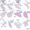
Representative images of Prussian Blue iron stain of maternal organs and fetus. Representative images (×4 magnification) of Prussian Blue iron stain and a nuclear fast counter stain of maternal organs and fetus comparing 150 mg NPs/kg SNARE treatment versus saline control. Arrows point to the accumulation of iron in the organs. Tissues were embedded, sectioned, and stained for iron after necropsy on GD 17. There was a general accumulation of iron at the 150 mg NPs/kg of SNAREs compared to the saline control for the different organs with a significant increase for the liver and the lungs compared to the saline control. (a) Fetus, (b) placenta, (c) kidneys, (d) spleen (E) liver (F) lungs
FIGURE 12
FIGURE 12
Representative images of Prussian Blue iron stain of maternal lungs and liver. Representative images (×4, ×10) of Prussian Blue iron stain and a nuclear fast counter stain of maternal organs comparing 150 mg NPs/kg SNARE treatment versus saline-treated control. Arrows point to the accumulation of iron in the organs. Tissues were embedded, sectioned, and stained for iron after necropsy on GD 17. There was a general accumulation of iron at the 150 mg NPs/kg of SNAREs compared to the saline-treated control for the different organs with a significant increase in the liver and the lungs. (a) ×4 Magnification liver, (b) ×4 magnification lungs, (c) ×10 magnification liver, (d) ×20 magnification lungs
FIGURE 13
FIGURE 13
Distribution of intravenously injected SNAREs in pregnant dams via MRI. Pregnant dams were administered with 30 and 100 mg NPs/kg SNAREs, slightly lower doses than the MTD (150 mg NPs/kg). Distribution of NPs in the maternal liver (a) and kidney (b) as well as fetuses (c) was investigated at 24, 48 hr and 5 days postinjection via MRI. The liver demonstrated significant accumulation of NPs at both doses by 4 hr and persisted up to 5 days. Kidneys exhibited significant deposition at only the higher dose by 4 hr and remained present up to 24 hr, but the large size of the fetuses by Day 5 blocked MRI acquisition of the kidneys. Data were analyzed using Slicer software and mean intensity normalized to a water capillary tube to ensure comparability across images. Data shown represent mean ± standard error (n ≥ 3 biological replicates). The mean of every treatment was compared to each other and the controls and significance is denoted by (*) for p < .05 and (****) for p < .0001
FIGURE 14
FIGURE 14
Clearance of intravenously injected SNAREs in pregnant dams. Tail vein blood was collected at 30 min, 1, 2, 4, 8, 24 hr time points and total serum iron concentration determined (a). SNARE-treated dams demonstrated higher total serum iron concentration and thus slower clearance of NPs compared to DIONP-injected at earlier time points up to 8 hr postinjection. By 24 hr, total iron concentration for saline controls were similar to SNARE treated dams. (b) Area under the curve (AUC) demonstrated larger area and thus slower clearance of SNARE compared to DIONP

References

    1. Ali Noori KP, Modaresi M, Messripour M, Yousefi MH, & Amiri GR (2011). Effect of magnetic iron oxide nanoparticles on pregnancy and testicular development of mice. African Journal of Biotechnology, 10(7), 1221–1227.
    1. Athie-Morales V, Smits HH, Cantrell DA, & Hilkens CM (2004). Sustained IL-12 signaling is required for Th1 development. Journal of Immunology, 172(1), 61–69. - PubMed
    1. Banobre-Lopez M, Teijeiro A, & Rivas J (2013). Magnetic nanoparticle-based hyperthermia for cancer treatment. Reports of Practical Oncology and Radiotherapy, 18(6), 397–400. - PMC - PubMed
    1. Biederer J, Beer M, Hirsch W, Wild J, Fabel M, Puderbach M, & van Beek EJR (2012). MRI of the lung (2/3). Why … when … how? Insights Imaging, 3(4), 355–371. - PMC - PubMed
    1. Bolandparvaz A, Harriman R, Alvarez K, Lilova K, Zang Z, Lam A, … Lewis J (2019). Towards a nanoparticle-based prophylactic for maternal autoantibody-related autism. Nanomedicine, 21, 102067. - PMC - PubMed

Publication types