. 2020 Feb 7;16(2):e1008597.
doi: 10.1371/journal.pgen.1008597. eCollection 2020 Feb.

Skp, Cullin, F-box (SCF)-Met30 and SCF-Cdc4-Mediated Proteolysis of CENP-A Prevents Mislocalization of CENP-A for Chromosomal Stability in Budding Yeast

Affiliations

Skp, Cullin, F-box (SCF)-Met30 and SCF-Cdc4-Mediated Proteolysis of CENP-A Prevents Mislocalization of CENP-A for Chromosomal Stability in Budding Yeast

Wei-Chun Au et al. PLoS Genet. .

Abstract

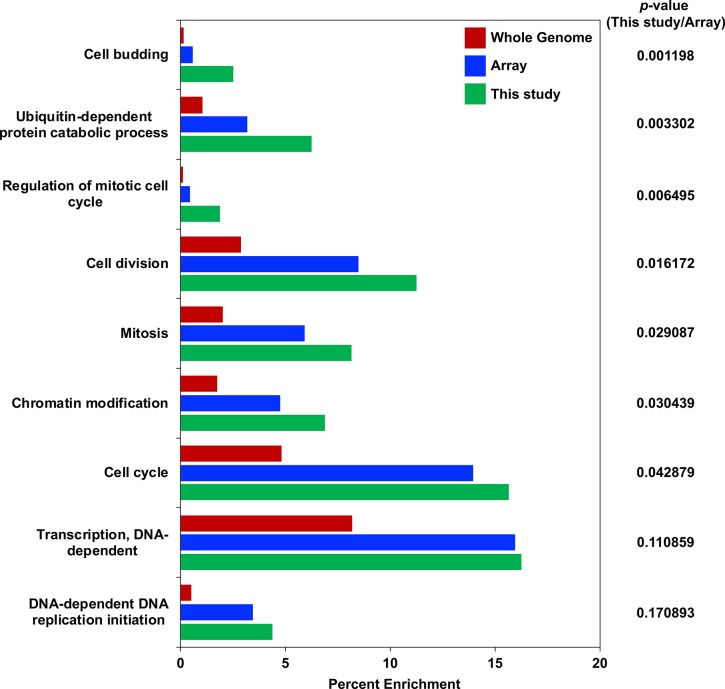
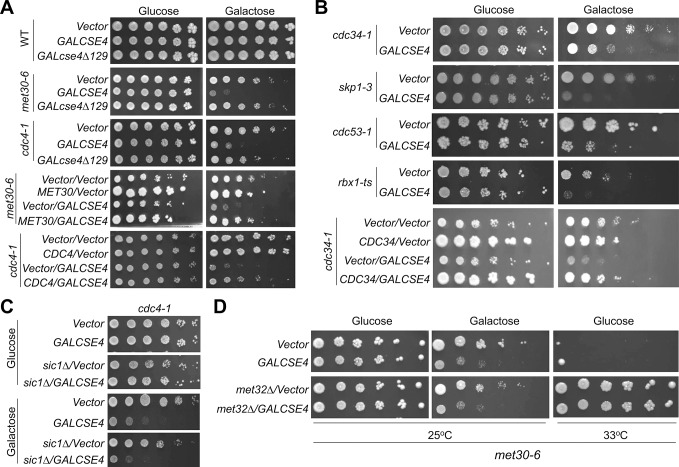
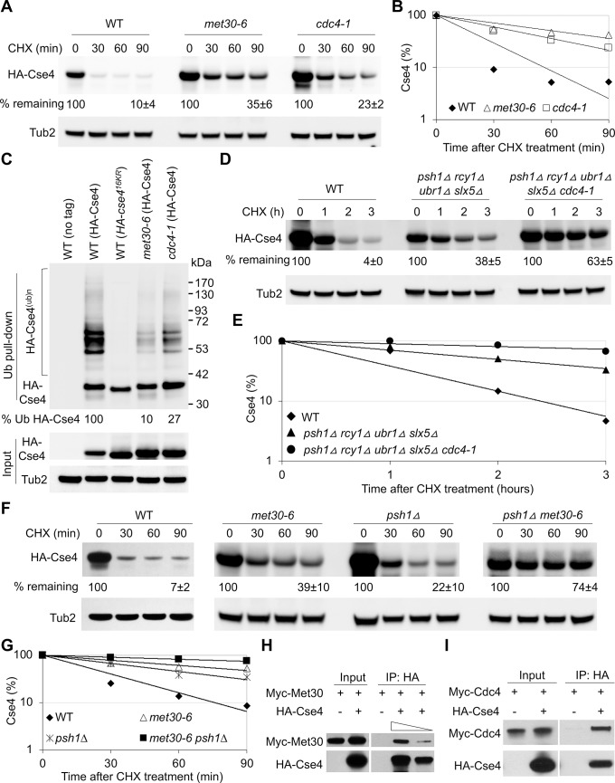
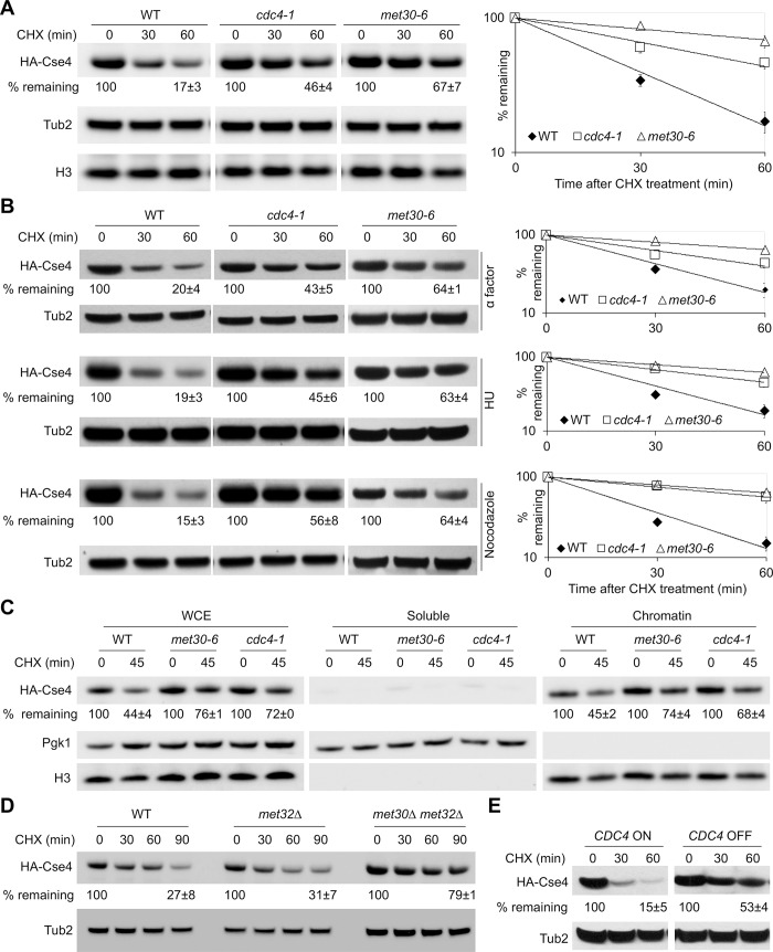
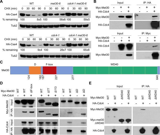
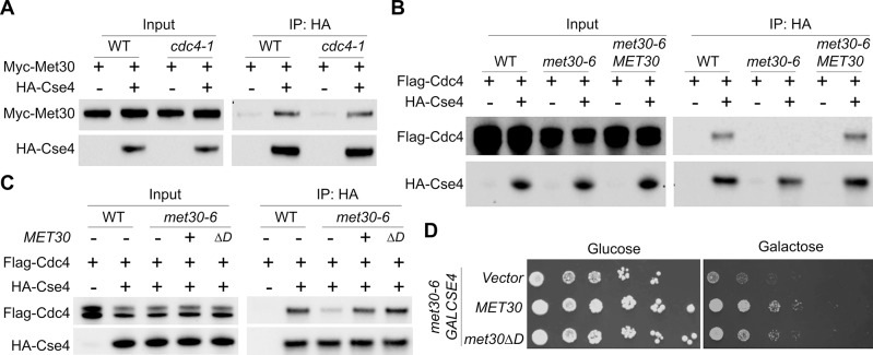
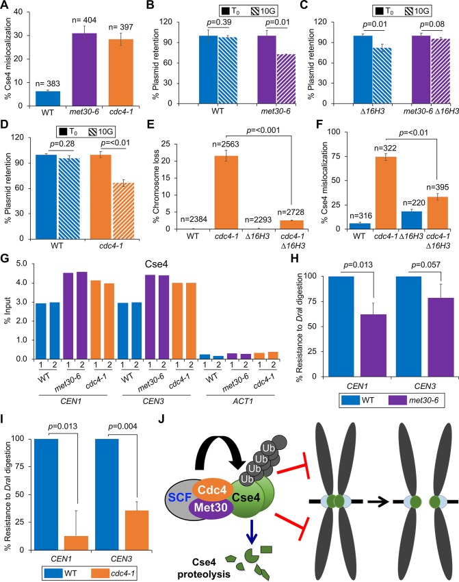
Restricting the localization of the histone H3 variant CENP-A (Cse4 in yeast, CID in flies) to centromeres is essential for faithful chromosome segregation. Mislocalization of CENP-A leads to chromosomal instability (CIN) in yeast, fly and human cells. Overexpression and mislocalization of CENP-A has been observed in many cancers and this correlates with increased invasiveness and poor prognosis. Yet genes that regulate CENP-A levels and localization under physiological conditions have not been defined. In this study we used a genome-wide genetic screen to identify essential genes required for Cse4 homeostasis to prevent its mislocalization for chromosomal stability. We show that two Skp, Cullin, F-box (SCF) ubiquitin ligases with the evolutionarily conserved F-box proteins Met30 and Cdc4 interact and cooperatively regulate proteolysis of endogenous Cse4 and prevent its mislocalization for faithful chromosome segregation under physiological conditions. The interaction of Met30 with Cdc4 is independent of the D domain, which is essential for their homodimerization and ubiquitination of other substrates. The requirement for both Cdc4 and Met30 for ubiquitination is specifc for Cse4; and a common substrate for Cdc4 and Met30 has not previously been described. Met30 is necessary for the interaction between Cdc4 and Cse4, and defects in this interaction lead to stabilization and mislocalization of Cse4, which in turn contributes to CIN. We provide the first direct link between Cse4 mislocalization to defects in kinetochore structure and show that SCF-mediated proteolysis of Cse4 is a major mechanism that prevents stable maintenance of Cse4 at non-centromeric regions, thus ensuring faithful chromosome segregation. In summary, we have identified essential pathways that regulate cellular levels of endogenous Cse4 and shown that proteolysis of Cse4 by SCF-Met30/Cdc4 prevents mislocalization and CIN in unperturbed cells.


Conflict of interest statement

NO authors have competing interests.

Figures

Fig 1
Fig 1. GO analysis of negative genetic interactor genes with GAL-CSE4 for biological processes.
Enrichment of genes in biological processes. The GO analysis for biological processes was performed (p-values ranging from 7.72e-09 to 1.74e-04). Displayed is the percentage of annotated genes in each category over the number of genes in the whole genome (Red bars), genes from the TS array in each category over the number of genes present on the TS array (Blue bars), and genes in each category identified from This study over the total number of negative genetic interactors (Green bars). The order of biological process groups is arranged based on the calculated p value which assesses the probability of having a genetic interaction with GAL-CSE4 in a given biological process from the genes available on the TS array (most significant on the top).
Fig 2
Fig 2. SCF-Cdc4 and SCF-Met30 mutants display SDL with GAL-CSE4.
(A) met30 and cdc4 mutants display SDL with GAL-CSE4. WT (BY4741) or the indicated mutant strains transformed with vector (pMB433), GAL-HA-CSE4 (pMB1597) or GAL-HA-cse4Δ129 (pMB1459) were grown to logarithmic phase, five-fold serial dilutions were prepared and plated on either glucose or galactose plates at 25°C. Complementation of GAL-CSE4 induced SDL of met30-6 and cdc4-1 was performed with met30-6 (TSA948) and cdc4-1 (TSA878) strains with or without GAL-CSE4 transformed with vector or plasmids expressing MET30 (pMB1619) or CDC4 (pMB1617) at 25°C. (B) Mutants of SCF components display SDL with GAL-CSE4. WT (BY4741) or the indicated mutant strains transformed with vector (pMB433) or GAL-HA-CSE4 (pMB1597) were grown to logarithmic phase; five-fold serial dilutions were prepared and plated on either glucose or galactose plates at 25°C except for the cdc53-1 strain, which was incubated at 33°C. Complementation of GAL-CSE4 induced SDL of cdc34-1 was performed with cdc34-1 with or without GAL-CSE4 transformed with vector or plasmid expressing CDC34 (pMB1618) at 25°C. (C) sic1Δ does not rescue the SDL of GAL-CSE4 cdc4-1. Growth assays were performed using cdc4-1 (YMB9571) and cdc4-1 sic1Δ (YMB9713) with or without GAL-CSE4 plated on glucose or galactose plates at 25°C. (D) met32Δ does not rescue the SDL of GAL-CSE4 met30-6 strain. Growth assays were performed using met30-6 (YMB8442) and met32Δ met30-6 (YMB10681) strains with or without GAL-CSE4 (pMB1597) plated on glucose or galactose medium at 25°C. The suppression of temperature sensitivity of met30-6 by met32Δ was tested on glucose medium at the non-permissive temperature of 33°C.
Fig 3
Fig 3. Met30 and Cdc4 interact with Cse4 and regulate ubiquitin-mediated proteolysis of Cse4.
(A) Increased stability of overexpressed Cse4 in met30-6 and cdc4-1 strains. Western blot analysis was performed with whole cell extracts (WCE) prepared from strains expressing GAL-HA-CSE4 (pMB1597) grown in galactose media for one hour for WT strain and 3 hours for met30-6 (TSA848) and cdc4-1 (TSA878) strains at 25°C and probed with anti-HA (HA-Cse4) and anti-Tub2 antibodies (loading control). Percentage of remaining HA-Cse4 (normalized to Tub2) at the 90 minutes after CHX treatment is shown. Results from three biological repeats are shown as mean ± standard deviation. (B) Line graph for results shown in (A). (C) Reduced levels of ubiquitinated Cse4 in met30-6 and cdc4-1 strains. Ub pull-down was performed with WCE prepared after growth of strains in galactose medium for one hour for WT (BY4741) and three hours for met30-6 (YMB9353), and cdc4-1 (YMB9571) strains carrying vector or GAL-HA-CSE4 (pMB1597) at 25°C. WT strains expressing non-tagged Cse4 (empty vector) or HA- cse4(16KR) (pMB1892) were used as negative control for laddering pattern of ubiquitinated Cse4. Western blots were probed with anti-HA antibody. The percentage of ubiquitinated Cse4 is calculated by normalizing the amount of ubiquitinated Cse4 from the Ub pull-down to the levels of non-modified Cse4 in the input where WT is set to 100%. (D) cdc4-1 increases the stability of overexpressed Cse4 in quadruple mutant (psh1Δ slx5Δ rcy1Δ ubr1Δ)(YHR333) strain. The stability of overexpressed Cse4 (pMB1458) was examined in WT, quadruple (YMB11244) and quintuple (psh1Δ slx5Δ rcy1Δ ubr1Δ cdc4-1) (YMB11245) mutant strains. Growth in galactose medium was for two hours for WT and quadruple strains and three hours for the quintuple strain. The average of percentage of remaining HA-Cse4 from two biological repeats at 90 min post CHX treatment is shown. (E) Line graph for result shown in (D). (F) met30-6 further increases the stability of overexpressed Cse4 in psh1Δ strain. Stability of overexpressed Cse4 is determined as in (A) for WT(BY4741), met30-6 (YMB9353), psh1Δ (YMB9352) and met30-6 psh1Δ (YMB9350) strains. WCE prepared after growth of strains in galactose medium for one hour for WT and psh1Δ strains and 3 hours for met30-6 and met30-6 psh1Δ strains. The results represent the average of two biological repeats. A shorter (non-saturated) exposure of Western blot results for met30-6 psh1Δ is shown and used for quantification. (G) Line graph for results shown in (F). (H) Cse4 interacts with Met30 in vivo. Protein extracts from a WT strain (BY4741) expressing Myc-Met30 (pK699) with either vector (pMB433) or GAL-HA-CSE4 (pMB1597) were prepared after transient induction of Cse4 in galactose medium for 3 hours at 25°C. Input or IP (anti-HA) samples were analyzed by Western blot and probed with anti-Myc and anti-HA antibodies. For quantification, IP samples in two concentrations (undiluted and diluted 1:3) were loaded (indicated by the triangle). (I) Cse4 interacts with Cdc4 in vivo. Protein extracts from Myc-CDC4 strain (YMB9674) with either vector (pMB433) or GAL-HA-CSE4 (pMB1597) were prepared after transient induction of Cse4 in galactose medium for 3 hours at 25°C. Input or IP (anti-HA) samples were analyzed by Western blot and probed with anti-Myc and anti-HA antibodies.
Fig 4
Fig 4. Met30 and Cdc4 regulate stability of endogenous Cse4 independent of cell cycle stage.
(A) Increased stability of endogenous HA-Cse4 but not histone H3 in met30-6 and cdc4-1 strains. Western blot analysis was performed using WCE from WT (YMB9673), cdc4-1 (YMB9571), and met30-6 (YMB8789) strains expressing endogenous HA-Cse4 grown at 25°C. Western blots were probed with anti-HA, anti-H3 and anti-Tub2 (loading control) antibodies. Percentage of remaining HA-Cse4 at 60 minutes after CHX treatment (50 μg/ml) is indicated. Line graphs of the results at different time points is shown on the right. Results from at least two biological experiments are shown as mean ± average deviation. (B) Defects in Cse4 proteolysis in cdc4-1 and met30-6 strains are cell cycle independent. Levels of endogenous HA-Cse4 were analyzed by Western blot analysis as described in (A) except WCE were prepared from cells arrested in either G1 phase (with alpha factor), S phase (with hydroxyurea; HU), or G2/M phase (with nocodazole) for 90 min (S2 Fig). Percentage of remaining HA-Cse4 at 60 minutes after CHX treatment (50 μg/ml) is indicated. Line graphs of the results at different time points are shown on the right. Results from two biological experiments are shown as mean ± average deviation. (C) Stabilized Cse4 is enriched in chromatin. Whole cell extracts, soluble and chromatin fractions from WT (YMB9673), cdc4-1 (YMB9571) and met30-6 (YMB8789) strains expressing endogenous HA-Cse4 grown at 25°C were analyzed by Western blot analysis using anti-HA (HA-Cse4), anti-Tub2, and anti-H3 antibodies. Tub2 and histone H3 were used as markers for soluble and chromatin fractions, respectively. Percentage of HA-Cse4 remaining after 45 minutes of CHX treatment are shown. Results from two biological experiments are shown as mean ± average deviation. (D) Deletion of MET32 does not suppress the defect in Cse4 proteolysis in met30Δ met32Δ strain. Western blot analysis was performed with WCE from WT (YMB9673), met32Δ (YMB10859) and met30Δ met32Δ (YMB10799) strains grown at 25°C. Western blots were probed with anti-HA or anti-Tub2 antibodies. Percentage of HA-Cse4 remaining at 90 minutes after CHX treatment (50 μg/ml) is indicated. Results from two biological experiments are shown as mean ± average deviation. (E) Endogenous HA-Cse4 is stabilized upon depletion of Cdc4. The CDC4 shut-off strain (YMB10212) expressing endogenous HA-Cse4 was grown in galactose at 25°C. CHX (50 ug/ml) treated cells were collected at indicated time points from galactose grown culture (CDC4-ON) or after shift to glucose medium (CDC4-OFF) for 60 min. Western blots were probed with anti-HA or anti-Tub2 (used as a loading control) antibodies. Percentage of HA-Cse4 remaining at 60 minutes after CHX treatment is indicated. Results of at least two biological experiments are shown as mean ± average deviation.
Fig 5
Fig 5. Met30 and Cdc4 interact in vivo and cooperatively regulate proteolysis of Cse4.
(A) Cdc4 and Met30 cooperatively regulate proteolysis of Cse4. Western blot analysis was performed with WCE prepared from WT (YMB9673), met30-6 (YMB8789), cdc4-1 (YMB9571) and cdc4-1 met30-1 (YMB10033) strains expressing endogenous HA-Cse4. The percentage of remaining HA-Cse4 at 90 minutes after CHX treatment (50 μg/ml) is indicated. Results from two biological experiments are shown as mean ± average deviation. (B) Met30 interacts with Cdc4 in vivo. Top panel: Co-IP was performed with anti-HA antibody using WCE from cdc4Δ::HA-CDC4 strain (YMB10217) with Myc-MET30 (pK699); control strains WT (BY4741) with either vector (pRS415) or Myc-MET30 (pK699) grown in selective glucose medium at 25°C. Western blot analysis of Input and IP (anti-HA) samples were analyzed using anti-HA and anti-Myc antibodies. Bottom Panel: Co-IP was performed with anti-Myc using WCE from cdc4Δ::HA-CDC4 strain (YMB10217) with Myc-MET30 (pK699); and control strains (YMB10217) with vector (pRS415) grown at 25°C. Western blot analysis of Input and IP (anti-HA) samples were analyzed using anti-HA and anti-Myc antibodies. All tagged proteins are expressed from their native promoter. (C) Schematic of Met30 domains. Homodimerization domain (D), F-box and WD40 domain with amino acid numbers indicated. (D) The N-terminus, homodimerization domain (D domain) and F-box of Met30 are dispensable for the interaction of Met30 and Cdc4. Co-IP experiments were performed with anti-HA using WCE from a cdc4Δ::HA-CDC4 strain (YMB10217) with Myc-MET30 (pK699), Myc-met30ΔF-box (Δ187–227 aa, pK680), Myc-met30Δ77 (Δ1–77 aa, pMB1835), Myc-met30Δ113 (Δ1–113 aa, pMB1837) or Myc-met30ΔD (Δ124–182 aa, pMB1830) and control WT strain (BY4741) with Myc-MET30 (pK699) or Myc-met30ΔD (Δ124–182 aa, pMB1830) grown at 25°C. Western blot analysis of Input and IP (anti-HA) samples were probed with anti-Myc and anti-HA antibodies. All tagged proteins are expressed from their native promoters. (E) The WD40 domain of Met30 is required for the interaction of Met30 and Cdc4. Co-IP experiments were performed with anti-HA using WCE from a cdc4Δ::HA-CDC4 strain (YMB10217) with Myc-MET30 (pK699) or Myc-met30ΔWD40 (Δ277–640 aa, pMB1861) and control WT strain (BY4741) with Myc-MET30 (pK699) or Myc-met30ΔWD40 (Δ277–640 aa, pMB1861) grown at 25°C. Western blot analysis of Input and IP (anti-HA) samples were analyzed using anti-HA and anti-Myc antibodies. All tagged proteins are expressed from their native promoters.
Fig 6
Fig 6. Met30 regulates the interaction of Cdc4 with Cse4.
(A) The interaction between Met30 and Cse4 is not affected in a cdc4-1 strain. Co-IP experiments were performed with anti-HA using WCE from WT strain (YMB9673) expressing Myc-MET30 (pK699) with vector (pRS426) or HA-CSE4 (pMB1831) and cdc4-1 (YMB9571) cells expressing Myc-MET30 (pK699) with vector (pRS426) or HA-CSE4 (pMB1831) grown in selective glucose medium at 25°C. Input and IP (anti-HA) samples were analyzed by Western blot analysis and probed with anti-Myc and anti-HA antibodies. All tagged proteins are expressed from their native promoter. (B) The interaction between Cdc4 and Cse4 is reduced in the met30-6 strain. Co-IP experiments were performed with anti-HA using WCE from a control WT strain (YMB9673) expressing Flag-CDC4 (pMB1840) with vector (pRS426) or HA-CSE4 (pMB1831) and met30-6 strain (YMB8789) expressing Flag-CDC4 (pMB1840) with vector (pRS426) or HA-CSE4 (pMB1831). To check the complementation effects on Cdc4/Cse4 interaction, Co-IP experiments were performed with anti-HA using WCE from met30-6 cells (YMB8789) expressing MET30 (pK699) and Flag-CDC4 (pMB1840) with vector (pRS426) or HA-CSE4 (pMB1831) grown in selective glucose medium at 25°C. Input and IP (anti-HA) samples were analyzed by Western blot analysis and probed with anti-Flag and anti-HA antibodies. All tagged proteins are expressed from their native promoter. (C) The homodimerization domain of Met30 is dispensable for the interaction of Cdc4 with Cse4. Co-IP experiments were performed with anti-HA using WCE from control WT strain (YMB9673) expressing Flag-CDC4 (pMB1840) with vector (pRS426) or HA-CSE4 (pMB1831). To check for the complementation of defects in interaction between Cdc4 and Cse4, Co-IP experiments were performed with anti-HA using WCE from met30-6 strain (YMB8789) expressing Flag-CDC4 (pMB1840) and HA-CSE4 (pMB1831) with vector (pRS413), MET30 (pK699) or met30ΔD (pMB1951). Input and IP (anti-HA) samples were analyzed by Western blot analysis and probed with anti-Flag and anti-HA antibodies. (D) The homodimerization domain of Met30 is dispensable for the GAL-CSE4-mediated lethality in a met30-6 strain. met30-6 (YMB8789) with pMB1807 (GAL-CSE4) was transformed with Vector (pRS416), MET30 (pP88) or met30ΔD (pMB1918) on a CEN plasmid. Strains were grown to logarithmic phase and five-fold serial dilutions were plated on either glucose- or galactose-containing plates and incubated at 25°C for 5–6 days.
Fig 7
Fig 7. Mislocalization of Cse4 contributes to defects in chromosome segregation in met30-6 and cdc4-1 strains.
(A) Endogenous Cse4 is mislocalized to non-centromeric regions in met30-6 and cdc4-1strains. Localization of Cse4 was examined by chromosome spreads in WT (YMB8788), met30-6 (YMB8789) and cdc4-1 (YMB9571) strains grown at 25°C. Cse4 localization was determined using Cy3-conjugated secondary antibody and DNA was stained with DAPI. Cse4 localization is restricted to 1–2 foci was scored as normal, mislocalization of Cse4 results in more than 3 foci or increased area of Cse4 localization within the nucleus (S6 Fig). Images were acquired with 63X objective with the same exposure time. Error bars represent the standard deviation of three biological experiments. n = number of cells scored. (B) Increased plasmid loss in met30-6 strain. WT (YMB9983) and met30-6 (YMB9984) strains transformed with CEN plasmid (pRS416) were grown in medium selective (SC-Ura) for the plasmid (denoted as T0) and then grown in non-selective medium (YPD) for 10 generations (10G). Equal number of cells from T0 and 10G were plated on YPD and SC-Ura plates at 25°C. Plasmid retention was measured by the ratio of colonies grown on SC-URA/YPD. The percentage of plasmid retention (SC-URA/YPD) at 10G is normalized to that at T0 where the percentage of plasmid retention is set to 100%. Error bars represent the standard deviation of three biological experiments. (C) Plasmid loss phenotype of met30-6 strains is suppressed by constitutive expression of histone H3 (Δ16H3). WT (YMB9985) and met30-6 (YMB9986) strains containing Δ16H3 were transformed with pRS416 and assayed for plasmid retention as described in (B) above. (D) Increased plasmid loss in cdc4-1 strain. Plasmid loss was determined as described in (B) with WT (BY4741) and cdc4-1 (TSA878) strains transformed with pRS416 plasmid. (E) Increased chromosome loss in cdc4-1 is suppressed by constitutive expression of histone H3 (Δ16H3). Loss of the reporter chromosome (RC) was measured using a colony color assay in which loss of the RC results in red sectors in an otherwise white colony. Log phase WT (YPH1015), cdc4-1 (YMB10365), Δ16H3 (YMB6331) and cdc4-1 Δ16H3 (YMB10366) strains grown in selective medium to maintain the RC, and then plated on complete synthetic medium with limiting adenine at 33°C to allow the loss of the RC. The frequency of chromosome loss was measured by the percentage of colonies that show loss of the RC in the first cell division resulting in a colony which is at least half-red. Three individual isolates were examined for each strain. The results show the average of three biological experiments. Error bars represent standard deviation. n: number of colonies examined. (F) Mislocalization of Cse4 is suppressed by constitutive expression of histone H3 (Δ16H3) in a cdc4-1 strain. Localization of endogenous HA-Cse4 was examined by chromosome spreads as in (A) using WT (YMB10436), cdc4-1 (YMB10437), Δ16H3 (YMB10438) and cdc4-1 Δ16H3 (YMB10439) strains expressing endogenous HA-CSE4 at 33°C. n = number of cells scored. (G) The CEN levels of Cse4 are not reduced in met30-6 and cdc4-1 strains. Wild type (WT, YMB9673), met30-6 (YMB8789) and cdc4-1 (YMB9571) strains expressing HA-Cse4 from its native promoter were grown in YPD at 25°C to the logarithmic phase. ChIP for HA-Cse4 was performed using anti-HA agarose beads (A2095, Sigma Aldrich. Enrichment of Cse4 at CEN1, CEN3 and ACT1 (negative control) was determined by qPCR and is shown as % input. Results of two biological replicates denoted as 1 and 2 are shown. (H) Defects in kinetochore integrity in met30-6 strains. Nuclei prepared from WT (YMB9673), and met30-6 (YMB8789) grown to logarithmic phase of growth at 25°C were treated with or without Dra1. The extent of Dra1-specific cleavage at CEN1 (302 bp, OMB426/427) and CEN3 (243bp, OMB244/245) loci was measured by qPCR using equal amount of genomic DNA (100 ng) from these strains. % Dra1 resistance was quantified as the ratio of CEN from uncut /cut samples normalized to that observed in WT set to 100%. Values represent mean±standard deviation of three biological repeats. (I) Defects in kinetochore integrity in cdc4-1 strains. Assays as described in (H) were done using nuclei prepared from WT (YMB9673), and cdc4-1 (YMB9571) grown at 33°C. (J) Schematic Model depicting a cooperative role of SCF-Met30 and SCF-Cdc4 in preventing the mislocalization of Cse4 for chromosomal stability. We propose that the interaction of a heterodimer of SCF-Met30/Cdc4 with Cse4 regulates ubiquitin-mediated proteolysis of Cse4, and this prevents stable maintenance of Cse4 at non-centromeric regions for faithful chromosome segregation.

References

    1. McKinley KL, Cheeseman IM. The molecular basis for centromere identity and function. Nat Rev Mol Cell Biol. 2016;17(1):16–29. Epub 2015/11/26. 10.1038/nrm.2015.5 . - DOI - PMC - PubMed
    1. Sharma AB, Dimitrov S, Hamiche A, Van Dyck E. Centromeric and ectopic assembly of CENP-A chromatin in health and cancer: old marks and new tracks. Nucleic Acids Res. 2018. Epub 2018/12/28. 10.1093/nar/gky1298 . - DOI - PMC - PubMed
    1. Athwal RK, Walkiewicz MP, Baek S, Fu S, Bui M, Camps J, et al. CENP-A nucleosomes localize to transcription factor hotspots and subtelomeric sites in human cancer cells. Epigenetics Chromatin. 2015;8:2 Epub 2015/03/20. 10.1186/1756-8935-8-2 - DOI - PMC - PubMed
    1. Au WC, Crisp MJ, DeLuca SZ, Rando OJ, Basrai MA. Altered dosage and mislocalization of histone H3 and Cse4p lead to chromosome loss in Saccharomyces cerevisiae. Genetics. 2008;179(1):263–75. Epub 2008/05/07. 10.1534/genetics.108.088518 - DOI - PMC - PubMed
    1. Lacoste N, Woolfe A, Tachiwana H, Garea AV, Barth T, Cantaloube S, et al. Mislocalization of the centromeric histone variant CenH3/CENP-A in human cells depends on the chaperone DAXX. Mol Cell. 2014;53(4):631–44. Epub 2014/02/18. 10.1016/j.molcel.2014.01.018 . - DOI - PubMed

Publication types

MeSH terms

Substances