Case Reports
. 2020 Feb 20;180(4):633-644.e12.
doi: 10.1016/j.cell.2020.01.027. Epub 2020 Feb 6.

Posttranslational Modifications Mediate the Structural Diversity of Tauopathy Strains

Affiliations
Case Reports

Posttranslational Modifications Mediate the Structural Diversity of Tauopathy Strains

Tamta Arakhamia et al. Cell. .

Erratum in

Abstract

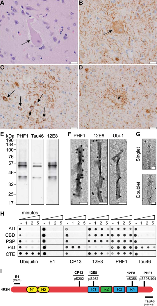
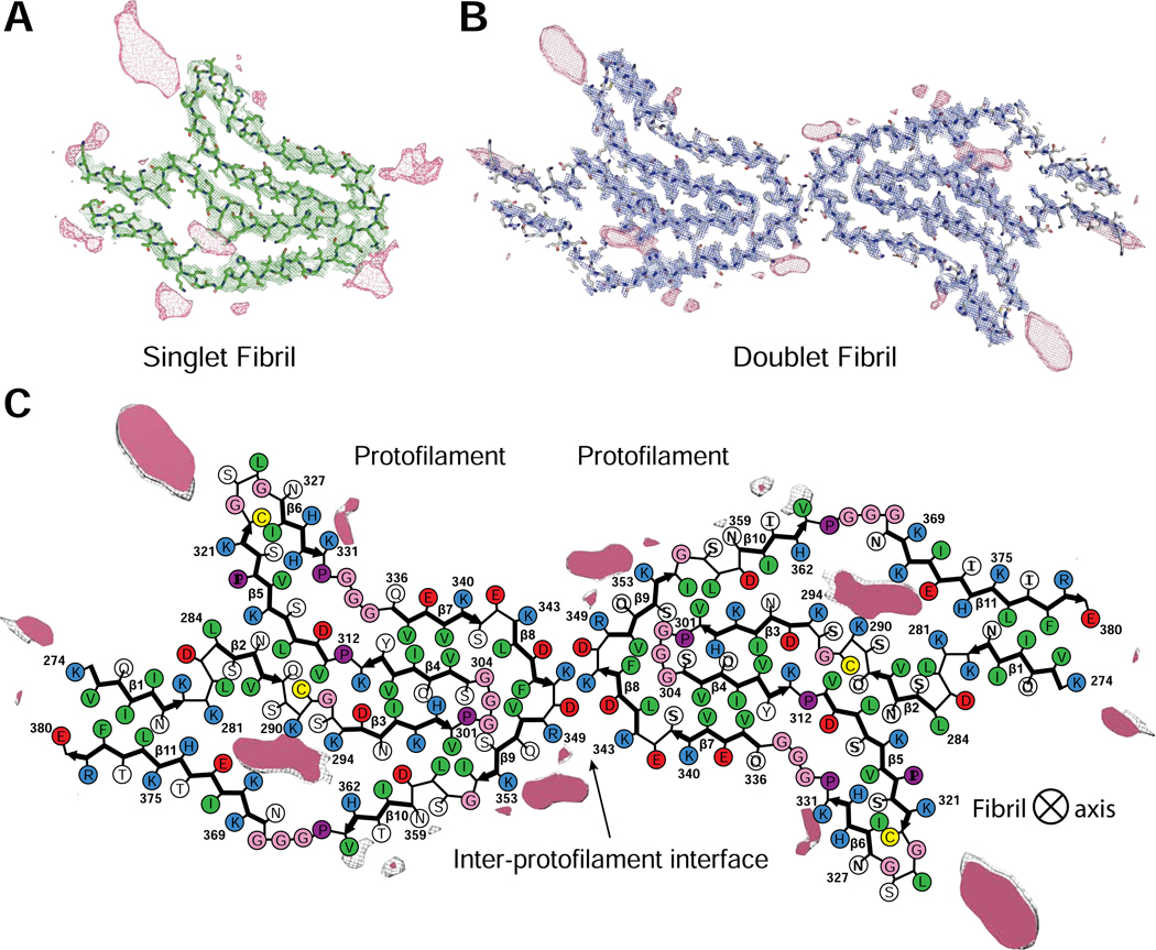
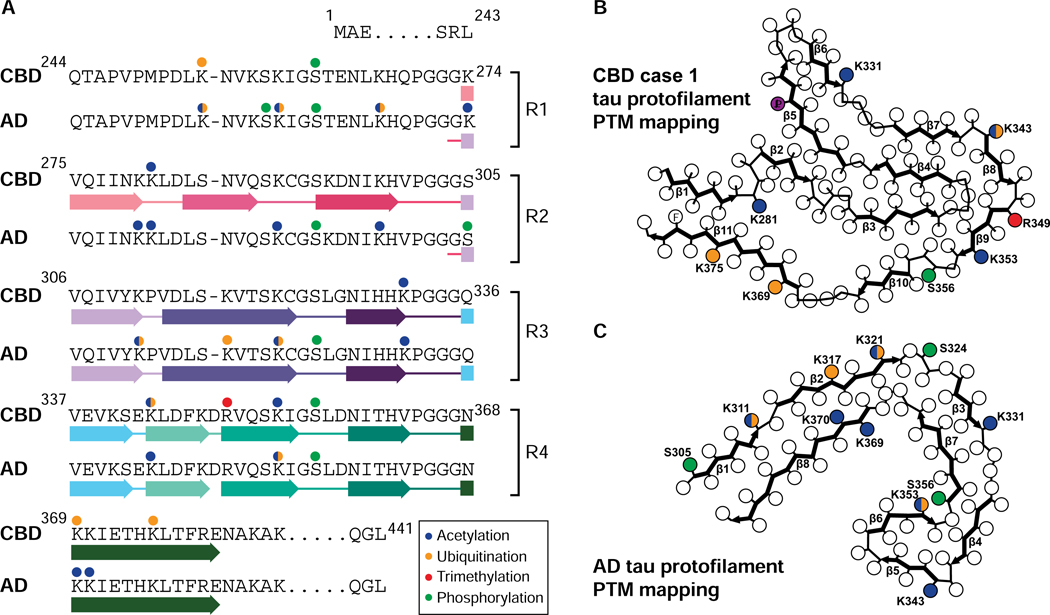
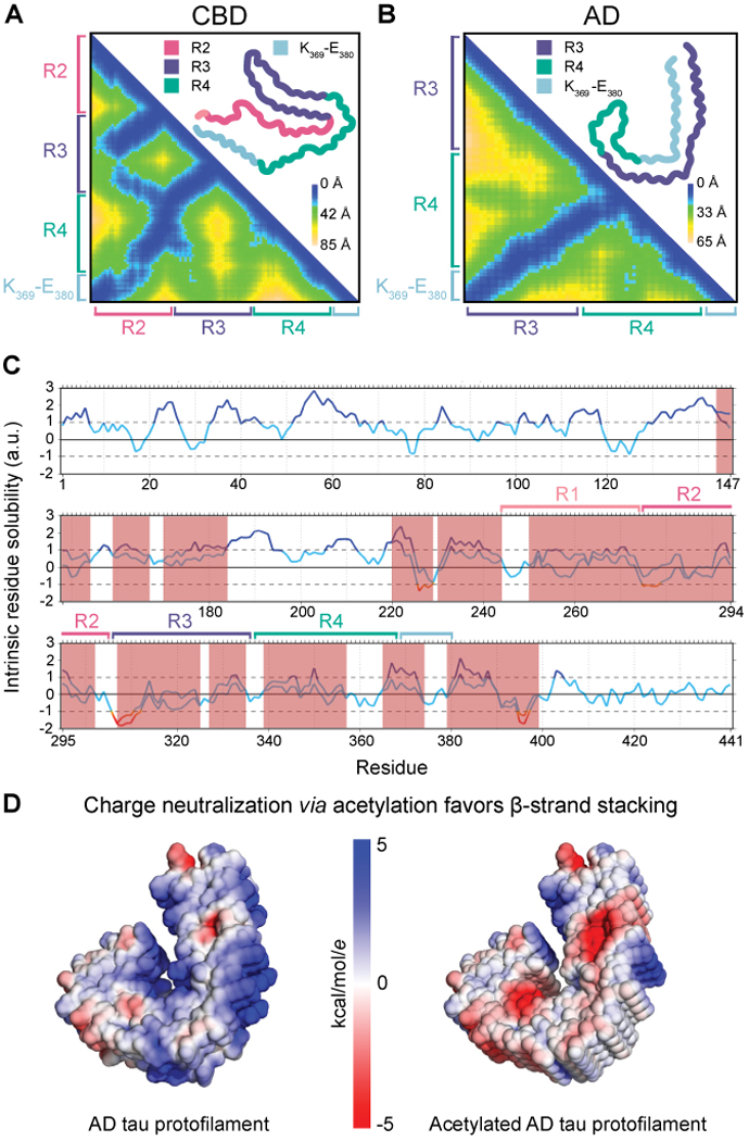
Tau aggregation into insoluble filaments is the defining pathological hallmark of tauopathies. However, it is not known what controls the formation and templated seeding of strain-specific structures associated with individual tauopathies. Here, we use cryo-electron microscopy (cryo-EM) to determine the structures of tau filaments from corticobasal degeneration (CBD) human brain tissue. Cryo-EM and mass spectrometry of tau filaments from CBD reveal that this conformer is heavily decorated with posttranslational modifications (PTMs), enabling us to map PTMs directly onto the structures. By comparing the structures and PTMs of tau filaments from CBD and Alzheimer's disease, it is found that ubiquitination of tau can mediate inter-protofilament interfaces. We propose a structure-based model in which cross-talk between PTMs influences tau filament structure, contributing to the structural diversity of tauopathy strains. Our approach establishes a framework for further elucidating the relationship between the structures of polymorphic fibrils, including their PTMs, and neurodegenerative disease.

Keywords: Alzheimer's disease; acetylation; corticobasal degeneration; cryo-EM; integrated structural biology; posttranslational modifications; tau strains; tauopathy; templated seeding; ubiquitination.


Conflict of interest statement

Declaration of Interests The authors declare no competing interests.

Figures

Figure 1.
Figure 1.. Neuropathological and biochemical characterization of CBD tau filaments.
(A-D) Representative neuropathological changes in the frontal cortex of a patient with CBD. Ballooned neurons revealed by H&E staining (A, arrow) and immunolabeling with CP13 (B, arrow). CP13 immunostaining also labels neuritic threads (B-D), as well as pleomorphic small neuronal inclusions (C, arrows) and astrocytic plaques (D, asterisk). Scale bar equal to 20 microns (A-D). (E) Sarkosyl-insoluble material from the frontal cortex of the CBD patient used for cryo-EM analysis was evaluated by Western blot, including the tau antibodies PHF1 (pS396/404), Tau46 (aa404–441) and 12E8 (pS262, pS356). (F) Representative immuno-electron microscopy images of sarkosyl-insoluble tau filaments purified from CBD brain and stained with tau phospho-specific antibodies PHF1 and 12E8, as well as ubiquitin (Ubi-1). Primary antibodies were visualized with secondary antibody conjugated with 6 nm gold particles. Scale bar equal to 30 nm. (G) Representative cryo-EM images of the singlet and doublet tau fibrils in CBD. Scale bar equal to 100 Å. (H) Pronase treatment eliminates ubiquitin immunoreactivity from sarkosyl-insoluble core. To examine the impact of pronase treatment on ubiquitination of the filament core in tauopathies, the sarkosyl insoluble fraction was obtained and incubated in the presence or absence of pronase (1, 2, or 5 minute treatment), and subsequently evaluated by dot blot (including anti-ubiquitin and the tau antibodies E1 [aa19–33], CP13 [pS202], 12E8, PHF1, and Tau46). (I) Schematic diagram depicting the location of the antibody epitope within the tau protein. Note the amount of each sarkosyl-insoluble fraction was normalized for total tau levels. See also Figures S1 and S5.
Figure 2.
Figure 2.. Cryo-EM structures of tau filaments from CBD.
Cryo-EM density (mesh) and atomic models (colored sticks) of (A) singlet and (B) doublet fibrils in CBD. Extra densities (pink mesh), which are directly connected, or in close proximity, to many sidechains in the cryo-EM maps, correspond to non-tau protein components. (C) Schematic view of the CBD doublet fibril. The extra, unknown densities are overlaid as capped pink mesh. See also Figure S2.
Figure 3.
Figure 3.. PTMs of tau filaments from CBD and AD detected by MS.
(A) Sequence alignment of the four microtubule-binding repeats (R1-R4) of tau and the sequence after R4 that is part of the CBD and AD fibril cores. The positions of filamentous β-strands in both diseases are shown. PTMs detected by MS in tau fibrils from CBD case 1 and AD, the structures of which were determined by cryo-EM in this work, are shown with acetylation, ubiquitination, trimethylation, and phosphorylation sites marked with blue, orange, red, and green balls, respectively. Sidechains with multiple PTMs detected are shown with two colours. Acetylation of K321 and K370 (Park, et al., 2018) and ubiquitination of K353 (Cripps et al., 2006) in the AD fibril core are included from the literature. PTMs are mapped onto schematics of the protofilament structures from (B) CBD case 1, and (C) AD. The same color scheme as described above is used to depict PTMs. See also Figures S4 and S7 and Tables S1, S2, S3, and S4.
Figure 4.
Figure 4.. Intra-protofilament contacts and effect of acetylation on the fibril core.
Heat maps of inter-residue Cα-Cα distances within the tau protofilaments found in (A) CBD and (B) AD, indicating the interactions between microtubule binding repeats (R1-R4) and K369–380 of tau. (C) Predicted effects of acetylation, as represented by an acetyllysine mimic by mutating lysine (K) to glutamine (Q), on the solubility of the tau protein extracted from AD brain. Regions of altered residue solubility are highlighted in red with positive values indicating solubility and negative values corresponding to aggregation-prone regions. Only acetylated residues implicated in AD brain (Park et al., 2018) were substituted. (D) Surface representation of (left) unmodified tau protofilament from AD and (right) acetylated coloured by electrostatic potential. See also Figure S3.
Figure 5.
Figure 5.. Visualization of PTMs of tau protofilaments from CBD and AD using a combination of cryo-EM and MS PTM mapping.
An average of 10 z-slices from the (A) CBD singlet fibril and (C) AD tau fibril cryo-EM 3D reconstructions reveals strong, large densities visibly attached to K321, K343, K353, and K369 on the CBD singlet map and K317 and K321 on the AD tau protofilament (red dashed circles). Many of these sites are detected to be ubiquitinated by MS, and the lysine-connected densities are much too large to be an acetyl group. A large, buried density proximal to K290, K294, H362, and K370 in the CBD singlet fibril is also shown (green dashed circle). Schematics shown to scale in (B) and (D) highlight the structural role mono- or poly-ubiquitinated chains at these lysines may play in the CBD singlet fibril and AD tau protofilament, respectively. Brackets surrounding ubiquitin indicate the possibility of a (poly)-ubiquitin chain. The position of the H362 sidechain is shown as a filled blue circle. Scale bar is equal to 25 Å.
Figure 6.
Figure 6.. Visualization of PTMs of tau fibrils from CBD and AD using a combination of cryo-EM and MS PTM mapping.
An average of 10 z-slices from the (A) CBD doublet fibril and (C) AD straight filament (EMD-3743) cryo-EM 3D reconstructions reveals strong, large densities visibly attached to K321 and K353 on the CBD doublet map and K317, K321, and K311 on the AD straight filament (red dashed circles). In particular, note the large, near-stoichiometric densities at the interface between protofilaments (A, C). Many of these sites are detected to be ubiquitinated by MS and the lysine-connected densities are much too large to be an acetyl group. A large, buried density proximal to K290, K294, H362 and K370 in the CBD doublet fibril and a large, solvent-exposed density next to K353, H362 and K369 in the AD straight filament are also shown (green dashed circle). Schematics shown to scale in (B) and (D) highlight the structural role mono- or poly-ubiquitinated chains at these lysines may play in the CBD doublet fibril and AD straight filament, respectively. Brackets surrounding ubiquitin indicate the possibility of a (poly)-ubiquitin chain. The position of the K353 and H362 sidechains are shown as filled blue circles. Scale bar is equal to 25 Å. See also Figure S6.
Figure 7.
Figure 7.. Proposed structure-based model of how interplay between PTMs influences tau filament structure.
Based on our cryo-EM maps and MS PTM mapping onto atomic models, we conclude that ubiquitination of tau can mediate inter-protofilament interfaces in the doublet CBD fibril and straight filament from AD. If sites on tau favoring the formation of doublet fibrils in CBD (K353) or straight filaments in AD (K311 and K317/321) are acetylated, or ubiquitinated with low occupancy, this inter-protofilament interface is less likely to form. The singlet fibril in CBD does not bind to a second protofilament and paired helical filaments in AD have structures that do not require mediation by non-tau components at their inter-protofilament interface. The outcome of this model is that the incorporation of ubiquitin into tau filaments in CBD and AD mediates inter-protofilament packing resulting in distinct ultrastructural polymorphs, tuning the ratio of fibril subtypes in tau inclusions.

Comment in

  • Tau intricacies in disease.
    Baumann K. Baumann K. Nat Rev Mol Cell Biol. 2020 Apr;21(4):180. doi: 10.1038/s41580-020-0233-0. Nat Rev Mol Cell Biol. 2020. PMID: 32127686 No abstract available.

References

    1. Abreha MH, Dammer EB, Ping L, Zhang T, Duong DM, Gearing M, Lah JJ, Levey AI, and Seyfried NT (2018). Quantitative Analysis of the Brain Ubiquitylome in Alzheimer’s Disease. Proteomics 18, e1800108. - PMC - PubMed
    1. Afonine PV, Poon BK, Read RJ, Sobolev OV, Terwilliger TC, Urzhumtsev A, and Adams PD (2018). Real-space refinement in PHENIX for cryo-EM and crystallography. Acta Crystallogr. D Struct. Biol 74, 531–544. - PMC - PubMed
    1. Bai B, Hales CM, Chen PC, Gozal Y, Dammer EB, Fritz JJ, Wang X, Xia Q, Duong DM, Street C, et al. (2013). U1 small nuclear ribonucleoprotein complex and RNA splicing alterations in Alzheimer’s disease. Proc. Natl. Acad. Sci. USA 110, 16562–16567. - PMC - PubMed
    1. Ballatore C, Lee VM, and Trojanowski JQ (2007). Tau-mediated neurodegeneration in Alzheimer’s disease and related disorders. Nat. Rev. Neurosci 8, 663–672. - PubMed
    1. Bloom GS (2014). Amyloid-beta and tau: the trigger and bullet in Alzheimer disease pathogenesis. JAMA Neurol. 71, 505–508. - PubMed

Publication types