Review
. 2020 Apr:193:107959.
doi: 10.1016/j.exer.2020.107959. Epub 2020 Feb 4.

The cereus matter of Bacillus endophthalmitis

Affiliations
Review

The cereus matter of Bacillus endophthalmitis

Md Huzzatul Mursalin et al. Exp Eye Res. 2020 Apr.

Abstract

Bacillus cereus (B. cereus) endophthalmitis is a devastating intraocular infection primarily associated with post-traumatic injuries. The majority of these infections result in substantial vision loss, if not loss of the eye itself, within 12-48 h. Multifactorial mechanisms that lead to the innate intraocular inflammatory response during this disease include the combination of robust bacterial replication, migration of the organism throughout the eye, and toxin production by the organism. Therefore, the window of therapeutic intervention in B. cereus endophthalmitis is quite narrow compared to that of other pathogens which cause this disease. Understanding the interaction of bacterial and host factors is critical in understanding the disease and formulating more rational therapeutics for salvaging vision. In this review, we will discuss clinical and research findings related to B. cereus endophthalmitis in terms of the organism's virulence and inflammogenic potential, and strategies for improving of current therapeutic regimens for this blinding disease.

Keywords: Bacillus; Bacteria; Endophthalmitis; Infection; Inflammation; Microbiology.


Conflict of interest statement

Declaration of competing interest The authors report no proprietary or commercial interest in any product mentioned or concept discussed in this article.

Figures

Figure 1:
Figure 1:
(A) Photograph of a B. cereus-infected eye after penetrating trauma. This image demonstrates chemosis, corneal opacification, and proptosis. Copyright © 2017 Zhisheng Ke, MD, The Eye Hospital of Wenzhou Medical University, Wenzhou, China. (B) Photograph of a patient’s left eye infected with B. cereus following cataract surgery. The patient had diffuse eyelid swelling, conjunctival chemosis, an intact corneal wound, a corneal ring abscess, an anterior chamber hypopyon and fibrin infiltration, and an obscure fundus view. Reproduced with permission from Oxford University Press.
Figure 2:
Figure 2:
(A) Gram staining of B. cereus. Magnification, 400X (B) B. cereus rods stained with Leifson Flagella Stain. Magnification, 1000X. (C) 1μL of B. cereus was pipetted onto trypticase soy agar (TSA) + 5% sheep blood agar and incubated for 18h at 37°C. Colonies with characteristics of double zone of hemolysis were observed at each inoculation point. (D) Transmission electron microscopy (TEM) of B. cereus. Flagella are denoted by black arrows, while white arrows denote pili.
Figure 3:
Figure 3:
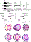
Comparative analysis of the cytotoxicity of bacterial secreted products on human retinal Muller cells (MIO-M1). Muller cells were treated with overnight culture supernatants from B. cereus, S. aureus, E. faecalis, S. epidermidis, or S. pneumoniae for 45 minutes. For E. faecalis, the cytotoxicity of supernatants from cytolysin component LL- and LS-producing strains were tested as previously described in LaGrow et al., 2017. The levels of LDH release from affected retinal Muller cells were measured using the Pierce LDH Cytotoxicity Assay Kit (Thermo Fisher Scientific) according to the manufacturer’s instructions. Data is represented as the mean ± SEM of % cytotoxicity of retinal Muller cells based on the positive LDH control. Statistical significance was determined by unpaired Student t-test (**P ≤ 0.01, N=3/group).
Figure 4:
Figure 4:
Comparative analysis of intraocular bacterial growth (A, B), quantification of myeloperoxidase (C), retinal function (D) and pathology (E) in Gram-positive experimental endophthalmitis in mice. 100 CFU/eye of B. cereus, E, faecalis, or S. pneumoniae, or 5000 CFU/eye of S. aureus or S. epidermidis were intravitreally injected into the posterior segment of the eyes of C57BL/6J mice. Infection parameters were measured at 12 hours postinfection. Figure 4A demonstrates the intraocular bacterial burden of these pathogens. The thick black bar indicates the initial bacterial inoculum at the time of infection. Figure 4B demonstrates intraocular bacterial growth rate in terms of hour−1 over 12 hours of infection. Figure 4C demonstrates the level of myeloperoxidase in those infected eyes. Figure 4D demonstrates the retinal A-wave and B-wave retention of these pathogens. Figure 4E illustrates hematoxylin and eosin staining of whole eyes. Quantitative data represents the mean ± SEM of N≥5 eyes per time point. Statistical significance was determined by unpaired Student t-test (ns P ≥ 0.05, * P ≤ 0.0159, ** P ≤ 0.0079). Sections are representative of three eyes per group.
Figure 5:
Figure 5:
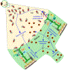
Model of B. cereus endophthalmitis. (A) Initiation of B. cereus infection in the eye after an ocular trauma or surgery. (B) Rapid bacterial growth in the vitreous and migration toward the retina. (C) Production bacterial toxins and enzymes. These bacterial products can cause dysfunction and permeability of the blood-retinal barrier. (D) Activation of TLR pathways by bacterial envelope constituents. (E) TLR pathway activation leads to the generation of proinflammatory mediators which recruit inflammatory cells (PMN) into the eye, beginning at the optic nerve head. These mediators may also disrupt the blood-retinal barrier. (F) Infiltrating PMNs may block light pathways to the retina and cause bystander damage to retinal cells, leading to vision loss. Vit: Vitreous, ON: Optic nerve.

References

    1. Aarthi P, Bagyalakshmi R, Therese KL, Malathi J, Mahalakshmi B, Madhavan HN, 2012. Optimization and application of a reverse transcriptase polymerase chain reaction to determine the bacterial viability in infectious endophthalmitis. Curr. Eye. Res 37, 1114–1120. 10.3109/02713683.2012.704476. - DOI - PubMed
    1. Abu el-Asrar AM, al-Amro SA, al-Mosallam AA, al-Obeidan S, 1999. Post-traumatic endophthalmitis: Causative organisms and visual outcome. Eur. J. Ophthalmol 9, 21–31. - PubMed
    1. Affeldt JC, Flynn HW Jr., Forster RK, Mandelbaum S, Clarkson JG, Jarus GD, 1987. Microbial endophthalmitis resulting from ocular trauma. Ophthalmology 94, 407–413. - PubMed
    1. Agaisse H, Gominet M, Okstad OA, Kolsto AB, Lereclus D, 1999. PlcR is a pleiotropic regulator of extracellular virulence factor gene expression in Bacillus thuringiensis. Mol. Microbiol 32, 1043–1053. - PubMed
    1. Agrawal R, 2012. Post-traumatic endophthalmitis. Eye (Lond) 26, 1277–1278. 10.1038/eye.2012.104. - DOI - PMC - PubMed

Publication types

MeSH terms

Substances