. 2020 Aug;146(2):390-405.
doi: 10.1016/j.jaci.2020.01.038. Epub 2020 Feb 4.

Optimal identification of human conventional and nonconventional (CRTH2-IL7Rα-) ILC2s using additional surface markers

Affiliations

Optimal identification of human conventional and nonconventional (CRTH2-IL7Rα-) ILC2s using additional surface markers

Sucai Liu et al. J Allergy Clin Immunol. 2020 Aug.

Abstract

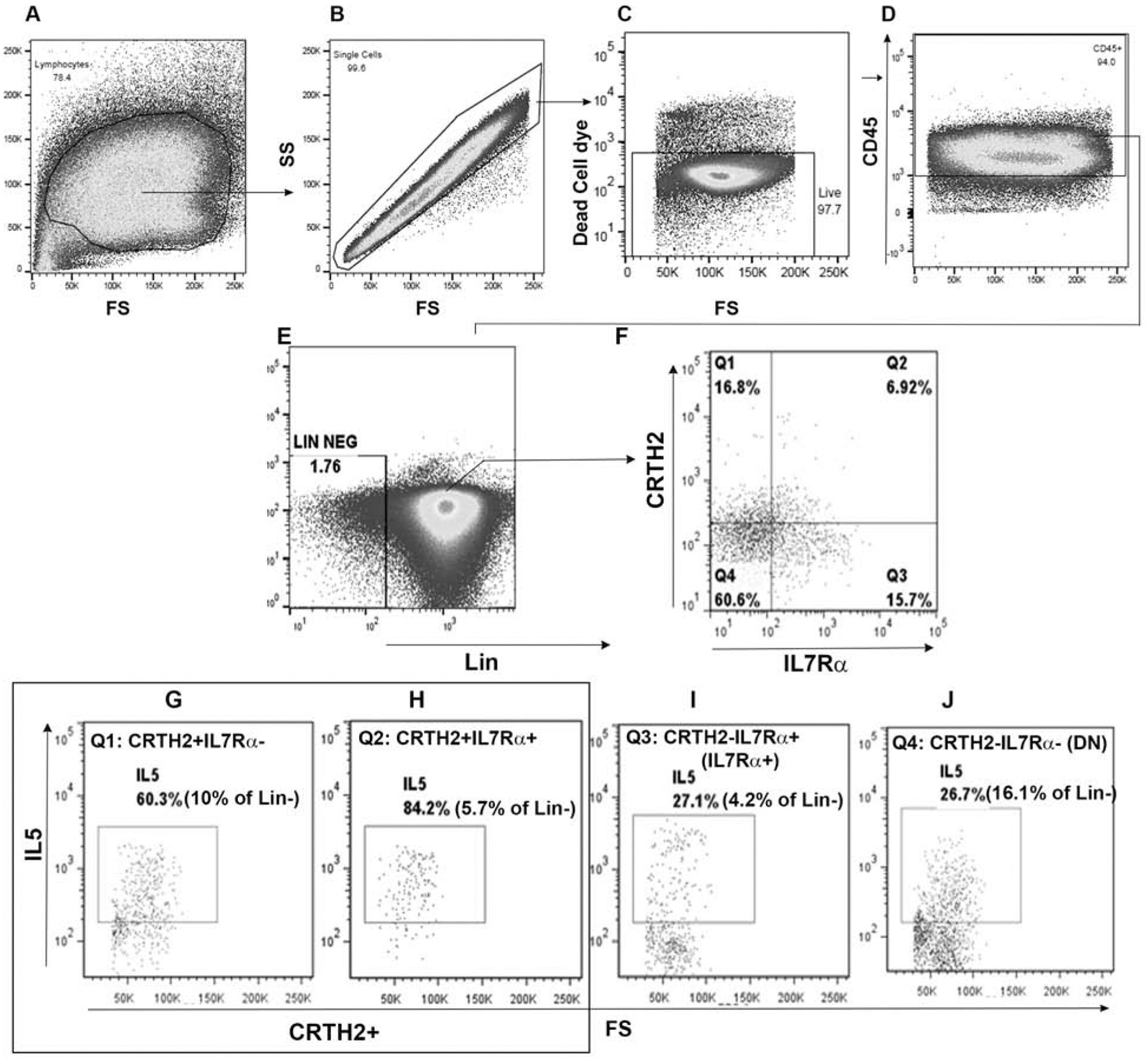
Background: Human type 2 innate lymphoid cells (ILC2s) are identified by coupled detection of CRTH2 and IL7Rα on lineage negative (Lin-) cells. Type 2 cytokine production by CRTH2-IL7Rα- innate lymphoid cells (ILCs) is unknown.

Objective: We sought to identify CRTH2-IL7Rα- type 2 cytokine-producing ILCs and their disease relevance.

Methods: We studied human blood and lung ILCs from asthmatic and control subjects by flow cytometry, ELISA, RNA sequencing, quantitative PCR, adoptive transfer to mice, and measurement of airway hyperreactivity by Flexivent.

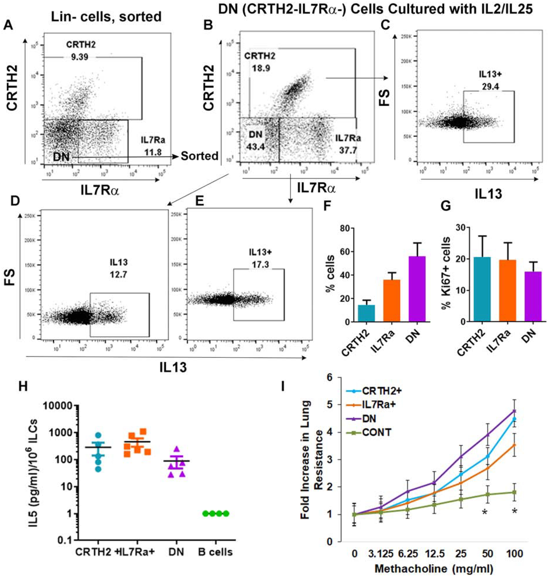
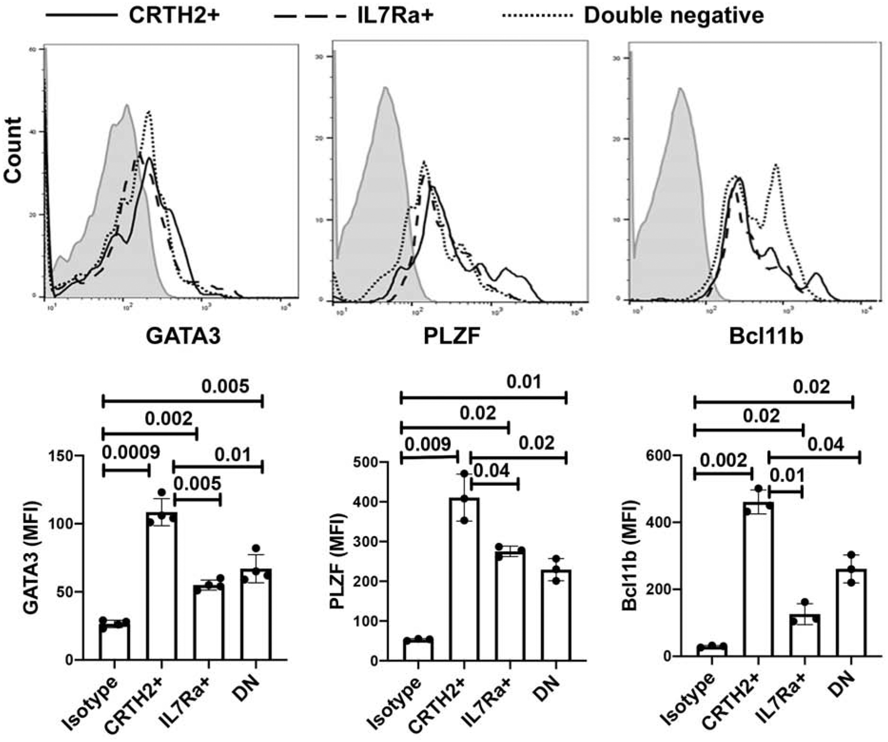
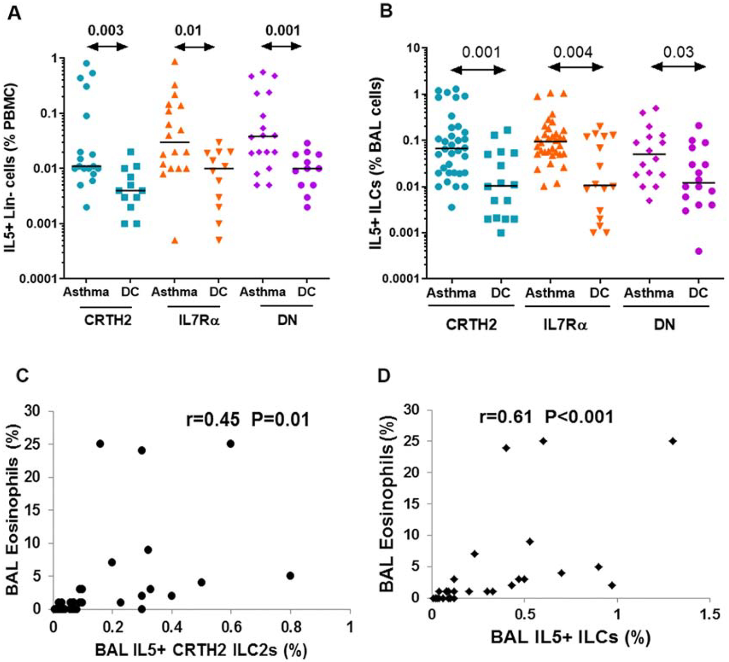
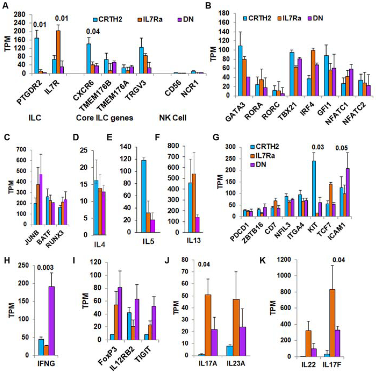
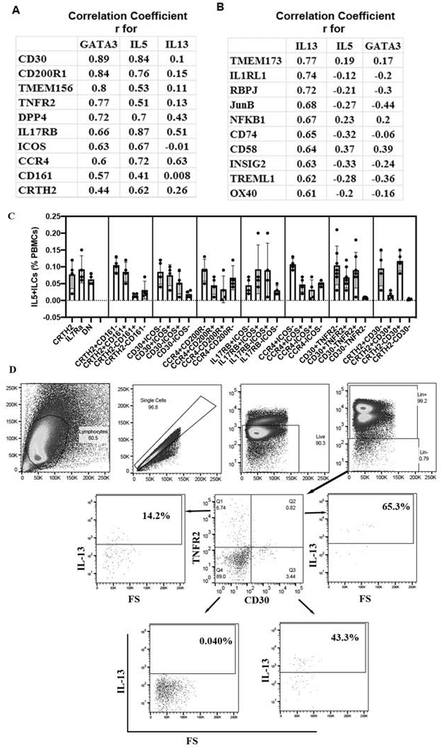
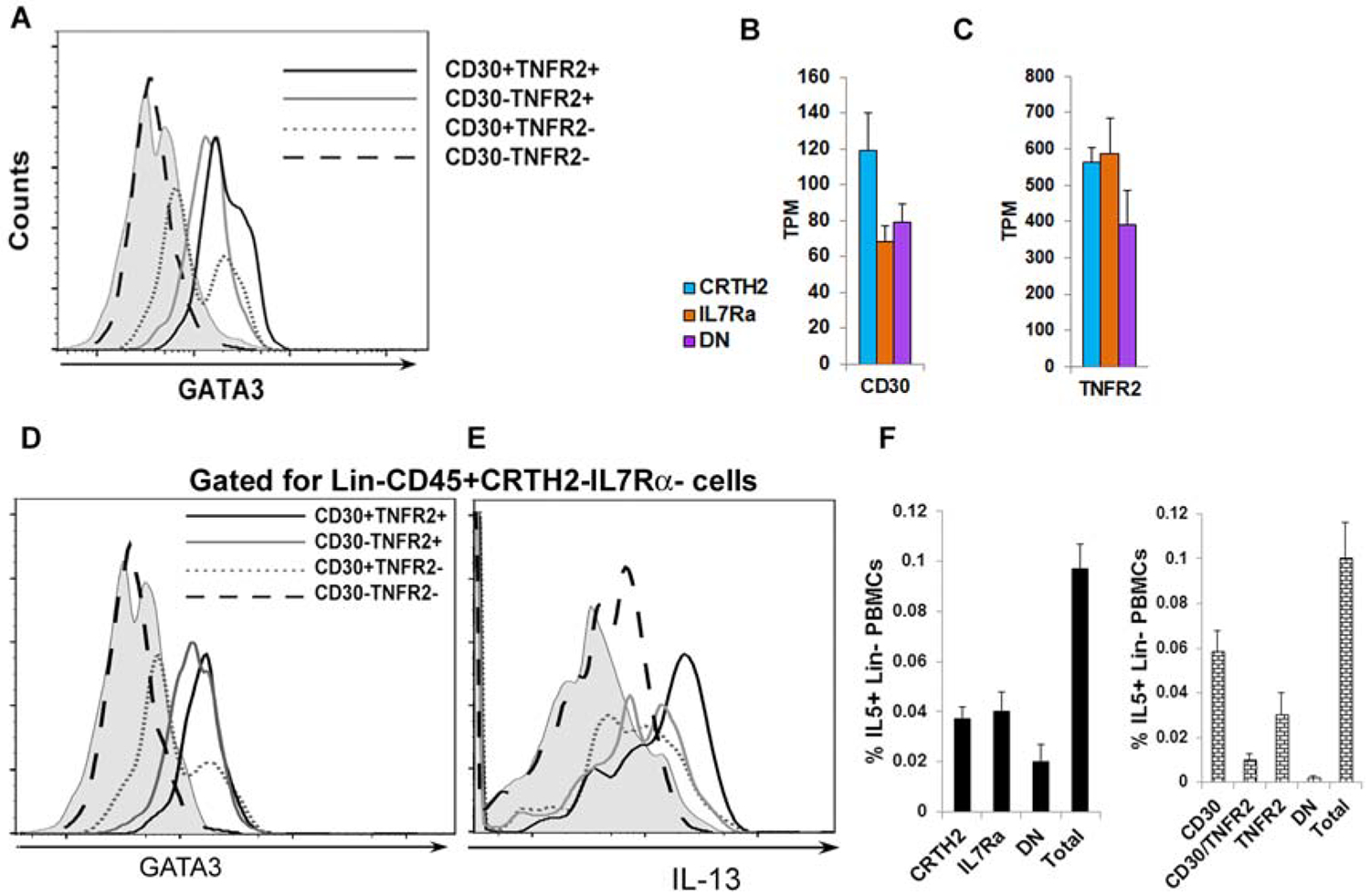
Results: We found that IL-5 and IL-13 were expressed not only by CRTH2+ but also by CRTH2-IL7Rα+ and CRTH2-IL7Rα- (double-negative [DN]) human blood and lung cells. All 3 ILC populations expressed type 2 genes and induced airway hyperreactivity when adoptively transferred to mice. The frequency of type 2 cytokine-positive IL7Rα and DN ILCs were similar to that of CRTH2 ILCs in the blood and lung. Their frequency was higher in asthmatic patients than in disease controls. Transcriptomic analysis of CRTH2, IL7Rα, and DN ILCs confirmed the expression of mRNA for type 2 transcription factors in all 3 populations. Unexpectedly, the mRNA for GATA3 and IL-5 correlated better with mRNA for CD30, TNFR2, ICOS, CCR4, and CD200R1 than for CRTH2. By using a combination of these surface markers, especially CD30/TNFR2, we identified a previously unrecognized ILC2 population.

Conclusions: The commonly used surface markers for human ILC2s leave a majority of type 2 cytokine-producing ILC2s unaccounted for. We identified top GATA3-correlated cell surface-expressed genes in human ILCs by RNA sequencing. These new surface markers, such as CD30 and TNFR2, identified a previously unrecognized human ILC2 population. This ILC2 population is likely to contribute to asthma.

Keywords: Asthma; cytokines; novel ILC2 population; type 2 innate lymphoid cells.


Conflict of interest statement

The authors declare no competing financial interests.

Figures

Figure 1.
Figure 1.
Expression of IL5 by ILC2 subpopulations. PBMCs from an allergic asthmatic subject were cultured with IL2/IL25 for 5 days and analyzed for IL5+ cells by flow cytometry. Mononuclear cells (A) were sequentially gated for single cells (B), CD45+ cells (C) and lineage (lin) – cells (D). Gated lin- cells were analyzed for expression of CRTH2 and IL7Rα (E). Each quadrant (Q1: CRTH2+IL7Rα−[F]; Q2: CRTH2+IL7Rα+ [G]; Q3: CRTH2-IL7Rα+ [H]; and Q4: CRTH2-IL7Rα- ([I]) was then analyzed for expression of IL5. The threshold for positive staining was determined by isotype antibody staining and FMO (fluorescence minus one) (Fig. S1).
Figure 2.
Figure 2.
Differentiation, proliferation, cytokine secretion and in vivo activity of DN ILCs. A-E: FACS-sorted DN (lin-CRTH2-IL7Rα-) cells from PBMCs obtained from an asthmatic patient (A) were cultured with IL2/IL25 for 7 days and their expression of CRTH2 and IL7Rα was analyzed (B). The newly emerged CRTH2+ (C), IL7Rα+ (E) and the remaining DN cells (D) were gated for expression of IL13. F: Results of experiments from 3 different asthmatic donors. G: FACS-sorted CRTH2+, IL7Rα+ and DN cells were cultured with IL2/IL25 for 7 days and then analyzed for expression ki67 by flow cytometry (N=3). (H) CD45+Lin- CRTH2+, IL7Ra+ and DN ILCs from the human lung were cultured with IL2/IL25 for 5 days and then supernatant was assayed for IL5 by ELISA. Culture supernatant from IL2/IL25-treated B cells (negatively selected from PBMCs) was used a control. Each symbol represents a single lung donor. (I) Human lung-derived ILC2 subpopulations were cultured with IL2 overnight and adoptively transferred (105 cells/mouse) to Rag2−/−:γc−/− mice. Lung-derived CD45+CD3+CD4 T cells were injected (106 cells/mouse) as a control. One-day latter the mice were challenged with the Alternaria allergen for 3 consecutive days before methacholine challenge. The increase in lung resistance was statistically significant (*=P<0.05, t test) for all ILC2 populations as compared to the control (CONT).
Figure 3.
Figure 3.
Expression of ILC2-related markers in ILC2 subpopulations. PBMCs from 3–4 donors were cultured with IL2/IL25 for 3 days and then analyzed for expression of GATA3, PLZF, and Bcl11b, in lin- CRTH2+, IL7Rα+ and DN ILCs by flow cytometry. Histograms of mean fluorescence intensity are shown in the upper panel and bar graphs with statistical analysis are shown in the lower panel. Each symbol represents a donor.
Figure 4.
Figure 4.
Comparison of blood and bronchoalveolar lavage (BAL) ILC2 populations in asthmatic patients and disease controls (DC). A: PBMCs from 18 asthmatic patients and 12 disease controls were cultured in the medium alone for 5 days and then analyzed for IL5+ ILC2 populations. The frequency of IL5+lin- cells is presented as % PBMCs. ILC B: BAL cells from 34 asthmatic patients and 16 disease controls were stained ex vivo for IL5+ (CD45+Lin-) ILC2 populations by flow cytometry. The frequency of IL5+ ILCs is presented as % BAL cells. Statistical significance (Mann-Whitney U test) is shown at the top of the dot plots. C& D: Correlation (r) between BAL eosinophils and BAL IL5+ CRTH2+ ILC2s (C) and BAL total IL5+ ILC2s (CRTH2+, IL7Rα+ and DN).
Figure 5.
Figure 5.
Expression profile of ILC-related genes in ILC2 subpopulations by RNA-seq. FACS-sorted CRTH2+IL7Rα+ (CRTH2), CRTH2-IL7Rα+ (IL7Ra) and DN lin- cells from the peripheral blood of 4 healthy donors were used to isolate RNA and perform RNA-seq. A: Expression of genes used for sorting of ILCs (PTGDR2, IL7R), and core ILC- and NK cell-related genes (TPM: transcripts per million). B and C: Expression of genes related to ILC1 (TBX21), ILC2 (, GATA3, RORA, IRF4, GFI1, NFATC1, NFATC2, JUNB, BATF), and ILC3 (RORC and RUNX3). D-F: Expression of type-2 cytokine (IL4, IL5, and IL13) genes. G. Expression of ILC2 precursor-related genes. H and I: Top immune response genes expressed by DN cells. J and K: Top cytokine genes expressed by IL7Rα+ cells. Gene expression values are normalized to transcripts per million (TPM). Statistical significance, when present, is shown on the top of the bars.
Figure 6.
Figure 6.
A list of the top genes that correlated with GATA3 and type-2 cytokines, and their surface expression on ILC2 populations. RNA-seq data was analyzed for Pearson correlation (r) with type-2 genes, and the top genes that correlated with GATA3/IL5 and IL13 are shown in panels A & B, respectively. C: PBMCs from asthmatic patients were cultured with IL2/IL25 for 5 days, stained for flow cytometry and then analyzed for expression of IL5 by CD45+lin- cells expressing various combination of surface markers (N=4 for CCR4/CD200R, CCR4/ICOS, IL17RB/ICOS, CD30/ICOS and CD30/CRTH2; N=7 for CD30/TNFR2). D: Representative flow cytometry plots from a single donor showing the gating strategy for CD30 and TNFR2 expressing ILCs and their expression of IL13.
Figure 7.
Figure 7.
CD30/TNFR2 as ILC2 surface markers. A: A histogram of expression of GATA3 in various populations of lin-CD30/TNFR2 +/− cells. PBMCs were cultured with IL2/IL25 for 5 days, stained for flow cytometry and then gated sequentially for live mononuclear cells, single cells, lin- cells, CD30+TNFR2+, CD30+TNFR2−, CD30-TNFR2+ and CD30-TNFR2- cells. The expression of GATA3 was analyzed in the latter 4 ILC populations (N=4). B & C: Expression of mRNA for CD30 and TNFR2 in ILC populations as measured by RNA-seq. D & E: Expression of GATA3 and IL13 in DN, CD30/TNFR2+/− cells. PBMCs were cultured with IL2/IL25 for 5 days, stained for flow cytometry and then gated sequentially for live mononuclear cells, single cells, CD45+, lin- cells, CRTH2-IL7Ra-, CD30+TNFR2+, CD30+TNFR2−, CD30-TNFR2+ and CD30-TNFR2- cells. The expression of GATA3 and IL13 in the latter 4 ILC populations is shown (N=3). F: Comparison of flow cytometric enumeration of IL5+ ILCs in PBMCs using the CRTH2/IL7Rα and CD30/TNFR2 antibody-based detection approaches. IL5+ cells were first analyzed in lin- cells (Total). The lin- cells were then gated for CRTH2/IL7Rα (left) or CD30/TNFR2 (right) and IL5 expression was analyzed in the indicated cell populations (N=4). CD30: CD30+TNFR2−; TNFR2: CD30-TNFR2+; DN: CD30-TNFR2−.

Comment in

  • ILC2s: Are they what we think they are?
    Cavagnero KJ, Doherty TA. Cavagnero KJ, et al. J Allergy Clin Immunol. 2020 Aug;146(2):280-282. doi: 10.1016/j.jaci.2020.05.042. Epub 2020 Jun 12. J Allergy Clin Immunol. 2020. PMID: 32535133 Free PMC article. No abstract available.

References

    1. Klose CS, Artis D. Innate lymphoid cells as regulators of immunity, inflammation and tissue homeostasis. Nat Immunol 2016; 17:765–74. - PubMed
    1. Vivier E, Artis D, Colonna M, Diefenbach A, Di Santo JP, Eberl G, et al. Innate Lymphoid Cells: 10 Years On. Cell 2018; 174:1054–66. - PubMed
    1. Mjosberg JM, Trifari S, Crellin NK, Peters CP, van Drunen CM, Piet B, et al. Human IL-25- and IL-33-responsive type 2 innate lymphoid cells are defined by expression of CRTH2 and CD161. Nat Immunol 2011; 12:1055–62. - PubMed
    1. Bjorklund AK, Forkel M, Picelli S, Konya V, Theorell J, Friberg D, et al. The heterogeneity of human CD127(+) innate lymphoid cells revealed by single-cell RNA sequencing. Nat Immunol 2016; 17:451–60. - PubMed
    1. Barlow JL, Peel S, Fox J, Panova V, Hardman CS, Camelo A, et al. IL-33 is more potent than IL-25 in provoking IL-13-producing nuocytes (type 2 innate lymphoid cells) and airway contraction. J Allergy Clin Immunol 2013; 132:933–41. - PubMed

Publication types

MeSH terms