. 2020 May:138:104791.
doi: 10.1016/j.nbd.2020.104791. Epub 2020 Feb 4.

The green tea polyphenol epigallocatechin-3-gallate (EGCG) restores CDKL5-dependent synaptic defects in vitro and in vivo

Affiliations

The green tea polyphenol epigallocatechin-3-gallate (EGCG) restores CDKL5-dependent synaptic defects in vitro and in vivo

L Trovò et al. Neurobiol Dis. 2020 May.

Abstract

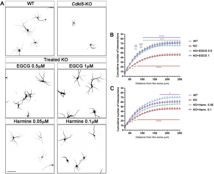
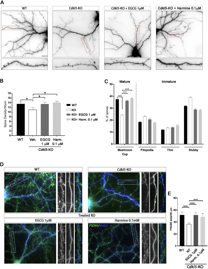
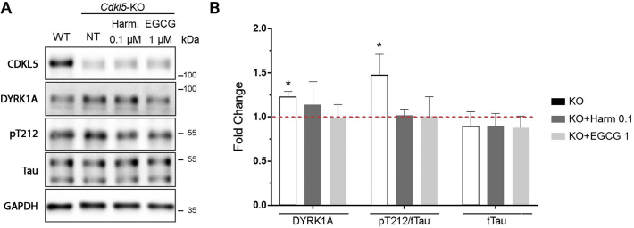
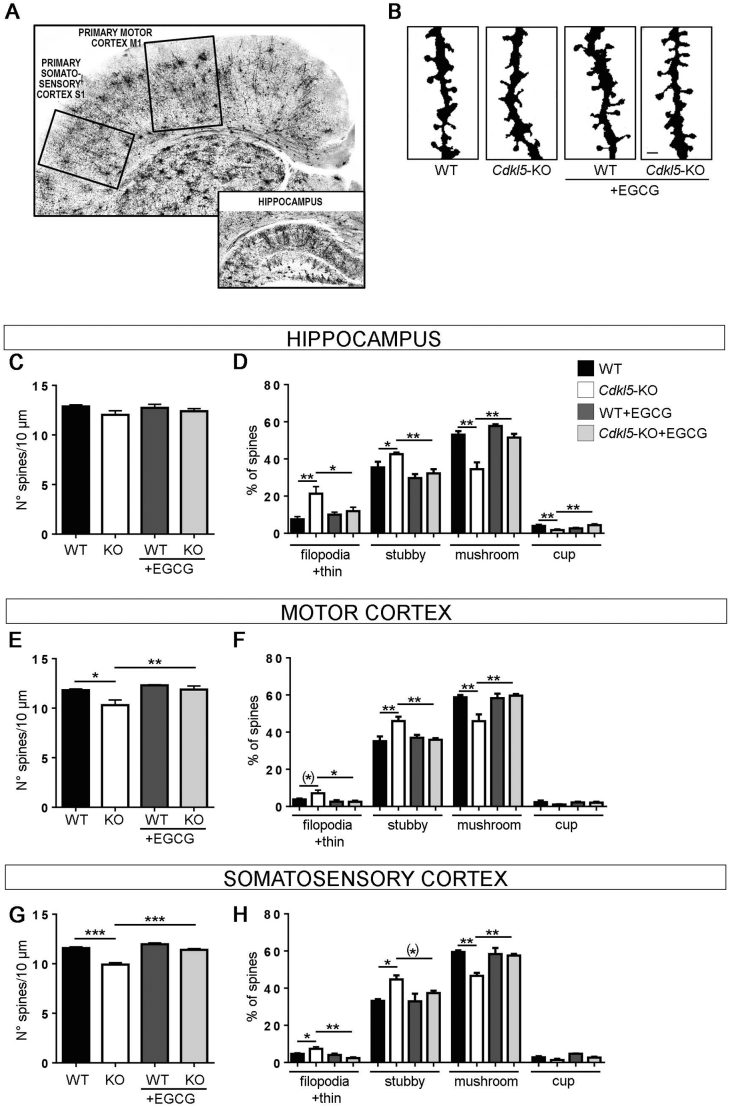
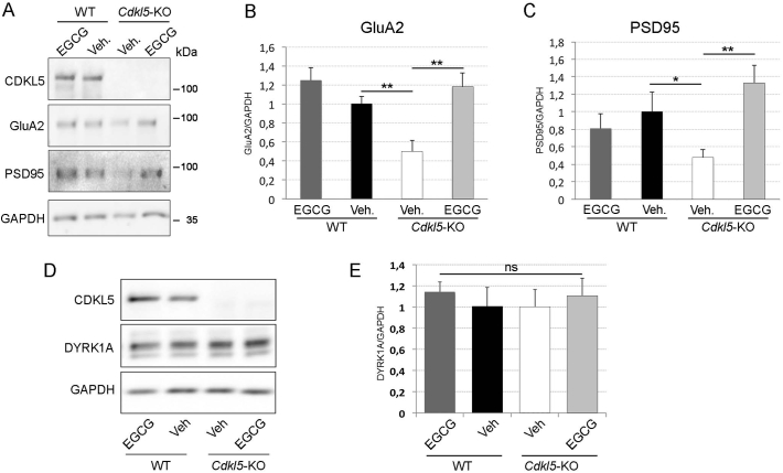
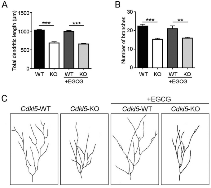
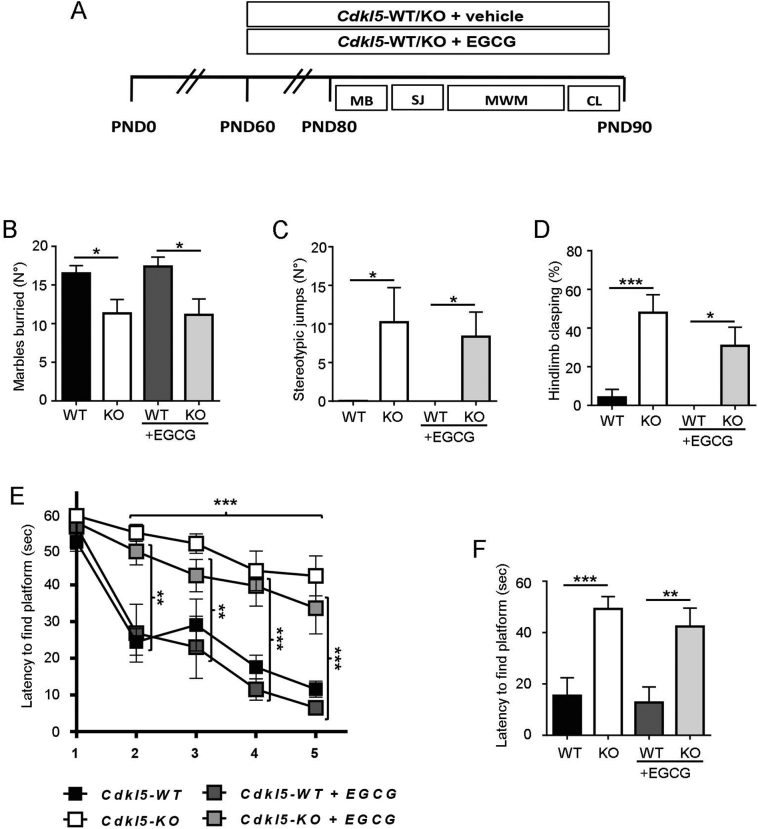
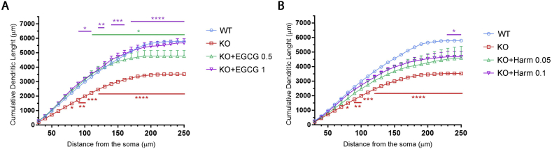
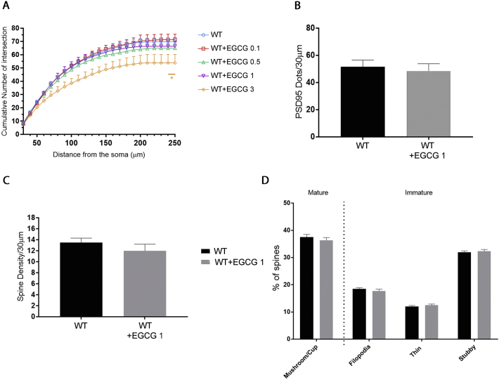
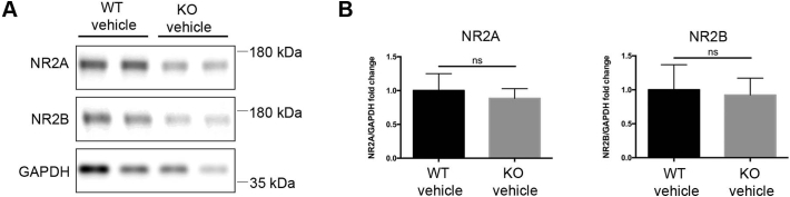
CDKL5 deficiency disorder (CDD) is a rare X-linked neurodevelopmental disorder that is characterised by early-onset seizures, intellectual disability, gross motor impairment, and autistic-like features. CDD is caused by mutations in the cyclin-dependent kinase-like 5 (CDKL5) gene that encodes a serine/threonine kinase with a predominant expression in the brain. Loss of CDKL5 causes neurodevelopmental alterations in vitro and in vivo, including defective dendritic arborisation and spine maturation, which most likely underlie the cognitive defects and autistic features present in humans and mice. Here, we show that treatment with epigallatocathechin-3-gallate (EGCG), the major polyphenol of green tea, can restore defects in dendritic and synaptic development of primary Cdkl5 knockout (KO) neurons. Furthermore, defective synaptic maturation in the hippocampi and cortices of adult Cdkl5-KO mice can be rescued through the intraperitoneal administration of EGCG, which is however not sufficient to normalise behavioural CDKL5-dependent deficits. EGCG is a pleiotropic compound with numerous cellular targets, including the dual-specificity tyrosine-phosphorylation-regulated kinase 1A (DYRK1A) that is selectively inhibited by EGCG. DYRK1A controls dendritic development and spine formation and its deregulation has been implicated in neurodevelopmental and degenerative diseases. Treatment with another DYRK1A inhibitor, harmine, was capable of correcting neuronal CDKL5-dependent defects; moreover, DYRK1A levels were upregulated in primary Cdkl5-KO neurons in concomitance with increased phosphorylation of Tau, a well-accepted DYRK1A substrate. Altogether, our results indicate that DYRK1A deregulation may contribute, at least in part, to the neurodevelopmental alterations caused by CDKL5 deficiency.

Keywords: CDKL5; DYRK1A; Epigallatocathechin-3-gallate; Synaptic defects.


Conflict of interest statement

Declaration of Competing Interest None. The following are the supplementary data related to this article. Supplementary data to this article can be found online at https://doi.org/10.1016/j.nbd.2020.104791.

Figures

Fig. 1
Fig. 1
Treatment with EGCG and harmine improves defective dendritic branching of Cdkl5-KO neurons in vitro. (A) Representative images of untreated WT/Cdkl5-KO neurons and KO neurons treated with EGCG (0.5 μM and 1 μM) or harmine (0.05 μM and 0.1 μM) from DIV7-DIV10. Neurons were stained against MAP2 to identify dendrites. Images were inverted for clarity. (B,C) Quantification of dendritic branching represented as cumulative number of intersections in untreated WT/Cdkl5-KO neurons or KO neurons treated with EGCG (A) or harmine (C). 15 branches/mouse, n = 5–6 mice/group from at least 3 different preparations. Statistical analysis: 2-WAY ANOVA followed by Dunnett's post-hoc test (*p < .05, ** p < .01, ***p < .001, ****p < .0001). Scale bar 100 μm.
Fig. 2
Fig. 2
Effect of EGCG and harmine on defective spine maturation of Cdkl5-KO neurons in vitro. (A) Representative images of untreated WT/Cdkl5-KO neurons or KO neurons treated with 1 μM EGCG or 0.1 μM harmine from DIV14-DIV17. The transfection with GFP at DIV15 allowed the visualization of spines. Scale bar 10 μm. (B) Quantification of the number of spine protrusions in 30 μm-long dendritic segments of WT/Cdkl5-KO neurons treated as indicated. Statistical analysis: 1-WAY ANOVA followed by Dunnett's post-hoc test (*p < .05). (C) Morphological classification of spines in WT/Cdkl5-KO treated neurons, represented as percentage of total number of spines. Statistical analysis: 2-WAY ANOVA followed by Dunnett's post-hoc test (***p < .001). 5 branches/mouse, n = 3 mice/group from 3 different preparations. Data are presented as mean ± SEM. (D) Representative images of untreated WT/Cdkl5-KO neurons or KO neurons treated as indicated and stained with PSD95 (green) and MAP2 (blue) to visualise excitatory spines and dendrites, respectively. (E) Quantification of the number of PSD95+ dots in 30 μm-long dendritic segments of WT/Cdkl5-KO neurons treated as indicated. 10 branches/mouse, n = 3–8 mice/group from at least 3 different preparations. Data are presented as mean ± SEM. Statistical analysis: 1-WAY ANOVA followed by Dunnett's post-hoc test (****p < .0001). Scale bar 10 μm. (For interpretation of the references to color in this figure legend, the reader is referred to the web version of this article.)
Fig. 3
Fig. 3
The absence of CDKL5 alters the DYRK1A pathway. (A) Representative WB showing the expression level of CDKL5, DYRK1A, total Tau and Tau phosphorylated on T212 in WT/Cdkl5-KO neurons or KO neurons treated with 1 μM EGCG or 0.1 μM harmine from DIV7-DIV10. GAPDH was used as internal standard. (B) Quantification of DYRK1A expression, the ratio between phosphorylated and total Tau (pT212/tTau) and total Tau levels in Cdkl5-KO neurons treated as indicated. Data are represented as fold change (mean ± SEM) compared to the untreated WT neurons indicated by the dotted horisontal line. Statistical analysis: 1-WAY ANOVA followed by Dunnett's post-hoc test (*p < .05). n = 3–10 mice/group from at least 3 different preparations.
Fig. 4
Fig. 4
EGCG supplementation corrects CDKL5-dependent synaptic defects in vivo. (A) Representative images of Golgi-stained coronal sections containing the primary motor (M1) and primary somatosensory (S1) cortex (upper image) and the hippocampus (lower image). (B) Representative images of dendritic protrusions of granule neurons in WT/Cdkl5-KO male mice treated with EGCG (25 mg/Kg, dialy i.p.) or vehicle for 30 days (PND60–90). Scale bar 1 μM. (C,E,G) Dendritic spine density (number of spines per 10 μm) in hippocampal granule neurons (C), and pyramidal neurons of layer II/III of the primary motor (E) and primary somatosensory cortex (G) in vehicle-treated (granule neurons: WT n = 6, KO n = 6; pyramidal neurons WT n = 3, KO n = 3) and EGCG-treated (granule neurons: WT n = 3, KO n = 4; pyramidal neurons WT n = 3, KO n = 3) WT/Cdkl5-KO male mice. (D,F,H) Morphological classification of spines in mice as in C,E,G. Data are presented as mean ± SEM. Statistical analysis: 2-WAY ANOVA followed by Fisher's LSD ((*)p = .06, *p < .05, **p < .01, ***p < .001).
Fig. 5
Fig. 5
EGCG supplementation corrects CDKL5-dependent synaptic defects in vivo. (A) Representative WB of hippocampal total lysates from mice treated as described in Fig. 4. (B,C) Quantification of GluA2 (B) and PSD95 (C) expression in hippocampi of WT/Cdkl5-KO mice upon normalisation with the internal standard GAPDH. n = 5 mice/group. Data are presented as mean ± SEM. Statistical analysis: 2-WAY ANOVA followed by Fisher's LSD. *p < .05, **p < .01, ***p < .001. (D,E) WB analysis (D) and relative quantification (E) of DYRK1A expression in hippocampal total lysates from PND90 mice treated as in A and normalised to GAPDH. n = 5 mice/group. Data are presented as mean ± SEM. Statistical analysis: 2-WAY ANOVA followed by Fisher's LSD.
Fig. 6
Fig. 6
EGCG supplementation has no effect on dendritic development in Cdkl5-KO mice. (A,B) Mean total dendritic length (A) and mean number of dendritic branches (B) of Golgi-stained granule neurons of vehicle-treated (WT n = 4, KO n = 3) and EGCG-treated (WT n = 3, KO n = 3) WT/Cdkl5-KO male mice. (C) Examples of the dendritic tree of Golgi-stained granule neurons of 1 animal from each experimental group. Data are presented as mean ± SEM. Statistical analysis: 2-WAY ANOVA followed by Fisher's LSD (**p < .01, ***p < .001).
Fig. 7
Fig. 7
EGCG supplementation has no effect on behavioural abnormalities in Cdkl5-KO mice. (A) Experimental protocol. Starting from PND 60, WT/Cdkl5-KO male mice were treated either with vehicle or EGCG (25 mg/Kg, daily i.p.) for 30 days (PND60 to 90). From PND80, after 20 days of treatment, mice were subjected to the following behavioural tests as indicated: MB, Marble burying; SJ, Stereotypical jumps, MWM, Morris water maze, CL, Hind-limb clasping. (B) Number of marbles buried in vehicle-treated (WT n = 8, KO n = 9) and EGCG-treated (WT n = 5, KO n = 7) WT/Cdkl5-KO male mice. (C) Number of stereotypical jumps in the corners of the open field arena during a 20 min trial in mice as in B. (D) Percentage of time spent hind-limb clasping during a 2 min interval in mice as in B. (E) Spatial learning (5-day learning period) assessed using the Morris water maze in mice as in B. (F) Spatial memory on day 6 (probe test) was assessed by evaluating the latency to enter the former platform zone. Data are presented as mean ± SEM. Statistical analysis: (C) Dunn's test after Kruskal-Wallis, (B, D-F) 2-WAY ANOVA followed by Fisher's LSD (*p < .05, **p < .01, ***p < .001).
Fig. S1
Fig. S1
Treatment with EGCG and harmine improves dendritic length of Cdkl5-null neurons in vitro. (A,B) Quantification of cumulative dendritic length in untreated WT/Cdkl5-KO neurons or KO neurons treated with 0.5 μM and 1 μM EGCG (A) or 0.05 μM and 0.1 μM harmine (B). 15 branches/mouse, n = 5–6 mice/group from at least 3 different preparations. Statistical analysis: 2-WAY ANOVA followed by Dunnett's post-hoc test (*p < .05, **p < .01, ***p < .001, ****p < .0001).
Fig. S2
Fig. S2
Treatment with EGCG and harmine has no detrimental effect on Cdkl5-WT neurons. (A) Quantification of the cumulative number of intersections in WT neurons treated with 0.1, 0.5, 1, or 3 μM of EGCG. 15 branches/mouse, n = 5–6 mice/group from at least 3 different preparations. Statistical analysis: 2-WAY ANOVA followed by Dunnett's post-hoc test. (B) Quantification of the number of spine protrusions in 30 μm-long dendritic segments of WT neurons treated with 1 μM EGCG or left untreated. 5 branches/mouse, n = 3 mice/group from 3 different preparations. Statistical analysis: Student's t-test. (C) Morphological classification of spines in WT neurons treated as in B, represented as percentage of total number of spines. 5 branches/mouse, n = 3 mice/group from 3 different preparations. Statistical analysis: 2-WAY ANOVA followed by Dunnett's post-hoc test. (D) Quantification of PSD95+ puncta of WT neurons treated with 1 μM EGCG or left untreated. 10 branches/mouse, n = 3–8 mice/group from at least 3 different preparations. Statistical analysis: Student's t-test. Data are presented as mean ± SEM.
Fig. S3
Fig. S3
Expression levels of NMDA-R subunits NR2A and NR2B are unaltered in hippocampal lysates of Cdkl5-KO mice. (A) Representative WB showing expression of NMDA-R subunits NR2A and NR2B in hippocampi of Cdkl5-WT and KO mice. (B) Quantification of NR2A and NR2B levels upon normalisation with the internal standard GAPDH. Data are expressed as fold change compared to Cdkl5-WT samples (mean ± SEM). Statistical analysis: Unpaired t-test. n = 5 mice/group. Of note, NMDA-R expression was previously found increased in post-synaptic density fractions of Cdkl5-KO hippocampi whereas levels were unaltered in the post-nuclear fraction (Okuda et al., 2017), which is the most comparable to the total lysates of our study.

References

    1. Amendola E., Zhan Y., Mattucci C., Castroflorio E., Calcagno E., Fuchs C., Lonetti G., Silingardi D., Vyssotski A.L., Farley D., Ciani E., Pizzorusso T., Giustetto M., Gross C.T. Mapping pathological phenotypes in a mouse model of CDKL5 disorder. PLoS One. 2014;9:5–16. - PMC - PubMed
    1. Arbones M.L., Thomazeau A., Nakano-Kobayashi A., Hagiwara M., Delabar J.M. DYRK1A and cognition: a lifelong relationship. Pharmacol. Ther. 2019 - PubMed
    1. Bertani I., Rusconi L., Bolognese F., Forlani G., Conca B., De Monte L., Badaracco G., Landsberger N., Kilstrup-Nielsen C. Functional consequences of mutations in CDKL5, an X-linked gene involved in infantile spasms and mental retardation. J. Biol. Chem. 2006;281:32048–32056. - PubMed
    1. Chen Q., Zhu Y.-C., Yu J., Miao S., Zheng J., Xu L., Zhou Y., Li D., Zhang C., Tao J., Xiong Z.-Q. CDKL5, a protein associated with Rett syndrome, regulates neuronal morphogenesis via Rac1 signaling. J. Neurosci. 2010;30:12777–12786. - PMC - PubMed
    1. Chen X., Nelson C.D., Li X., Winters C.A., Azzam R., Sousa A.A., Leapman R.D., Gainer H., Sheng M., Reese T.S. PSD-95 is required to sustain the molecular organization of the postsynaptic density. J. Neurosci. 2011 - PMC - PubMed

Publication types

Supplementary concepts