Review
. 2020 Feb 4;10(2):264.
doi: 10.3390/nano10020264.

Biocompatibility and Carcinogenicity of Carbon Nanotubes as Biomaterials

Affiliations
Review

Biocompatibility and Carcinogenicity of Carbon Nanotubes as Biomaterials

Kaoru Aoki et al. Nanomaterials (Basel). .

Abstract

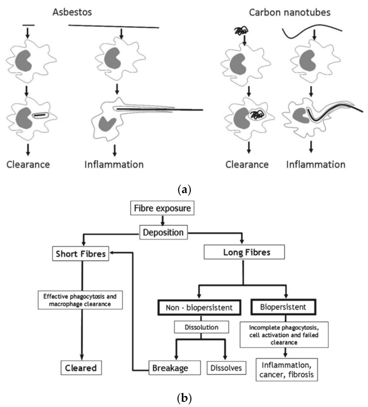
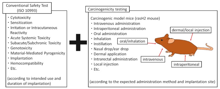
With the development of nanotechnology in recent years, there have been concerns about the health effects of nanoparticles. Carbon nanotubes (CNTs) are fibrous nanoparticles with a micro-sized length and nano-sized diameter, which exhibit excellent physical properties and are widely studied for their potential application in medicine. However, asbestos has been historically shown to cause pleural malignant mesothelioma and lung cancer by inhalation exposure. Because carbon nanotubes are also fibrous nanotubes, some have raised concerns about its possible carcinogenicity. We have reported that there is no clear evidence of carcinogenicity by local and intravenous administration of multi-walled CNTs to cancer mice models. We firmly believe that CNTs can be a safe, new, and high-performance biomaterials by controlling its type, site of administration, and dosage.

Keywords: biocompatibility; carbon nanotubes; carcinogenicity.


Conflict of interest statement

The authors declare no conflict of interest.

Figures

Figure 1
Figure 1
(a) Scanning electron microscopy (SEM) image of MWCNTs. Scale bar, 2 µm. (b) SEM image of asbestos (chrysotile). Scale bar, 20 µm. Reproduced with permission from [10]. Elsevier, 2010.
Figure 2
Figure 2
A histopathological image of the skin of ddY mice, subcutaneously administered with MWCNTs. (a) Group administered with MWCNT; (b) group administered with tattoo ink. In both groups, carbon particles are observed in the macrophages at one week following administration with slight inflammatory cell infiltration around them. At four weeks, carbon particles have remained in the macrophages, and the inflammatory reaction has subsided. Scale bar, 20 μm. Reproduced with permission from [16]. Elsevier, 2011.
Figure 3
Figure 3
(a) Histopathology of ddY mice administered with MWCNTs in the subperiosteum of the skull. MWCNTs were incorporated into macrophages, and no osteolysis was observed in the skull. Scale bar, 20 μm; (b) Histopathology of ddY mice, in which MWCNTs were administered into a tibial bone tunnel. One week after administration, woven bone is formed around MWCNTs, and after 4 weeks, bony cortex is regenerated with MWCNTs incorporated into the bone matrix. Scale bar, 20 μm. Reproduced with permission from [41]. John Wiley and Sons, 2008.
Figure 4
Figure 4
Survival curve of rasH2 mice with local subcutaneous administration of MWCNTs in the dorsum. The survival rate at 26 weeks after administration was 60% in the group that received MNU, a carcinogen, compared to 100% in the group that received MWCNTs. The survival rate of the carbon black group is almost equivalent to tattoo ink at 90%, but the carcinogenic case is likely a coincidence. Image is modified from a study by Takanashi et al. [58].
Figure 5
Figure 5
(a) Formation of Mesothelioma in the lung of rasH2 mice intravenously administered with MWCNTs. The right is an enlarged image of the square in the left image; (b) histological image of the pancreas in rasH2 mice administered intravenously with MWCNTs. Particles of MWCNTs are deposited in the tissue. There is no tumor formation. The right is an enlarged image of the square in the left image. Images are modified from a study by Sobajima et al. [59].
Figure 6
Figure 6
(a) When nanofibers such as asbestos (left) and CNTs (right) are phagocytosed by macrophages, inflammation may persist and tumors may be formed if long fibers trigger frustrated phagocytosis. Image is modified from a study by Donaldson et al. [60]. (b) Long, biopersistent nanofibers may induce inflammation, cancer, and fibrosis. Reproduced with permission from [61]. Elsevier, 2013.
Figure 7
Figure 7
Bio-safety testing of nano-sized biomaterials. Carcinogenicity tests are carried out by using carcinogenic model mice, in addition to conventional safety tests according to the intended application and implantation period of each biomaterial according to ISO 10993-1. The method of administration is selected according to the expected administration method of nano-biomaterials to humans and the implantation site.

Similar articles

Cited by

References

    1. Lu P.J., Cheng W.L., Huang S.C., Chen Y.P., Chou H.K., Cheng H.F. Characterizing titanium dioxide and zinc oxide nanoparticles in sunscreen spray. Int. J. Cosmet. Sci. 2015;37:620–626. doi: 10.1111/ics.12239. - DOI - PubMed
    1. McSweeney P.C. The safety of nanoparticles in sunscreens: An update for general practice. Aust. Fam. Phys. 2016;45:397–399. - PubMed
    1. Morita T., Takami N. Nano Si cluster-SiOx-C composite material as high-capacity anode material for rechargeable lithium batteries. J. Electrochem. Soc. 2006;153:A425–A430. doi: 10.1149/1.2142295. - DOI
    1. Aagaard J. The Carbomedics aortic heart valve prosthesis: A review. J. Cardiovasc. Surg. (Torino) 2004;45:531–534. - PubMed
    1. Morice M.C., Bestehorn H.P., Carrie D., Macaya C., Aengevaeren W., Wijns W., Dubois C., de Winter R., Verheye S., Hoffmann S., et al. Direct stenting of de novo coronary stenoses with tacrolimus-eluting versus carbon-coated carbostents. The randomized JUPITER II trial. EuroIntervention. 2006;2:45–52. - PubMed

LinkOut - more resources