Review
. 2020 Feb 5;13(3):705.
doi: 10.3390/ma13030705.

Surface Characterization of Electro-Assisted Titanium Implants: A Multi-Technique Approach

Affiliations
Review

Surface Characterization of Electro-Assisted Titanium Implants: A Multi-Technique Approach

Stefania Cometa et al. Materials (Basel). .

Abstract

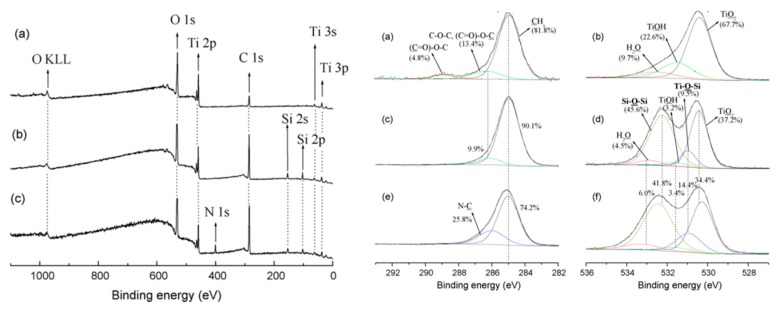
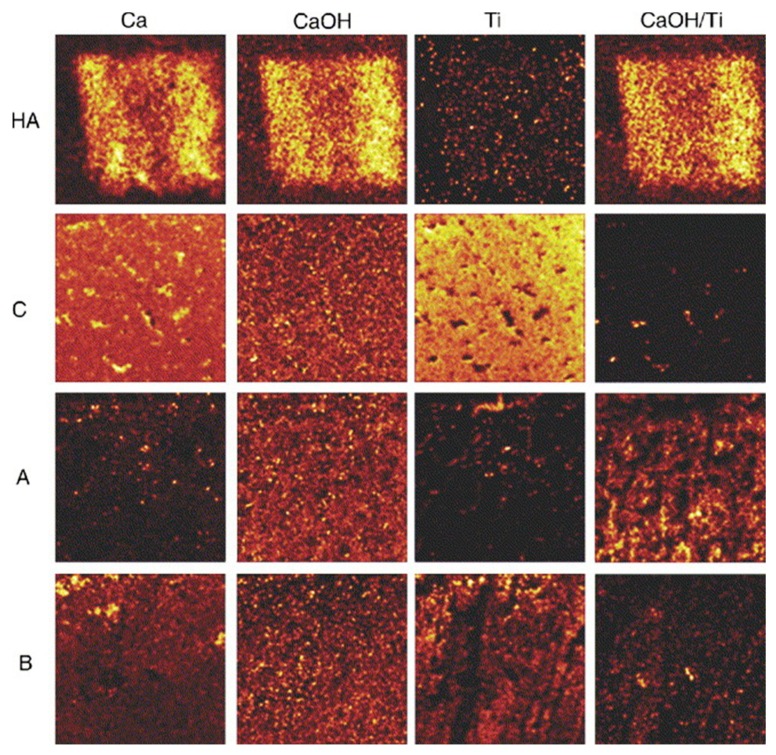
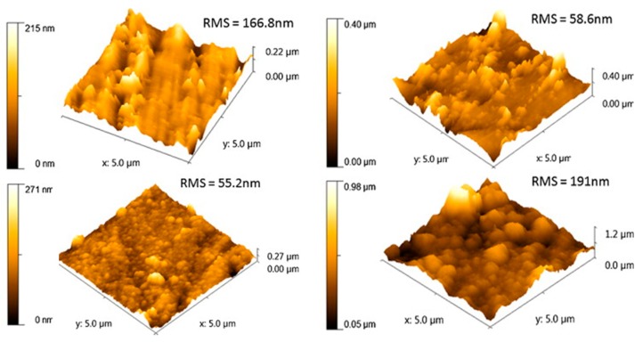
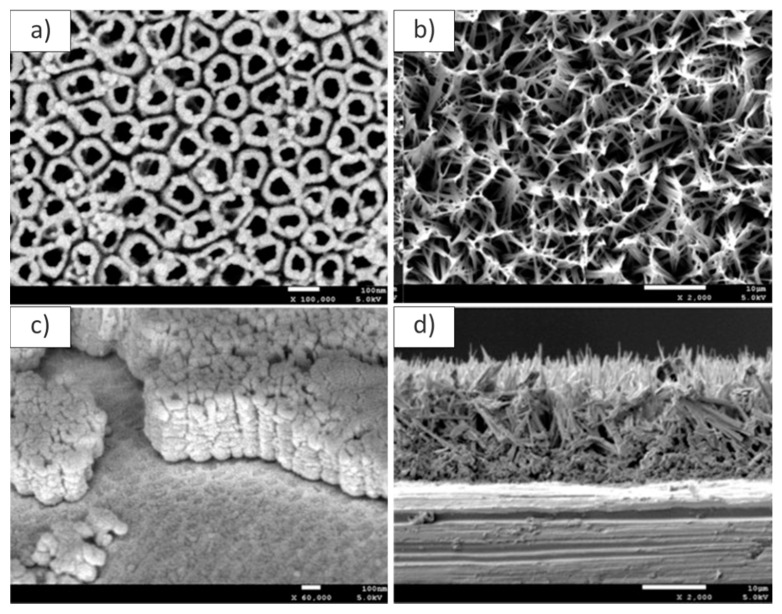
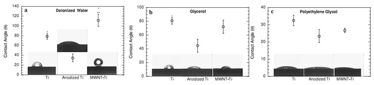
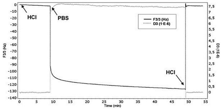
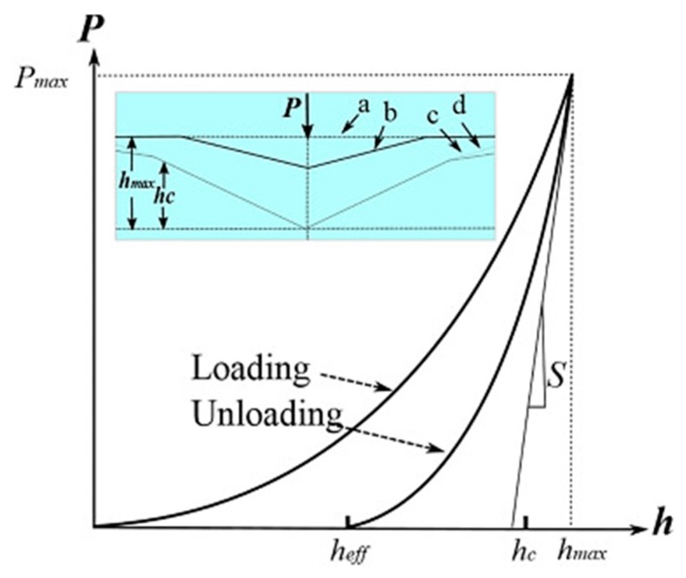
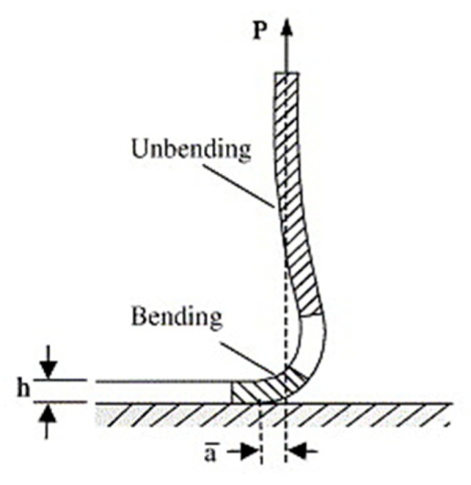
The understanding of chemical-physical, morphological, and mechanical properties of polymer coatings is a crucial preliminary step for further biological evaluation of the processes occurring on the coatings' surface. Several studies have demonstrated how surface properties play a key role in the interactions between biomolecules (e.g., proteins, cells, extracellular matrix, and biological fluids) and titanium, such as chemical composition (investigated by means of XPS, TOF-SIMS, and ATR-FTIR), morphology (SEM-EDX), roughness (AFM), thickness (Ellipsometry), wettability (CA), solution-surface interactions (QCM-D), and mechanical features (hardness, elastic modulus, adhesion, and fatigue strength). In this review, we report an overview of the main analytical and mechanical methods commonly used to characterize polymer-based coatings deposited on titanium implants by electro-assisted techniques. A description of the relevance and shortcomings of each technique is described, in order to provide suitable information for the design and characterization of advanced coatings or for the optimization of the existing ones.

Keywords: analytical characterization; mechanical tests; morphology; physico-chemical study; polymeric coatings; surface properties; titanium.


Conflict of interest statement

The authors declare no conflict of interest.

Figures

Figure 1
Figure 1
On the left: X-ray photoelectron spectroscopy (XPS) survey spectra of (a) titanium substrates and titanium substrates modified (2 h of reaction) by (b) HTCS and (c) PyHTCS. On the right: XPS of C1s and O1s high resolution spectra of titanium substrates (a,b) and titanium substrates modified (2 h of reaction) by HTCS (c,d) PyHTCS (e,f). (Reprinted with permission from [35], Elsevier 2008 copyright n. 4710120673412).
Figure 2
Figure 2
Time-of-Flight Secondary Ion Mass Spectrometry (TOF-SIMS) images showing the secondary ion distributions for the hydroxyapatite control (HA), not anodized titanium control after implantation (C), and the implanted porous surfaces A and B. The figure shows the results for Ca2+, CaOH, Ti, and a normalization image calculated from CaOH and Ti images of the sample. All images were normalized to the total ion count. The field of view is 200 μm × 200 μm, except for HA, which is 500 μm × 500 μm. (Reprinted with permission from [47], Elsevier 2006 copyright n. 4710120857878).
Figure 3
Figure 3
3D atomic force microscopy (AFM) images for Ti/24PDA and Ti/72PDA. Below, Ti/24PDA–PPy and Ti/72PDA–PPy. (Reprinted with permissions from [73], Elsevier 2014 copyright n. 4710121133177).
Figure 4
Figure 4
(a) FESEM image of TiO2 nanotubes and (b) GelGOHA composite coating on titanium. (c) Cross-section of TiO2 nanotubes and (d) GelGOHA coating on titanium. (Reprinted with permission from [40], Elsevier 2015 copyright n. 4710121336953).
Figure 5
Figure 5
Contact angle measurements with three liquids using an EasyDrop model system (Kr¨uss): (a) double-deionized water, (b) glycerol, and (c) polyethylene glycol (PEG), all on conventional pure Ti, anodized Ti, and MWNT–Ti. Data = mean ± SEM; n = 3; * p < 0.01 compared to anodized Ti and ** p < 0.01 compared to Ti. (Reprinted with permissions from [38], IOP Publishing Group 2011 copyright n. 1003986).
Figure 6
Figure 6
QCM-D study of the swelling–deswelling of PEGDA-co-AA hydrogel coating at different pH solutions. Arrows and labels indicate injections of HCl (pH 2.2) solution, PBS (pH 7.4), and the final HCl (pH 2.2) solution, respectively. (Reprinted with permission from [25], Whiley 2009 copyright n. 4710141377265).
Figure 7
Figure 7
A Schematic representation of a load–displacement curve in indentation testing. (a) Initial surface; (b) surface after load removal; (c) indenter; (d) surface profile under load. P is the peak indentation load; hmax is the indenter displacement at peak load; heff is the final depth of contact impression after unloading; and S is the initial unloading stiffness. (Reprinted with permission from [113], Materials Research Society 1992 copyright n. 4710150228702).
Figure 8
Figure 8
Peel test mechanism. (Adapted and reprinted with permission from [125], Elsevier 2005 copyright n. 4710150365138).
Figure 9
Figure 9
SEM micrograph of the crack growth in a Halar®-coated Ti specimen. (Reprinted with permission from [61], Elsevier 2018 copyright n. 4710150493083).

References

    1. Cometa S., Bonifacio M.A., Mattioli-Belmonte M., Sabbatini L., De Giglio E. Electrochemical strategies for titanium implant polymeric coatings: The why and how. Coatings. 2019;9:268–287. doi: 10.3390/coatings9040268. - DOI
    1. Kumar A.M., Adesina A.Y., Hussein M.A., Ramakrishna S., Al-Aqeeli N., Akhtar S., Saravanan S. PEDOT/FHA nanocomposite coatings on newly developed Ti-Nb-Zr implants: Biocompatibility and surface protection against corrosion and bacterial infections. Mater. Sci. Eng. C. 2019;98:482–495. doi: 10.1016/j.msec.2019.01.012. - DOI - PubMed
    1. Kokubo T., Yamaguchi S. Simulated body fluid and the novel bioactive materials derived from it. J. Biomed. Mater. Res. Part A. 2019;107:968–977. doi: 10.1002/jbm.a.36620. - DOI - PubMed
    1. Gil F.J., Rodriguez A., Espinar E., Llamas J.M., Padullés E., Juàrez A. Effect of oral behavior of Titanium Dental Implants. Int. J. Oral Maxillofac. Implants. 2012;27:64–68. - PubMed
    1. Su Y., Cockerill I., Zheng Y., Tang L., Qin Y.X., Zhu D. Biofunctionalization of metallic implants by calcium phosphate coatings. Bioact. Mater. 2019;4:196–206. doi: 10.1016/j.bioactmat.2019.05.001. - DOI - PMC - PubMed

LinkOut - more resources