. 2020 Feb 5;13(3):706.
doi: 10.3390/ma13030706.

VEGF/VEGF-R/RUNX2 Upregulation in Human Periodontal Ligament Stem Cells Seeded on Dual Acid Etched Titanium Disk

Affiliations

VEGF/VEGF-R/RUNX2 Upregulation in Human Periodontal Ligament Stem Cells Seeded on Dual Acid Etched Titanium Disk

Francesca Diomede et al. Materials (Basel). .

Abstract

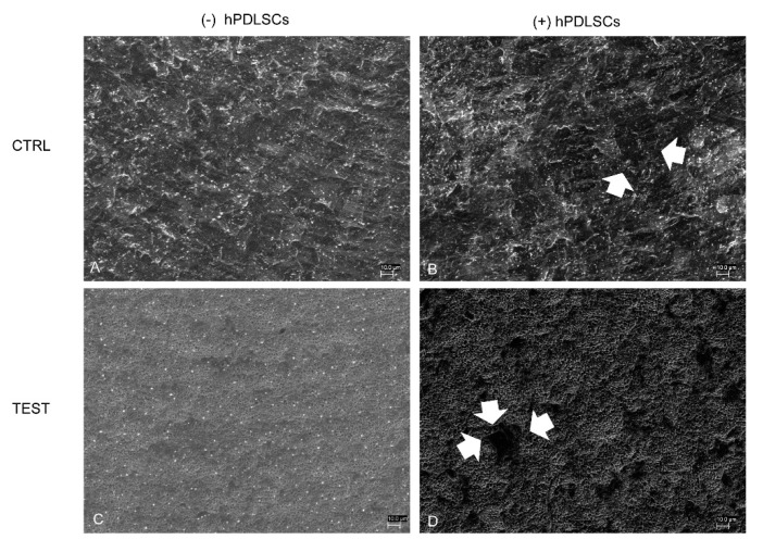
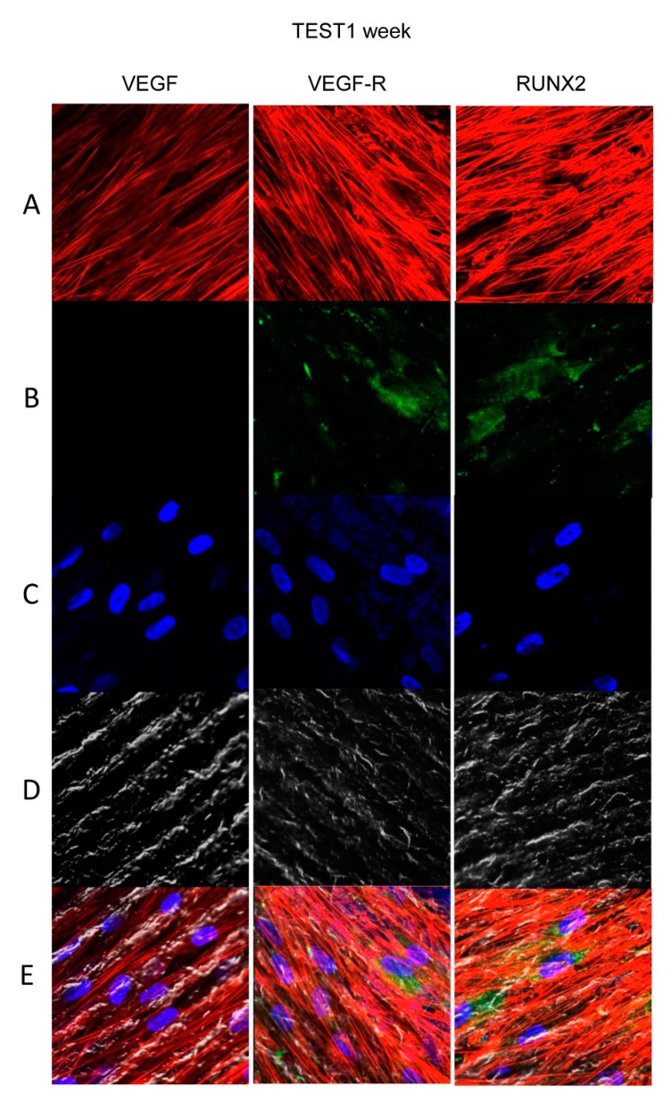
In restorative dentistry, the main implants characteristic is the ability to promote the osseointegration process as the result of interaction between angiogenesis and osteogenesis events. On the other hand, implants cytocompatibility remains a necessary feature for the success of surgery. The purpose of the current study was to investigate the interaction between human periodontal stem cells and two different types of titanium surfaces, to verify their cytocompatibility and cell adhesion ability, and to detect osteogenic and angiogenic markers, trough cell viability assay (MTT), Confocal Laser Scanning Microscopy (CLSM), scanning electron microscopy (SEM), and gene expression (RT-PCR). The titanium surfaces, machined (CTRL) and dual acid etched (TEST), tested in culture with human periodontal ligament stem cells (hPDLSCs), were previously treated in two different ways, in order to evaluate the effects of CTRL and TEST and define the best implant surface. Furthermore, the average surface roughness (Ra) of both titanium surfaces, CTRL and TEST, has been assessed through atomic force microscopy (AFM). The vascular endothelial growth factor (VEGF) and Runt-related transcription factor 2 (RUNX2) expressions have been analyzed by RT-PCR, WB analysis, and confocal laser scanning microscopy. Data evidenced that the different morphology and topography of the TEST disk increased cell growth, cell adhesion, improved osteogenic and angiogenic events, as well osseointegration process. For this reason, the TEST surface was more biocompatible than the CTRL disk surface.

Keywords: angiogenesis; cytocompatibility; osseointegration; osteogenesis.


Conflict of interest statement

The authors declare no conflict of interest.

Figures

Figure 1
Figure 1
(A,C) Topographical and (B,D) Three-dimensional Atomic Force Miscroscopy (AFM) pictures of machined titanium surface (CTRL, upper line) and dual acid etched titanium surface (TEST, bottom line).
Figure 2
Figure 2
Scanning electron microscopy (SEM) analysis of the human periodontal ligament stem cells (hPDLSCs) cultured on the titanium implant surfaces. (A) CTRL surface without cells. (B) TEST surface without cells. (C) Human PDLSCs adhere on CTRL surface. (D) Human PDLSCs adhere on TEST surface. Mag: 1000×.
Figure 3
Figure 3
Cell viability assay of hPDLSCs cultured on CTRL and TEST titanium surfaces. Trough cell viability assay (MTT) assay was performed at 24, 48, 72 h, and 1 week on both CTRL and TEST titanium surfaces.
Figure 4
Figure 4
Human PDLSCs cultured on CTRL titanium implant surface were observed after 1 week of being cultured. (A) Cytoskeleton actin was dyed in red fluorescence; (B) specific markers (vascular endothelial growth factor [VEGF], VEGF-R, and Runt-related transcription factor 2 [RUNX2]) were stained in green fluorescence; (C) nuclei were dyed in blue fluorescence. (D) PDLSCs with CTRL; (E) TL, transmission light: gray. Scale bar: 10 µm.
Figure 5
Figure 5
Human PDLSCs cultured on CTRL titanium implant surface were observed after 8 weeks of being cultured. (A) Cytoskeleton actin was dyed in red fluorescence; (B) specific markers (VEGF, VEGF-R, and RUNX2) were stained in green fluorescence; (C) nuclei were dyed in blue fluorescence. (D) PDLSCs with CTRL; (E) TL, transmission light: gray. Scale bar: 10 µm.
Figure 6
Figure 6
Human PDLSCs cultured on TEST titanium implant surface were observed after 1 week of incubation. (A) Cytoskeleton actin was stained in red fluorescence; (B) specific markers (VEGF, VEGF-R, and RUNX2) were dyed in green fluorescence; (C) nuclei were dyed in blue fluorescence. (D) PDLSCs with TEST; (E) TL, transmission light: gray. Scale bar: 10 µm.
Figure 7
Figure 7
Human PDLSCs cultured on TEST titanium implant surface were observed after 8 weeks of incubation. (A) Cytoskeleton actin was dyed in red fluorescence; (B) specific markers (VEGF, VEGF-R, and RUNX2) were stained in green fluorescence; (C) nuclei were dyed in blue fluorescence. (D) PDLSCs with TEST; (E) TL, transmission light: gray. Scale bar: 10 µm.
Figure 8
Figure 8
(A) Graph bar of RT-PCR of VEGF and RUNX2 in cells cultured on CTRL and TEST surface ** p < 0.01. (B) Protein-level expression of VEGF and RUNX2 in cells cultured on CTRL and TEST surface. (C) Graphs report densitometric measurements of proteins bands expressed as integrated optical intensity (IOI) mean of three separate analysis.

Similar articles

Cited by

References

    1. Rajan T.S., Scionti D., Diomede F., Grassi G., Pollastro F., Piattelli A., Cocco L., Bramanti P., Mazzon E., Trubiani O. Gingival Stromal Cells as an In Vitro Model: Cannabidiol Modulates Genes Linked With Amyotrophic Lateral Sclerosis. J. Cell. Biochem. 2017;118:819–828. doi: 10.1002/jcb.25757. - DOI - PubMed
    1. Yang B., Qiu Y., Zhou N., Ouyang H., Ding J., Cheng B., Sun J. Application of Stem Cells in Oral Disease Therapy: Progresses and Perspectives. Front. Physiol. 2017;8:197. doi: 10.3389/fphys.2017.00197. - DOI - PMC - PubMed
    1. Fellows C.R., Matta C., Zakany R., Khan I.M., Mobasheri A. Adipose, Bone Marrow and Synovial Joint-Derived Mesenchymal Stem Cells for Cartilage Repair. Front. Genet. 2016;7:213. doi: 10.3389/fgene.2016.00213. - DOI - PMC - PubMed
    1. Sanz A.R., Carrion F.S., Chaparro A.P. Mesenchymal stem cells from the oral cavity and their potential value in tissue engineering. Periodontology 2000. 2015;67:251–267. doi: 10.1111/prd.12070. - DOI - PubMed
    1. Di Nisio C., De Colli M., di Giacomo V., Rapino M., Di Valerio V., Marconi G.D., Gallorini M., Di Giulio M., Cataldi A., Zara S. A dual role for beta1 integrin in an in vitro Streptococcus mitis/human gingival fibroblasts co-culture model in response to TEGDMA. Int. Endod. J. 2015;48:839–849. doi: 10.1111/iej.12379. - DOI - PubMed

LinkOut - more resources