. 2020 Feb 5;12(2):411.
doi: 10.3390/nu12020411.

Inhibitory Effect of Centella asiatica Extract on DNCB-Induced Atopic Dermatitis in HaCaT Cells and BALB/c Mice

Affiliations

Inhibitory Effect of Centella asiatica Extract on DNCB-Induced Atopic Dermatitis in HaCaT Cells and BALB/c Mice

Yonghyeon Lee et al. Nutrients. .

Abstract

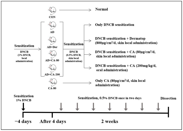
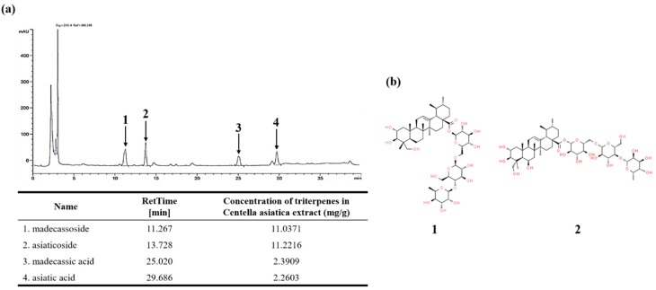
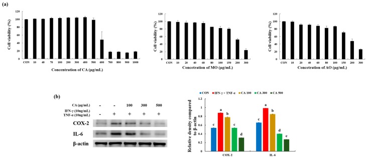
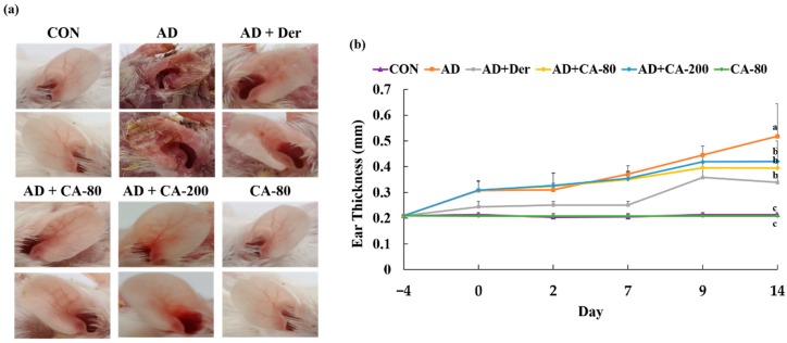
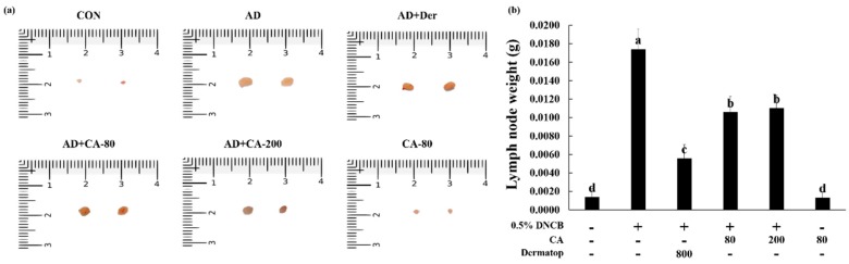
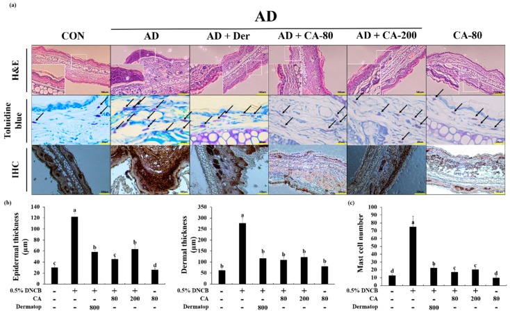
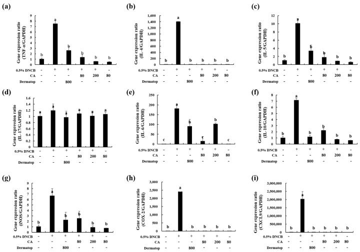
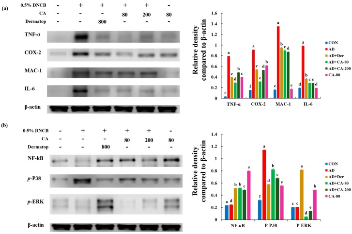
Atopic dermatitis (AD) is a chronic inflammatory skin disease caused mainly by immune dysregulation. This study explored the anti-inflammatory and immunomodulatory effects of the Centella asiatica ethanol extract (CA) on an AD-like dermal disorder. Treatment with CA inhibited the expression of interleukin-6 (IL-6) and tumor necrosis factor-α (TNF-α) in a dose-dependent manner in inflammatory stimulated HaCaT cells by interferon-γ (IFN-γ) and TNF-α-triggered inflammation. Eight-week-old BALB/c mice treated with 2,4-dinitrochlorobenzene (DNCB) were used as a mouse model of AD. In AD induce model, we had two types treatment of CA; skin local administration (80 µg/cm2, AD+CA-80) and oral administration (200 mg/kg/d, AD+CA-200). Interestingly, the CA-treated groups exhibited considerably decreased mast cell infiltration in the ear tissue. In addition, the expression of IL-6 in mast cells, as well as the expression of various pathogenic cytokines, such as TNF-α, IL-4, IL-5, IL-6, IL-10, IL-17, iNOS, COX-2, and CXCL9, was reduced in both AD+CA-80 and AD+CA-200 groups. Collectively, our data demonstrate the pharmacological role and signaling mechanism of CA in the regulation of allergic inflammation of the skin, which supports our hypothesis that CA could potentially be developed as a therapeutic agent for AD.

Keywords: Centella asiatica ethanol extract; anti-inflammation; asiaticoside; atopic dermatitis; madecassoside.


Conflict of interest statement

The authors declare no conflict of interest.

Figures

Figure 1
Figure 1
Experimental design of atopic dermatitis (AD) -like skin lesions mice model. BALB/c mice were randomly divided into six groups (n = 8 per group). AD-like skin lesions were evoked by administering 1% 2,4-dinitrochlorobenzene (DNCB). 200 µL of 0.5% DNCB was applied on each ear, and after 4 days. DNCB was alternatively repeatedly treated once in two days for 2 weeks. Centella asiatica ethanol extract (CA) was administrated by skin local administration (80 µg/cm2/d) and oral administration (200 mg/kg/d). The Dermatop was skin local administrated (800 µg/cm2/d). Without inducing AD, CA (80 µg/cm2/d) was skin locally administrated for 2 weeks.
Figure 2
Figure 2
(a) High-performance liquid chromatography (HPLC) chromatography of CA; (b) Chemical structures of major components. The HPLC analysis on CA was performed with a reverse-phase column (SunFire C18, 4.6 × 250 mm, 5-um diameter; Waters, Milford, MA, USA). The column was preserved under 40 °C at the flow rate of 1 mL/min and the injection volume of 10 µL. 1: madecassoside (MO), 2: asiaticoside (AO), 3: madecassic acid (MA), 4: asiatic acid (AA).
Figure 3
Figure 3
Polyphenol and flavonoid content, and antioxidant capacities of CA. (a) Total polyphenol and flavonoid; (b) 2,2’-azinobis-(3-ethylbenzothiazoline-6-sulfonic acid) (ABTS) and 2,2-diphenyl-1-picrylhydrazyl (DPPH) scavenging activity. Values represent the mean ± SD. Values with different letters were significantly different according to Duncan’s multiple range test (p< 0.05). GAE: gallic acid equivalents, CHE: (+)-catechin hydrate equivalents.
Figure 4
Figure 4
(a) 3-(4,5-dimethylthiazol-2-yl)-2,5-diphenyltetrazolium bromide (MTT) assay for the cell toxicity of CA, MO, and AO in HaCaT cells treated with different concentrations of CA at 37 °C for 24 h under 5% CO2; (b) Effects of CA on the expression of inflammation-related proteins in HaCaT cells stimulated by tumor necrosis factor-α (TNF-α) and interferon-γ (IFN-γ) in a CA dose-dependent manner. Values represent the mean ± SD. Values with different letters were significantly different according to Duncan’s multiple range test (p < 0.05).
Figure 5
Figure 5
Histopathological analysis to assess the effect of CA on mice ear thickness. (a) Photographs of mice ears from each group on day 14; (b) The ear thickness was measured 24 h after DNCB application with a dial thickness gauge. Values represent the mean ± SD. Values with different letters were significantly different according to Duncan’s multiple range test (p < 0.05).
Figure 6
Figure 6
(a,b) Photographs represent the size and weight of lymph nodes. Values represent the mean ± SD. Values with different letters were significantly different according to Duncan’s multiple range test (p < 0.05).
Figure 7
Figure 7
(a) Histological analysis to assess the effect of CA on epidermal and dermal thickness, and mast cell infiltration. Representative photomicrographs of ear sections stained with hematoxylin and eosin (H&E) or toluidine blue. In the toluidine blue staining panel, black arrows denote mast cells. In immunohistochemistry (IHC), a strong difference in staining intensity for NF-κB between atopic dermatitis model and CA treatment model was noted; (b) Photographs were taken under a regular light microscope at a magnification of 100× (H&E and IHC) and 400× (toluidine blue staining); (c) The epidermal and dermal thickness was gauged using microphotographs of hematoxylin and eosin stained tissue. The number of infiltrated mast cells was counted on the basis of toluidine blue staining. Values represent the mean ± SD. Values with different letters were significantly different according to Duncan’s multiple range test (p< 0.05).
Figure 8
Figure 8
Effect of CA on the expression of diverse pathogenic factors in the ear. (a), TNF-α; (b), IL-4; (c), IL-5; (d), IL-17; (e), IL-6; (f), IL-10 (g), iNOS; (h), COX-2; (i), CXCL9. The total RNA was isolated from the ear tissue. Quantitative real-time PCR was performed as described in the Materials and Methods. Values represent the mean ± SD. Values with different letters were significantly different according to Duncan’s multiple range test (p < 0.05).
Figure 9
Figure 9
Alleviation of increased expression of (a) various pathogenic cytokines and (b) NF-κB and mitogen-activated protein kinase (MAPK) proteins in AD-induced mice model, following treatment with CA-80 and CA-200. Values represent the mean ± SD. Values with different letters were significantly different according to Duncan’s multiple range test (p < 0.05).

References

    1. Biedermann T., Skabytska Y., Kaesler S., Volz T. Regulation of T Cell Immunity in Atopic Dermatitis by Microbes: The Yin and Yang of Cutaneous Inflammation. Front. Immunol. 2015;6:353. doi: 10.3389/fimmu.2015.00353. - DOI - PMC - PubMed
    1. Peng W., Novak N. Pathogenesis of atopic dermatitis. Clin. Exp. Allergy. 2015;45:566–574. doi: 10.1111/cea.12495. - DOI - PubMed
    1. Avena-Woods C. Overview of Atopic Dermatitis. Am. J. Manag. Care. 2017;23:115–123. - PubMed
    1. Nutten S. Atopic Dermatitis: Global Epidemiology and Risk Factors. Ann. Nutr. Metab. 2015;66:8–16. doi: 10.1159/000370220. - DOI - PubMed
    1. Park J.H., Choi J.Y., Son D.J., Park E.K., Song M.J., Hellström M.J., Hong J.T. Anti-Inflammatory Effect of Titrated Extract of Centella asiatica in Phthalic Anhydride-Induced Allergic Dermatitis Animal Model. Int. J. Mol. Sci. 2017;18:738. doi: 10.3390/ijms18040738. - DOI - PMC - PubMed