. 2020 Feb 5;9(2):428.
doi: 10.3390/jcm9020428.

Chronic Oral Palmitoylethanolamide Administration Rescues Cognitive Deficit and Reduces Neuroinflammation, Oxidative Stress, and Glutamate Levels in A Transgenic Murine Model of Alzheimer's Disease

Affiliations

Chronic Oral Palmitoylethanolamide Administration Rescues Cognitive Deficit and Reduces Neuroinflammation, Oxidative Stress, and Glutamate Levels in A Transgenic Murine Model of Alzheimer's Disease

Sarah Beggiato et al. J Clin Med. .

Abstract

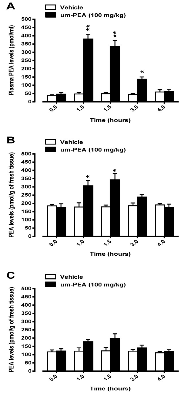
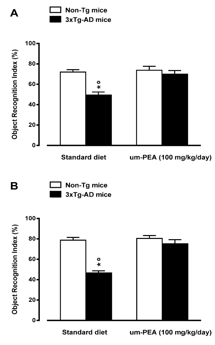
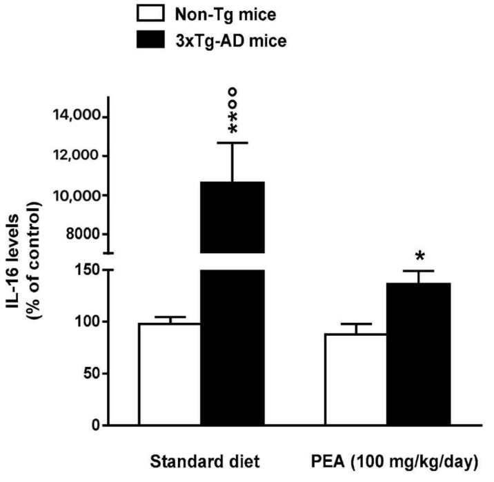
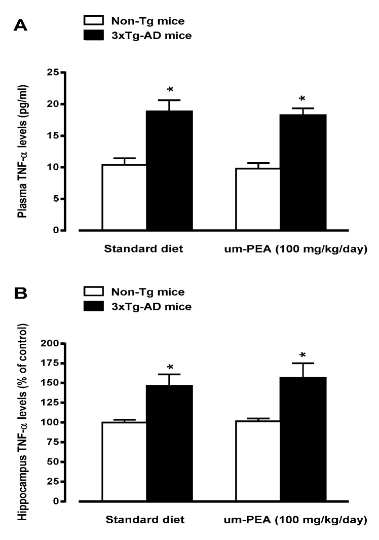
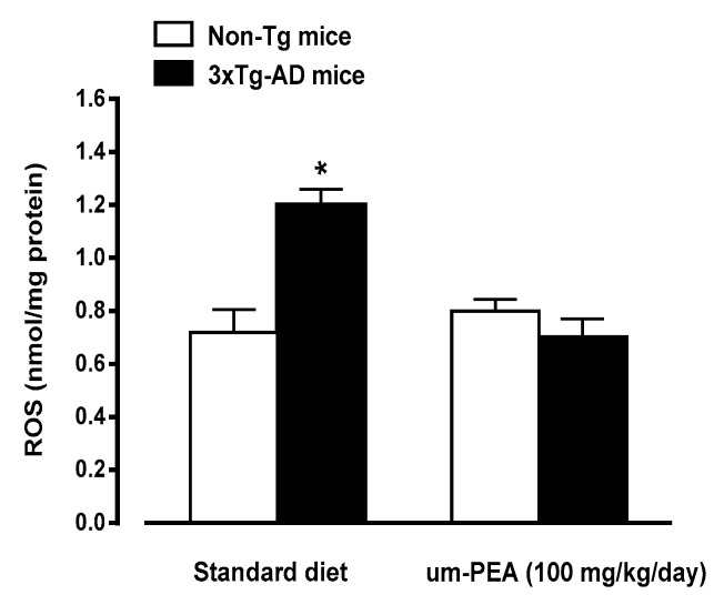
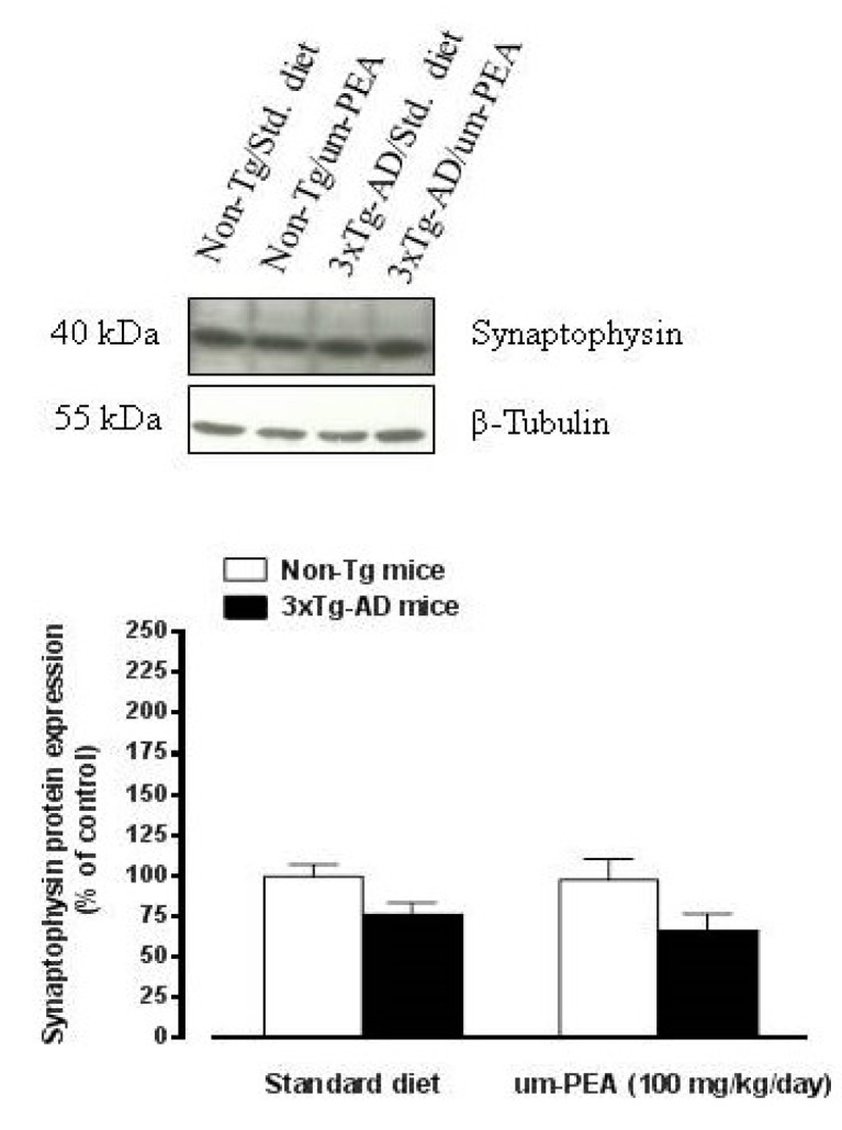
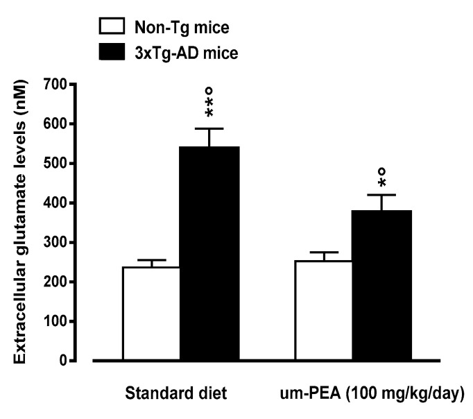
N-palmitoylethanolamide (PEA) is a lipid mediator belonging to the class of the N-acylethanolamine. Products containing PEA, also in ultramicronized formulation (um-PEA), are already licensed for use in humans for its analgesic and anti-inflammatory properties, and demonstrated high safety and tolerability. Preclinical studies indicate that PEA, especially in the ultramicronized form, could be a potential therapeutic agent for Alzheimer's disease (AD). In this study, we evaluated the neuroprotective and antioxidant effects of chronic (three months) um-PEA administration in an animal model of AD (3×Tg-AD mice). For translation purposes, the compound has been orally administered. Cognitive performance as well as biochemical markers [(interleukin-16 (IL-16) and tumor necrosis factor- (TNF-)] levels, reactive oxygen species (ROS) production, synaptophysin and glutamate levels) have been evaluated at the end of um-PEA treatment. The results indicate that orally administered um-PEA was adsorbed and distributed in the mice brain. The chronic treatment with um-PEA (100 mg/kg/day for three months) rescued cognitive deficit, restrained neuroinflammation and oxidative stress, and reduced the increase in hippocampal glutamate levels observed in 3×Tg-AD mice. Overall, these data reinforce the concept that um-PEA exerts beneficial effects in 3×Tg-AD mice. The fact that PEA is already licensed for the use in humans strongly supports its rapid translation in clinical practice.

Keywords: Alzheimer’s disease; cognitive dysfunctions; extracellular glutamate levels; hippocampus; reactive oxygen species; synaptophysin.


Conflict of interest statement

No conflict of interest.

Figures

Figure 1
Figure 1
Levels of palmitoylethanolamide (PEA) in plasma (panel A), hippocampus (panel B) and prefrontal cortex (panel C) of non-Tg mice 1, 1.5, 3, and 4 hours after oral administration (by gavage) of 100 mg/kg ultramicronized palmitoylethanolamide (um-PEA) or its vehicle [water/PEG/Tween-80 (90/5/5 ratio), v/v)]. Data are means ± SEM of five animals for each group. * p < 0.05; ** p < 0.01 significantly different from the respective vehicle time-point (Student t-test).
Figure 2
Figure 2
Oral ultramicronized palmitoylethanolamide (um-PEA) treatment rescues short-(3 h; panel A) and long-term (24 h; panel B) memory deficits in the 3×Tg-AD mice, as evaluated by novel object recognition test. Age-matched non-Tg mice and 3×Tg-AD mice (2 months ± 2 weeks of age) have been orally treated for 3 months with the compound (100 mg/kg/day, added to the animal food), while control animals were fed with a standard diet during the treatment period. Novel object recognition test was performed at the end of the treatment period. The data are presented as means ± SEM of 9–11 animals for each group. * p < 0.05 significantly different from the respective non-Tg mouse group; p < 0.05 significantly different from the respective PEA-treated group according to two-way ANOVA followed by Tukey multiple-comparison test.
Figure 3
Figure 3
Effects of oral ultramicronized palmitoylethanolamide (um-PEA) treatment on interleukin 16 (IL-16) levels in the hippocampus of non-Tg and 3×Tg-AD mice. Age-matched non-Tg mice and 3×Tg-AD mice (2 months ± 2 weeks of age) have been orally treated for 3 months with the compound (100 mg/kg/day, added to the animal food), while control animals were fed with a standard diet during the treatment period. Biochemical analyses were performed on hippocampus tissues taken from mouse brains at the end of the treatment period. The data are expressed as percentage ± SEM of control (standard diet fed non-Tg mice) (n = 4, in triplicate). * p < 0.05, ** p < 0.01 significantly different from the respective non-Tg mouse group; p < 0.01 significantly different from the respective PEA-treated group according to two-way ANOVA followed by Tukey multiple-comparison test.
Figure 4
Figure 4
Effects of oral ultramicronized palmitoylethanolamide (um-PEA) treatment on plasma (panel A) and hippocampus (panel B) tumor necrosis factor alpha (TNF-α) levels in non-Tg and 3×Tg-AD mice. Age-matched non-Tg mice and 3×Tg-AD mice (2 months ± 2 weeks of age) have been orally treated for 3 months with the compound (100 mg/kg/day, added to the animal food), while control animals were fed with a standard diet during the treatment period. Biochemical analyses were performed on hippocampus tissues taken from mouse brains at the end of the treatment period. The data are presented as absolute values (panel A) or as percentage ± SEM of control (standard diet-fed non-Tg mice; Panel B) (n = 4, in triplicate). * p < 0.05 significantly different from the respective non-Tg mouse group according to two-way ANOVA followed by Tukey multiple- comparison test.
Figure 5
Figure 5
Effects of oral ultramicronized palmitoylethanolamide (um-PEA) treatment on reactive oxygen species (ROS) production in hippocampus homogenates from non-Tg and 3×Tg-AD mice. Age-matched non-Tg mice and 3×Tg-AD mice (2 months ± 2 weeks of age) have been orally treated for 3 months with the compound (100 mg/kg/day, added to the animal food), while control animals were fed with a standard diet during the treatment period. ROS production was measured in hippocampus tissues taken from mouse brains at the end of the treatment period. The data are presented as mean ± SEM (n = 5, in triplicate). * p < 0.05 significantly different from the other groups according to two-way ANOVA followed by Tukey multiple-comparison test.
Figure 6
Figure 6
Oral ultramicronized palmitoylethanolamide treatment did not affect synaptophysin levels in the hippocampus of non-Tg and 3×Tg-AD mice. Age-matched non-Tg mice and 3×Tg-AD mice (2 months ± 2 weeks of age) have been orally treated for 3 months with the compound (100 mg/kg/day, added to the animal food), while control animals were fed with a standard diet during the treatment period. Representative western blot densitometric analysis of synaptophysin is reported above the graph. Western blot analysis was performed on hippocampus tissues taken from mouse brains at the end of the treatment period. Experiments were performed three times in triplicate and β-Tubulin was used as loading control. Results are expressed as percentage of the mean ± SEM of control value (i.e., standard diet fed non-Tg mice).
Figure 7
Figure 7
Oral ultramicronized palmitoylethanolamide (um-PEA) treatment rescues increased glutamate levels in the hippocampus of 3×Tg-AD mice. Age-matched non-Tg mice and 3×Tg-AD mice (2 months ± 2 weeks of age) have been orally treated for 3 months with the compound (100 mg/kg/day, added to the animal food), while control animals were fed with a standard diet during the treatment period. Microdialysis experiments were performed at the end of the treatment period. The data are presented as mean ± SEM of 6–7 animals/group. * p < 0.05, ** p < 0.01 significantly different from the respective non-Tg mouse group; p < 0.05 significantly different from the respective PEA-treated group according to two-way ANOVA followed by Tukey multiple-comparison test.

References

    1. Ballard C., Gauthier S., Corbett A., Brayne C., Aarsland D., Jones E. Alzheimer’s disease. Lancet. 2011;377:1019–1031. doi: 10.1016/S0140-6736(10)61349-9. - DOI - PubMed
    1. Cortés N., Andrade V., Maccioni R.B. Behavioral and Neuropsychiatric Disorders in Alzheimer’s Disease. J. Alzheimers Dis. 2018;63:899–910. doi: 10.3233/JAD-180005. - DOI - PubMed
    1. Zvěřová M. Clinical aspects of Alzheimer’s disease. Clin. Biochem. 2019;72:3–6. doi: 10.1016/j.clinbiochem.2019.04.015. - DOI - PubMed
    1. Selkoe D.J., Hardy J. The amyloid hypothesis of Alzheimer’s disease at 25 years. EMBO Mol. Med. 2016;8:595–608. doi: 10.15252/emmm.201606210. - DOI - PMC - PubMed
    1. Edison P., Donat C.K., Sastre M. In vivo Imaging of Glial Activation in Alzheimer’s Disease. Front. Neurol. 2018;9:625. doi: 10.3389/fneur.2018.00625. - DOI - PMC - PubMed