. 2020 Feb 5;21(3):1056.
doi: 10.3390/ijms21031056.

Regulation of Osteoclast Differentiation at Multiple Stages by Protein Kinase D Family Kinases

Affiliations

Regulation of Osteoclast Differentiation at Multiple Stages by Protein Kinase D Family Kinases

Amanda C Leightner et al. Int J Mol Sci. .

Abstract

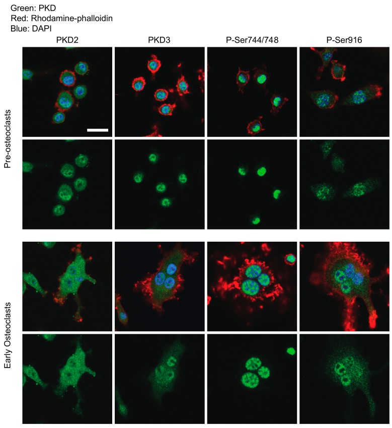
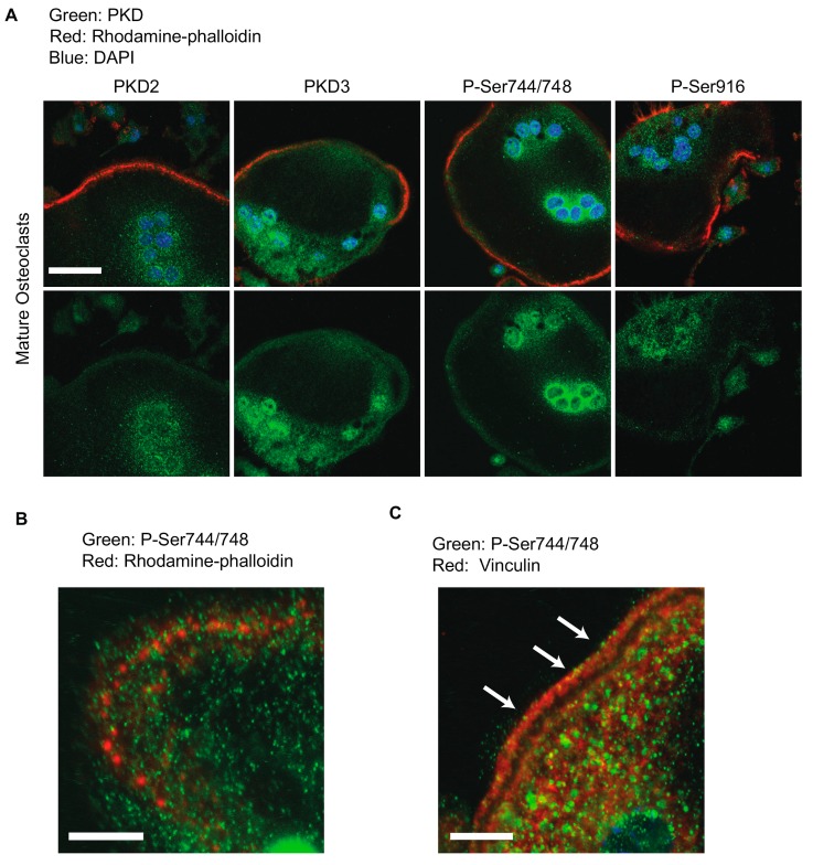
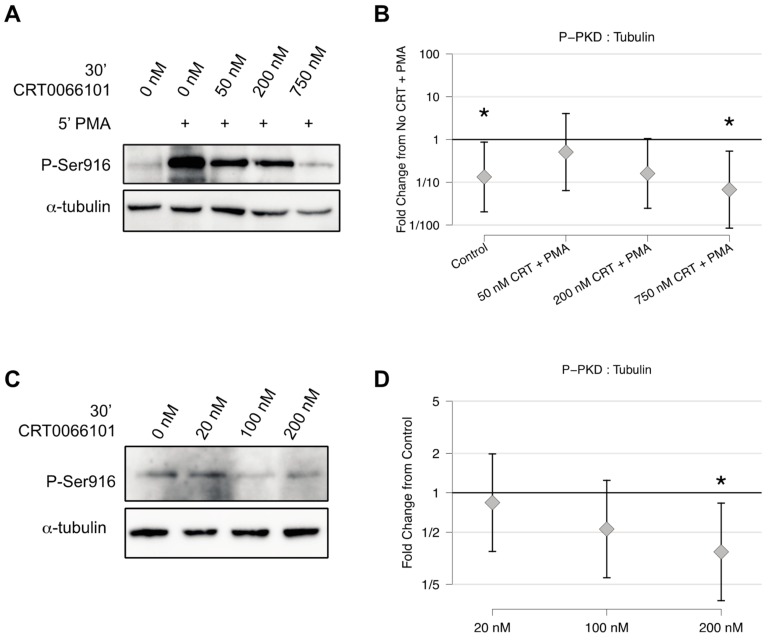
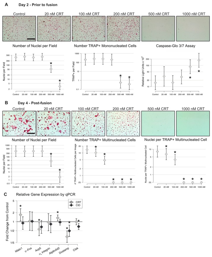
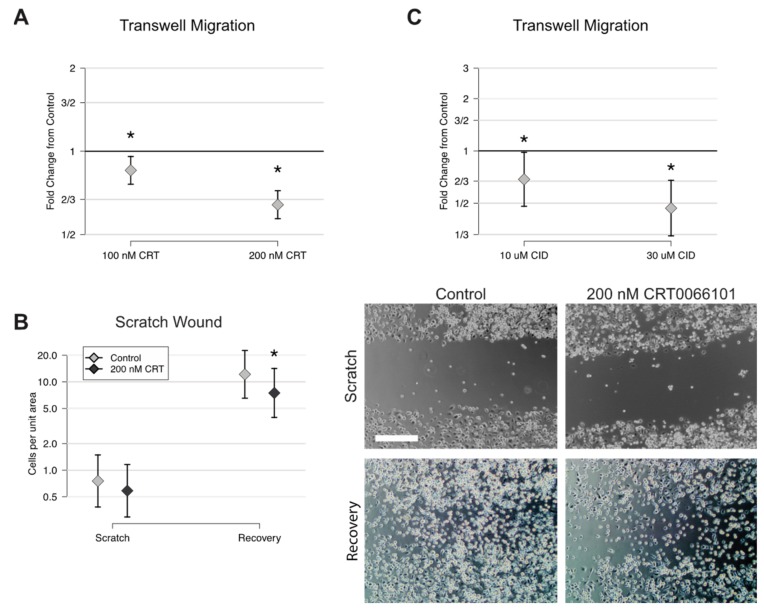
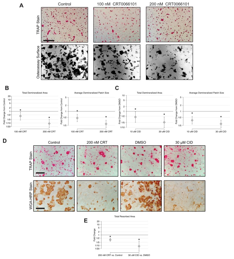
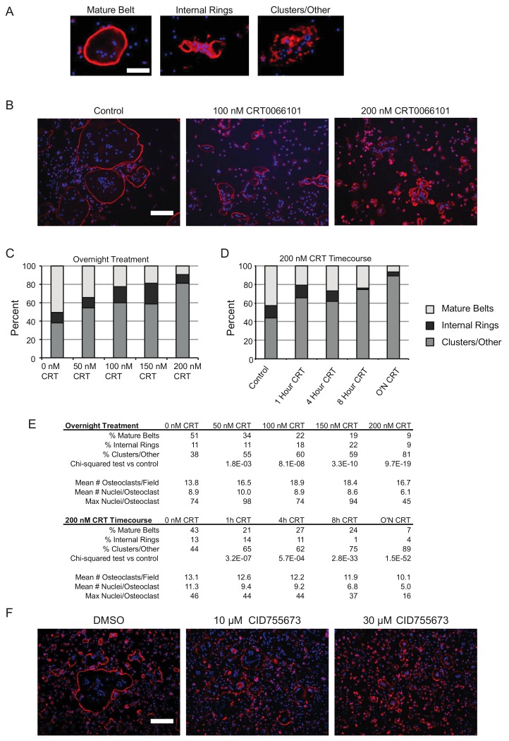
Balanced osteoclast and osteoblast activity is necessary for skeletal health, whereas unbalanced osteoclast activity causes bone loss in many skeletal conditions. A better understanding of pathways that regulate osteoclast differentiation and activity is necessary for the development of new therapies to better manage bone resorption. The roles of Protein Kinase D (PKD) family of serine/threonine kinases in osteoclasts have not been well characterized. In this study we use immunofluorescence analysis to reveal that PKD2 and PKD3, the isoforms expressed in osteoclasts, are found in the nucleus and cytoplasm, the mitotic spindle and midbody, and in association with the actin belt. We show that PKD inhibitors CRT0066101 and CID755673 inhibit several distinct aspects of osteoclast formation. Treating bone marrow macrophages with lower doses of the PKD inhibitors had little effect on M-CSF + RANKL-dependent induction into committed osteoclast precursors, but inhibited their motility and subsequent differentiation into multinucleated mature osteoclasts, whereas higher doses of the PKD inhibitors induced apoptosis of the preosteoclasts. Treating post-fusion multinucleated osteoclasts with the inhibitors disrupted the osteoclast actin belts and impaired their resorptive activity. In conclusion, these data implicate PKD kinases as positive regulators of osteoclasts, which are essential for multiple distinct processes throughout their formation and function.

Keywords: actin cytoskeleton; bone resorption; cellular differentiation; osteoclasts; protein kinase D.


Conflict of interest statement

The authors declare no conflict of interest. The funders had no role in the design of the study; in the collection, analyses, or interpretation of data; in the writing of the manuscript, or in the decision to publish the results.

Figures

Figure 1
Figure 1
Immunofluorescence localization of PKD2 and PKD3 in pre-osteoclasts and early post-fusion osteoclasts. Osteoclast cultures at preosteoclast and early multinucleated osteoclast stages of differentiation were stained with the indicated PKD antibodies (green), rhodamine-phalloidin (for F-actin, red) and DAPI (for DNA, blue) and imaged by confocal microscopy. Merged images are shown above and the corresponding green channel below. Scale bar = 20 µm. Each of these antibody stainings and timepoints has been performed three independent times with similar results.
Figure 2
Figure 2
Immunofluorescence localization of PKD2 and PKD3 in mature osteoclasts. (A) Mature multinucleated osteoclasts were stained with the indicated PKD antibodies (green), rhodamine-phalloidin (for F-actin, red) and DAPI (for DNA, blue) and imaged by confocal microscopy. Scale bar = 20 µm. (B) Higher magnification image showing podosomes assembling into a ring or belt with P-Ser744-positive foci (green) surrounding the F-actin core (red). Scale bar = 10 µm. (C) Higher magnification image showing P-Ser744-positive foci (green) co-localizing with vinculin-rich regions (red) adjacent to the actin belt. Arrows highlight examples of these foci. Scale bar = 10 µm. Each of these antibody stainings has been repeated at least three independent times with similar results.
Figure 3
Figure 3
CRT0066101 inhibits PKD activity in osteoclasts. (A) Preosteoclasts were pre-treated with increasing doses of CRT0066101 for 30 minutes prior to stimulation with 50 mM phorbol 12-myristate 13-acetate (PMA), indicated by ‘+’ for 5 minutes. (B) Quantitation of P-Ser916 levels relative to the level from PMA-alone set as 1.0. * p < 0.05 versus PMA alone. (C) Treatment of preosteoclasts with the indicated CRT0066101 doses for 30 minutes and blotted against endogenous P-Ser916. (D) Quantitation of experiments from (C) with the level from untreated cells set as 1.0. * p < 0.05 versus control. Each experiment was performed four times. Representative Western blots from single experiments are shown, while the graphs represent the collective results from the independent experiments analyzed together as described in Section 4.12.
Figure 4
Figure 4
CRT0066101 inhibits the formation of multinucleated osteoclasts. BMMs were stimulated with M-CSF and RANKL in the absence or presence of CRT0066101 for 2 days (A) or 4 days (B). Cells were fixed, stained for TRAP and DAPI, and counted. The total number of DAPI-positive nuclei, TRAP-positive mononucleated preosteoclasts, TRAP-positive multinucleated osteoclasts, and mean number of nuclei per multinucleated osteoclast are graphed. Scale bars in (A) and (B) = 300 µm. To measure apoptosis, preosteoclasts were treated with CRT0066101 for 3 hours and assayed using the Caspase-Glo3/7 kit (A—rightmost graph). (C) qPCR measurement of the indicated genes in the presence of 200 nM CRT0066101 or 30 µM CID755673. Data are graphed as fold change relative to untreated control (set as 1.0). * p < 0.05, versus control.
Figure 5
Figure 5
CRT0066101 and CID755673 inhibit preosteoclast cell motility. (A) Transwell migration assay showing a proportion of preosteoclasts migrating through the filter in the presence of CRT0066101, graphed relative to control set as 1.0. (B) Scratch wound migration assay comparing number of cells present immediately after scratching (Scratch) and 24 hours later (Recovery). Representative photos are shown at right. Scale bar = 300 µm (C) Transwell migration assay showing cells treated with CID755673. Each experiment was performed three times with similar results. * p < 0.05 versus control.
Figure 6
Figure 6
CRT0066101 and CID755673 reduce osteoclast resorptive activity. (A) TRAP staining and dark field microscopy of osteoassay plates cultured with mature osteoclasts in the presence of CRT0066101 for 2 days. (B–C) Quantitative analysis of average demineralized patch size and total demineralized area relative to the untreated control group (set as 1.0). (D) TRAP and wheat germ agglutinin-HRP staining of osteoclasts differentiated on bone slices and then treated with CRT0066101 or CID755673 for 4 days. The experiment was performed three times with similar results. (E) Total resorbed area on bone slices relative to the control groups. * p < 0.05. Scale bars in A = 300 µm. Scale bar in D = 250 µm.
Figure 7
Figure 7
CRT0066101 and CID755673 disrupt the actin ring in mature osteoclasts. (A) Typical examples of rhodamine-phalloidin-stained osteoclasts classified as mature actin belt, internal rings, or podosome clusters/other actin cytoskeletal morphologies. Scale bar = 200 µm (B) representative photos of rhodamine-phalloidin staining of mature osteoclast cultures treated overnight with CRT0066101. (C–D) Relative proportion of each class of actin cytoskeletal morphology after CRT0066101 treatment for the indicated doses and times. Eight random fields containing 150–200 multinucleated cells total per group were analyzed. (E) Quantitative analysis of the data presented in (BD). (F) Rhodamine-phalloidin staining of mature osteoclasts treated overnight with CID755673 or DMSO vehicle. Scale bar = 500 µm.

Similar articles

Cited by

References

    1. Boyce B.F. Advances in the regulation of osteoclasts and osteoclast functions. J. Dent. Res. 2013;92:860–867. doi: 10.1177/0022034513500306. - DOI - PMC - PubMed
    1. Weilbaecher K.N., Guise T.A., McCauley L.K. Cancer to bone: A fatal attraction. Nat. Rev. Cancer. 2011;11:411–425. doi: 10.1038/nrc3055. - DOI - PMC - PubMed
    1. Al-Rashid M., Ramkumar D.B., Raskin K., Schwab J., Hornicek F.J., Lozano-Calderon S.A. Paget Disease of Bone. Orthop. Clin. N. Am. 2015;46:577–585. doi: 10.1016/j.ocl.2015.06.008. - DOI - PubMed
    1. Vallet M., Ralston S.H. Biology and Treatment of Paget’s Disease of Bone. J. Cell. Biochem. 2016;117:289–299. doi: 10.1002/jcb.25291. - DOI - PubMed
    1. Hienz S.A., Paliwal S., Ivanovski S. Mechanisms of Bone Resorption in Periodontitis. J. Immunol. Res. 2015;2015:615486. doi: 10.1155/2015/615486. - DOI - PMC - PubMed

MeSH terms

LinkOut - more resources