Review
. 2021 May-Jun;50(3):303-307.
doi: 10.1067/j.cpradiol.2020.01.011. Epub 2020 Jan 8.

Imaging Features of Triple Negative Breast Cancer and the Effect of BRCA Mutations

Affiliations
Review

Imaging Features of Triple Negative Breast Cancer and the Effect of BRCA Mutations

Niloofar Karbasian et al. Curr Probl Diagn Radiol. 2021 May-Jun.

Abstract

Objective: The purpose of this study is to review the mammographic and the ultrasound features of triple negative breast cancer (TNBC) patients and to investigate the potential effect of BRCA mutations on the imaging features of these patients.

Methods: One hundred and seven patients with TNBC were enrolled in a retrospective study following IRB approval and approval of waiver of informed consent. BRCA mutations were assessed using genetic testing. Imaging features on mammography and ultrasound (US) as well as pathology and clinical information were retrospectively reviewed and characterized according to the BI-RADS lexicon (fifth edition). The relationships between BRCA mutations and the imaging findings were examined.

Results: TNBC commonly presented as an irregular mass with obscured margins on mammography and as an irregular hypoechoic mass with microlobulated or angular margins on US. Approximately two thirds of TNBC cases had a parallel orientation and approximately one third had posterior enhancement, features often associated with benign masses. There was no statistically significant difference in the mammographic and the US features of BRCA positive and BRCA negative triple negative tumors.

Conclusion: TNBC may have a parallel orientation and posterior enhancement, which are features often seen with benign masses. BRCA mutations do not affect the imaging features of triple negative breast tumors.


Conflict of interest statement

Conflict of Interest: The authors declare that they have no conflict of interest.

Figures
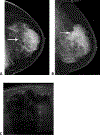
Fig. 1.
Fig. 1.. 38-year-old woman with a palpable anterior left breast mass.
A) The left craniocaudal mammogram shows an irregular mass with indistinct margins (arrow). B) The left lateromedial mammogram shows the mass (arrow) in the upper breast. C) Left breast extended field of view ultrasound shows an irregular hypoechoic mass with posterior shadowing. The patient was a BRCA1 mutation carrier. Ultrasound-guided biopsy of the left breast mass showed poorly differentiated triple negative invasive ductal carcinoma.
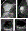
Fig. 2.
Fig. 2.. 36-year-old woman with a palpable abnormality in the upper outer left breast.
A) The left craniocaudal mammogram shows an irregular mass (arrow) with indistinct margins, corresponding to the palpable abnormality (triangle). B) The left mediolateral oblique mammogram shows an irregular high density mass (arrow) with spiculated margins, corresponding to the palpable abnormality. C) Transverse left breast ultrasound in the 1:00 region shows an irregular hypoechoic mass corresponding to the palpable abnormality. The patient was BRCA negative. Ultrasound-guided biopsy of the left breast 1:00 mass revealed triple negative high grade invasive ductal carcinoma.
Fig. 3.
Fig. 3.. 37-year-old woman with a palpable abnormality in the upper outer right breast.
Transverse right breast ultrasound shows an irregular hypoechoic mass in the 10:00 region with some posterior shadowing. The patient had a BRCA1 mutation. Ultrasound-guided biopsy in the right breast at 10:00 showed triple negative intermediate and high grade invasive ductal carcinoma along with ductal carcinoma in situ.
Fig. 4.
Fig. 4.. 37-year-old woman with a palpable abnormality in the upper outer right breast.
A) The right craniocaudal spot compression mammogram shows an oval high density mass (arrow) B) The right lateromedial mammogram shows the oval mass (arrow) with circumscribed margins. C) Transverse right breast ultrasound at 10:00 shows the oval mass with internal cystic spaces, circumscribed margins and posterior enhancement. D) Longitudinal right breast ultrasound at 10:00 shows the circumscribed oval hypoechoic mass with an isoechoic posterior component and posterior enhancement. The patient had a BRCA1 mutation. Ultrasound-guided biopsy of the right breast 10:00 mass revealed triple negative poorly differentiated invasive ductal carcinoma.
Fig. 5.
Fig. 5.. 37-year-old woman undergoing ultrasound to evaluate a mass seen in the upper outer right breast on mammography.
A) Longitudinal right breast ultrasound in the 10:00 region shows a hypoechoic oval mass (arrow) with a parallel orientation, anterior to a prepectoral silicone implant, correlating with a mass seen on mammography. The sonographic appearance of the mass simulates that of a benign process, such as a fibroadenoma. The patient had a BRCA1 mutation. B) Right breast ultrasound-guided core biopsy was performed on the 10:00 hypoechoic oval mass (short arrow) with a parallel orientation and posterior enhancement. The core biopsy was performed with an 18-gauge needle (long arrow). Pathology showed poorly differentiated triple negative invasive ductal carcinoma.

References

    1. Fulford LG, Reis-Filho JS, Ryder K, et al. Basal-like grade III invasive ductal carcinoma of the breast: patterns of metastasis and long-term survival. Breast Cancer Res 2007; 9:R4. - PMC - PubMed
    1. Dent R, Trudeau M, Pritchard KI, et al. Triple-negative breast cancer: clinical features and patterns of recurrence. Clin Cancer Res 2007; 13:4429–4434 - PubMed
    1. Dogan BE, Turnbull LW. Imaging of triple-negative breast cancer. Ann Oncol 2012; 23 Suppl 6:vi23–29 - PubMed
    1. Atchley DP, Albarracin CT, Lopez A, et al. Clinical and pathologic characteristics of patients with BRCA-positive and BRCA-negative breast cancer. J Clin Oncol 2008; 26:4282–4288 - PMC - PubMed
    1. Ko ES, Lee BH, Kim HA, Noh WC, Kim MS, Lee SA. Triple-negative breast cancer: correlation between imaging and pathological findings. Eur Radiol 2010; 20:1111–1117 - PubMed

LinkOut - more resources