. 2020 Apr;39(4):379-388.
doi: 10.1016/j.healun.2019.12.009. Epub 2020 Jan 21.

Respiratory viral infection in lung transplantation induces exosomes that trigger chronic rejection

Affiliations

Respiratory viral infection in lung transplantation induces exosomes that trigger chronic rejection

Muthukumar Gunasekaran et al. J Heart Lung Transplant. 2020 Apr.

Abstract

Background: Respiratory viral infections can increase the risk of chronic lung allograft dysfunction after lung transplantation, but the mechanisms are unknown. In this study, we determined whether symptomatic respiratory viral infections after lung transplantation induce circulating exosomes that contain lung-associated self-antigens and assessed whether these exosomes activate immune responses to self-antigens.

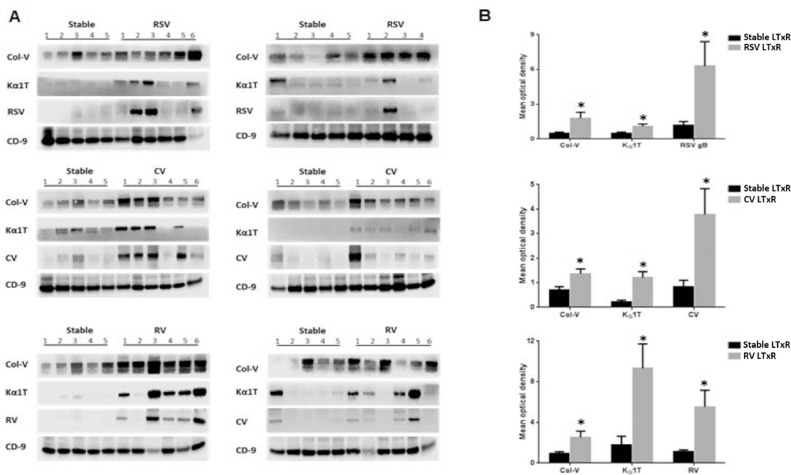
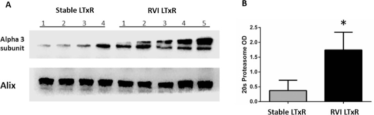
Methods: Serum samples were collected from lung transplant recipients with symptomatic lower- and upper-tract respiratory viral infections and from non-symptomatic stable recipients. Exosomes were isolated via ultracentrifugation; purity was determined using sucrose cushion; and presence of lung self-antigens, 20S proteasome, and viral antigens for rhinovirus, coronavirus, and respiratory syncytial virus were determined using immunoblot. Mice were immunized with circulating exosomes from each group and resulting differential immune responses and lung histology were analyzed.

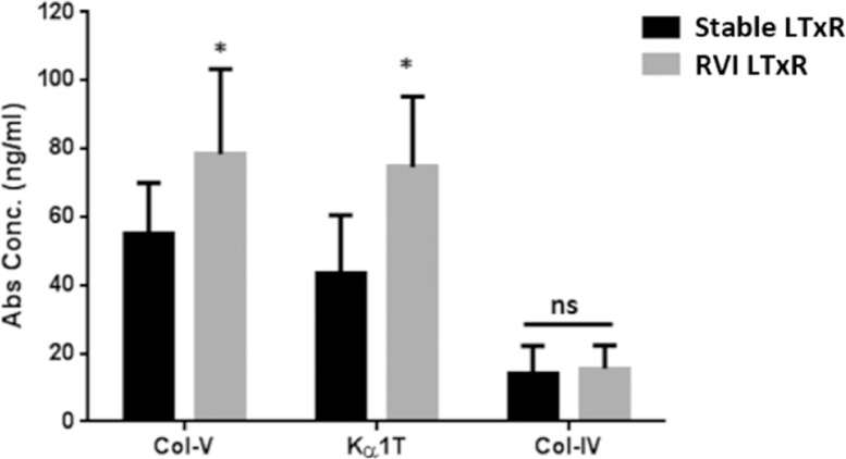
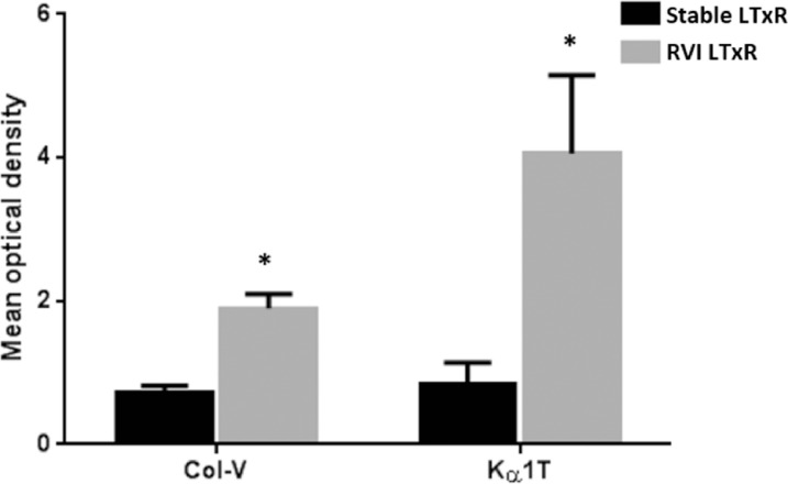
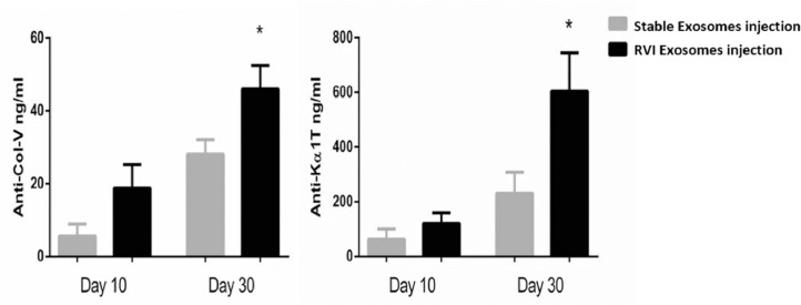
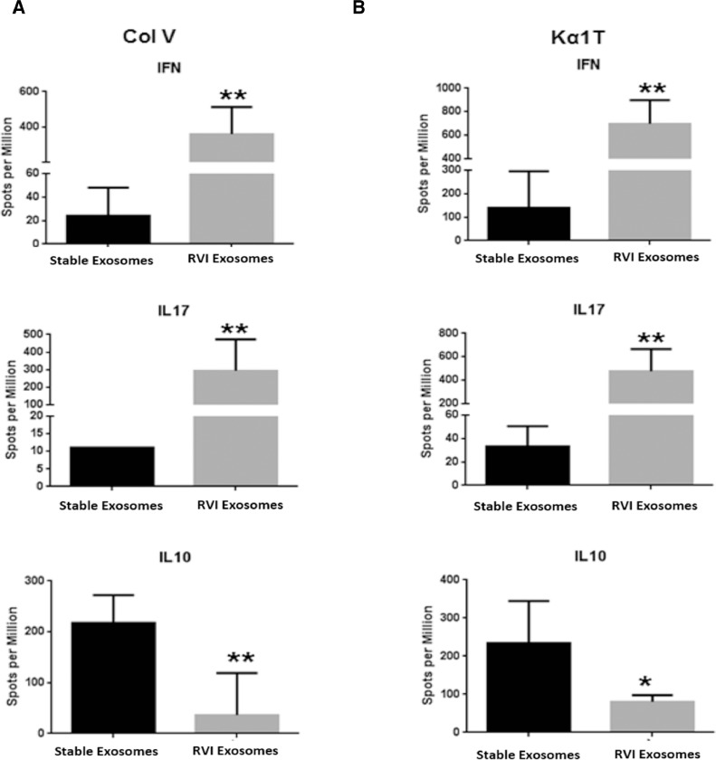
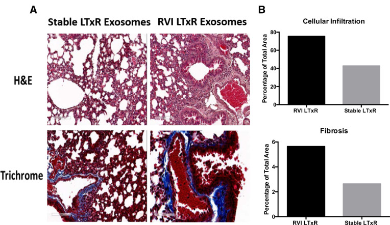
Results: Exosomes containing self-antigens, 20S proteasome, and viral antigens were detected at significantly higher levels (p < 0.05) in serum of recipients with symptomatic respiratory viral infections (n = 35) as compared with stable controls (n = 32). Mice immunized with exosomes from recipients with respiratory viral infections developed immune responses to self-antigens, fibrosis, small airway occlusion, and significant cellular infiltration; mice immunized with exosomes from controls did not (p < 0.05).

Conclusions: Circulating exosomes isolated from lung transplant recipients diagnosed with respiratory viral infections contained lung self-antigens, viral antigens, and 20S proteasome and elicited immune responses to lung self-antigens that resulted in development of chronic lung allograft dysfunction in immunized mice.

Keywords: antibodies; antigens; chronic rejection; exosomes; graft rejection; lung transplantation; respiratory viral infection.


Figures

Figure 1
Figure 1
Abs to lung-associated SAgs in LTxRs diagnosed with RVI and stable LTxRs. Serum samples collected from LTxRs diagnosed with RVI (n = 35) and from stable LTxRs (n = 32) were used to measure Abs to lung-associated SAgs (Col-V and Kα1T). Abs to Col-V (78.3 ± 25.1 vs 54.9 ± 15.1, p = 0.0169) and Kα1T (74.7 ± 20.6 vs 43.3 ± 17.2, p = 0.0145) were significantly higher in LTxRs with RVI compared with stable LTxRs. Further, Abs to kidney-associated antigen Col-IV were not significantly different between stable LTxRs and LTxRs with RVI (14.2 ± 8 vs 15.6 ± 6.7, p = 0.248). The antibody development to lung SAgs was compared between the stable and RVI LTxR using Mann–Whitney test. Asterisk indicates statistically significant. Ab, antibody; Col-IV, collagen-IV; Col-V, collagen-V; Kα1T, K-alpha-1 tubulin; LTxR, lung transplant recipient; RVI, respiratory viral infection; SAg, self-antigen.
Figure 2
Figure 2
Exosomes containing lung-associated SAgs in LTxRs diagnosed with RVI and stable LTxRs. Exosomes isolated from serum samples of LTxRs with RVI (n = 34) and stable LTxRs (n = 30) were analyzed for the presence of lung-associated SAgs (Col-V and Kα1T) by immunoblot. The mean relative optical densities of Col-V (1.9 ± 0.2 vs 0.73 ± 0.09, p = 0.0003) and Kα1T (4.06 ± 1.09 vs 0.83 ± 0.31, p = 0.009) were significantly higher in LTxRs with RVI than stable LTxRs. Optical density was measured using ImageJ software and the OD value of SAgs were calculated in LTxRs with RVI and stable LTxRs after normalization with CD9 OD value. CD9 also served as loading control and exosome-specific markers. The presence of lung SAgs in the exosomes was compared between the cohorts using Mann–Whitney test. Asterisk indicates statistically significant. Col-V, collagen-V; Kα1T, K-alpha-1 tubulin; LTxR, lung transplant recipient; OD, optical density; RVI, respiratory viral infection; SAg, self-antigen.
Figure 3
Figure 3
Lung-associated SAgs and viral antigens were demonstrable in exosomes isolated from patients with RVI. Exosomes isolated from serum samples of patients with RVI and from stable LTxRs were used to detect the presence of lung-associated SAgs and viral antigens using immunoblot. The results showed a significant increase in lung-associated antigens and viral antigens. (A) RSV (n = 10), coronavirus (n = 12), and rhinovirus (n = 12) in respective patients with viral infection compared with stable LTxRs (n = 30). (B) Graphical representation shows the optical density of lung-associated SAgs and viral antigens measured in RVI and stable LTxRs using ImageJ software. Optical density of lung SAgs and viral antigens were normalized with exosomes specific marker CD9. The presence of lung SAgs and viral antigens in the exosomes was compared between the cohorts using Mann–Whitney test. Asterisk indicates statistically significant. Col-V, collagen-V; CV, coronavirus; Kα1T, K-alpha-1 tubulin; LTxR, lung transplant recipient; RSV, respiratory syncytial virus; RV, rhinovirus; RVI, respiratory viral infection; SAg, self-antigen.
Figure 4
Figure 4
Exosomes containing 20S proteasome core in LTxRs with RVI and stable LTxRs. Circulatory exosomes isolated from LTxRs with RVI (n = 5) and stable LTxRs (n = 4) were used to detect the presence of 20S proteasome subunit α3 using immunoblot. (A) The exosomes isolated from patients with RVI showed a significant increase in 20S proteasome compared with exosomes from stable LTxRs (Mean optical density: 1.74 ± 0.6 vs 0.37 ± 0.35, p = 0.0317). Alix served as loading control and exosome-specific marker. (B) Graphical representation shows optical intensity of 20S proteasome α3 subunit abundance in LTxRs with viral infection and stable LTxRs. The presence of 20S proteasome was compared between stable LTxRs and LTxRs with RVI using Student's t-test. LTxR, lung transplant recipient; RVI, respiratory viral infection.
Figure 5
Figure 5
Exosomes from LTxRs with RVI induce a humoral immune response to lung SAgs. Serum samples collected on Days 10 and 30 from C57BL/6 mice immunized with exosomes isolated from LTxRs with RVI (n = 5) and from stable LTxRs (n = 5) were utilized to measure Abs to lung SAgs by ELISA. Serum samples collected on Day 30 from mice immunized with exosomes from LTxRs with RVI showed significantly increased Abs to SAgs (Col-V, 28.1 ± 4.0 vs 45.9 ± 6.5, p = 0.04; Kα1T, 230.4 ± 77.1 vs 604.6 ± 140, p = 0.04) when compared with mice injected with exosomes from stable LTxRs. The antibody development was compared between the cohorts using Student's t-test. Asterisk indicates statistically significant. Ab, antibody; Col-V, collagen-V; ELISA, enzyme-linked immunosorbent assay; Kα1T, K-alpha-1 tubulin; LTxR, lung transplant recipient; RVI, respiratory viral infection; SAg, self-antigen.
Figure 6
Figure 6
Exosomes from LTxRs with RVI induce cytokine-producing T cells to lung SAgs. Spleens were collected on Day 30 from C57BL/6 mice immunized with exosomes of LTxRs with RVI (n = 5) and from stable LTxRs (n = 5) were used to measure cytokine-producing T cells against lung SAgs by ELISPOT. Mice immunized with exosomes of RVI showed significant increase in T cells producing IL-17 and interferon gamma to SAgs. Mice injected with exosomes isolated from stable LTxRs showed increased frequency of IL-10–producing T cells compared with mice immunized with RVI exosomes. The cytokine levels were compared between the cohorts using Mann–Whitney test. Asterisks indicate statistically significant. ELISPOT, enzyme-linked ImmunoSpot assay; IL, interleukin; LTxR, lung transplant recipient; RVI, respiratory viral infection; SAgs, self-antigens; ** statistically significant.
Figure 7
Figure 7
Fibrosis and cellular infiltration were demonstrable in mice injected with exosomes isolated from LTxRs with RVI. Mice were killed on Day 30 and their lungs were collected and analyzed using hematoxylin and eosin and trichrome staining. (A) Interstitial and inflammatory infiltrates and fibrosis was more prominent in mice injected with exosomes from LTxRs with RVI compared with mice injected with exosomes from stable LTxRs. Images were obtained on a Leica microscope at × 40 and morphometric analysis was performed using Aperio ImageScope software (Leica). (B) The morphometric data are given for the representative images. LTxR, lung transplant recipient; RVI, respiratory viral infection.

References

    1. Arcasoy SM, Kotloff RM. Lung transplantation. N Engl J Med. 1999;340:1081–1091. - PubMed
    1. Glanville AR. Bronchoscopic monitoring after lung transplantation. Semin Respir Crit Care Med. 2010;31:208–221. - PubMed
    1. Sato M, Waddell TK, Wagnetz U. Restrictive allograft syndrome (RAS): a novel form of chronic lung allograft dysfunction. J Heart Lung Transplant. 2011;30:735–742. - PubMed
    1. Witt CA, Gaut JP, Yusen RD. Acute antibody-mediated rejection after lung transplantation. J Heart Lung Transplant. 2013;32:1034–1040. - PMC - PubMed
    1. Stewart S, Fishbein MC, Snell GI. Revision of the 1996 working formulation for the standardization of nomenclature in the diagnosis of lung rejection. J Heart Lung Transplant. 2007;26:1229–1242. - PubMed

Publication types

MeSH terms

LinkOut - more resources