

Review
doi: 10.1104/pp.19.01396.
Epub 2020 Feb 7.
In Vitro Analytical Approaches to Study Plant Ligand-Receptor Interactions
Affiliations
- PMID: 32034053
- PMCID: PMC7140929
- DOI: 10.1104/pp.19.01396
Item in Clipboard
Review
In Vitro Analytical Approaches to Study Plant Ligand-Receptor Interactions
Plant Physiol.
2020 Apr.
Abstract
State-of-the-art in vitro methods characterize receptor-ligand interactions, highlighting experiment strategies, advantages and limitations.
Figures
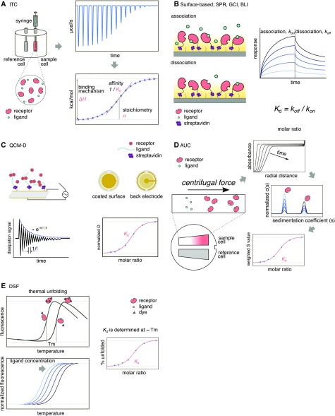
Cartoon of overview of label-free methods. A, Schematic of an ITC instrument and an ITC thermogram (raw data), which provides information about affinity, stoichiometry, and thermodynamics. B, Schematic representation of surface-based optics methods (SPR, GCI, and BLI). At least one of the receptor or ligand needs to be immobilized. Recent developments allow for preferential orientation of molecules on the chip surface. Typical curves of a titration experiment are shown in the sensogram (low and high concentrations of ligand are represented in light blue and black, respectively). C, Schema of QCM-D principle. One of the biomolecules is immobilized on a quartz surface and voltage is applied through to the quartz disc to make it oscillate. The frequency of oscillation (f) is very sensitive to small fluctuations of mass on the surface. Structural properties are measured as changes in dissipation (D) after cutting the electrical power. D, Illustration of the principle of AUC. Buffer and a sample containing known concentrations of receptor and ligand are placed in a double-sector cell and high centrifugation speed is used to sediment the components of the sample while a radial scanner monitors sedimentation. In sedimentation velocity, the data obtained are modeled using hydrodynamics theory to calculate the size and proportions of the sedimented molecules. Fluorescence tags can be used to improve detection. E, In a DSF assay, the thermal unfolding of a receptor is monitored in the presence of increasing concentrations of ligand. The Kd is obtained at the ∼Tm of the free receptor. The method can use intrinsic Trp fluorescence or compatible dyes.
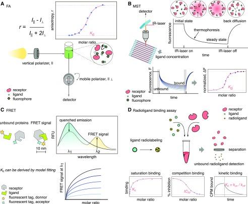
Cartoon of overview of labeled methods. A, Depiction of FA principle. The sample is irradiated with polarized light (vertical) and the detector can filter the resulting light with a second polarizer that changes from vertical to horizontal. The rotational speed of molecules correlates with the depolarization of light. The anisotropy value, r, is related to the movement of the molecules in the sample: the bigger the molecule (i.e. receptor/ligand complex), the slower the rotation speed compared to the free ligand, which will lead to less depolarized light, increasing the r value. The method can use intrinsic fluorescence of molecules as well as fluorophores. B, Representation of MST. A focus IR-laser generates a temperature gradient spot in the sample, inducing molecule displacement (thermophoresis). A detector monitors the sample spot, tracking the displacement of the sample. C, In FRET, a compatible donor/acceptor pair is required. The reduction in the signal of the donor (quenched) is used to generate a dose response graph. The donor and acceptor molecules need to be in close proximity (10 nm or less) for efficient energy transfer. D, Representation of a typical radioligand binding assay. In competition binding, the receptor is mixed in the presence of label-free and radiolabeled ligand. After incubation, the receptor is removed from the sample and the amount of unbound radioligand is used to calculate binding affinities.
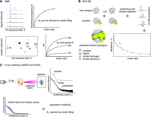
Cartoon of overview of structure-based methods. A, Scheme of NMR binding assays. The one-dimensional chemical shift uses labeled substrates, the height of the NMR ligand signal decreases upon binding with the receptor. In a two-dimensional chemical shift assay (bottom), the receptor is labeled with two isotopes and the NMR spectra are plotted. R1, R2, and R3 represent receptor amino acids that move upon ligand binding and are proportional to the amount of ligand. B, In an HDX-MS assay, the free receptor is incubated with heavy water, allowing amide hydrogen exchange with deuterium. The samples are digested and analyzed by MS to obtain the rates of deuterium uptake in different peptides. When the ligand is added to the receptor prior to deuterium exchange, some parts of the receptor will be “protected,” preventing HDX. The amount of deuterium obtained for some peptides will be inversely proportional to the amount of ligand. C, Principle of the light scattering techniques SAXS and WAXS. The scattering profile of samples containing increasing concentrations of ligand are compared and processed using the scattering profile of the buffer and that of the free receptor. Population modeling and model fitting are used to calculate the Kd.
References
Publication types
MeSH terms
Substances
LinkOut - more resources
Full Text Sources
