Meta-Analysis
. 2020 Apr;31(4):829-840.
doi: 10.1681/ASN.2019080851. Epub 2020 Feb 7.

Anti-Factor B Antibodies and Acute Postinfectious GN in Children

Affiliations
Meta-Analysis

Anti-Factor B Antibodies and Acute Postinfectious GN in Children

Sophie Chauvet et al. J Am Soc Nephrol. 2020 Apr.

Abstract

Background: The pathophysiology of the leading cause of pediatric acute nephritis, acute postinfectious GN, including mechanisms of the pathognomonic transient complement activation, remains uncertain. It shares clinicopathologic features with C3 glomerulopathy, a complement-mediated glomerulopathy that, unlike acute postinfectious GN, has a poor prognosis.

Methods: This retrospective study investigated mechanisms of complement activation in 34 children with acute postinfectious GN and low C3 level at onset. We screened a panel of anticomplement protein autoantibodies, carried out related functional characterization, and compared results with those of 60 children from the National French Registry who had C3 glomerulopathy and persistent hypocomplementemia.

Results: All children with acute postinfectious GN had activation of the alternative pathway of the complement system. At onset, autoantibodies targeting factor B (a component of the alternative pathway C3 convertase) were found in a significantly higher proportion of children with the disorder versus children with hypocomplementemic C3 glomerulopathy (31 of 34 [91%] versus 4 of 28 [14%], respectively). In acute postinfectious GN, anti-factor B autoantibodies were transient and correlated with plasma C3 and soluble C5b-9 levels. We demonstrated that anti-factor B antibodies enhance alternative pathway convertase activity in vitro, confirming their pathogenic effect. We also identified crucial antibody binding sites on factor B, including one correlated to disease severity.

Conclusions: These findings elucidate the pathophysiologic mechanisms underlying acute postinfectious GN by identifying anti-factor B autoantibodies as contributing factors in alternative complement pathway activation. At onset of a nephritic syndrome with low C3 level, screening for anti-factor B antibodies might help guide indications for kidney biopsy to avoid misdiagnosed chronic glomerulopathy, such as C3 glomerulopathy, and to help determine therapy.

Keywords: complement; glomerulonephritis; pediatrics.


Figures

None
Graphical abstract
Figure 1.
Figure 1.
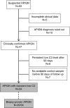
Thirty-four children with APIGN were included in the study. Among the 62 patients with suspected APIGN, five patients had incomplete data for making a final diagnosis and in ten patients APIGN diagnosis was ruled out. Six patients had anamnestic, clinical, and biologic data that supported the diagnosis of APIGN but plasma C3 levels remained low for >90 days. In seven other cases with APIGN, normalization of C3 levels was not assessed within 90 days after the diagnosis.
Figure 2.
Figure 2.
Anti-FB antibodies at acute phase of the disease are specific of APIGN and correlate with C3 and sC5b-9 levels. (A) Reactivity of autoantibodies against FB was tested by ELISA in plasma samples from 34 patients with APIGN, 28 patients with H-C3G (collected at acute phase of disease), and 84 healthy donors. Results of antibodies are expressed in arbitrary units (UA). The cutoff of positivity was determined with plasma samples from the 84 healthy donors (HD). (B) Receiving operater characteristic curve of the positivity of anti-FB autoantibodies in APIGN. The area under the receiving operater characteristic curve for the use of anti-FB antibodies titer to discriminate APIGN from H-C3G patients was 0.93 (95% CI, 0.88 to 0.99). The optimal cutoff point was 88.5, which had a sensitivity of 0.91 (95% CI, 0.76 to 0.98) and a specificity of 0.85 (95% CI, 0.71 to 0.94). (C and D) Correlation between titers of anti-FB antibodies and C3 levels and sC5b9 investigated in 34 and 27 tested patients, respectively. (C) Plasma C3 levels inversely correlate with titers of anti-FB antibodies (P=0.01; 95% CI, −0.68 to −0.142; R=−0.46). (D) Soluble C5b9 levels in plasma correlate with anti-FB antibody titers (P=0.001; 95% CI, 0.35 to 0.82; R=0.64). AUC, area under the curve.
Figure 3.
Figure 3.
Anti-FB antibodies in APIGN patients are transient. (A) Kinetics of anti-FB antibody positivity, C3 levels, and renal symptoms in patient with APIGN according to renal symptoms. Bars in gray indicate the presence of renal symptoms (high BP and/or hematuria and/or proteinuria and/or renal failure). Samples positive for anti-FB antibodies and low C3 levels are indicated by a red asterisk. Samples positive for anti-FB antibodies with normal C3 levels are indicated by a blue asterisk. Samples negative for anti-FB antibodies with low C3 levels are indicated by a yellow asterisk. Samples negative for anti-FB antibodies and/or normal C3 levels are indicated by a green asterisk. In patients 2, 9, 11, 20, 21, and 27, measurements of C3 levels confirming normalization of C3 within 90 days after renal diagnosis were performed in an external biologic center and were not indicated on the figure. (B) Titer of anti-FB autoantibodies at acute phase and on the last available sample. In 31 of 34 patients with anti-FB antibodies at diagnosis, control of anti-FB reactivity was available in 29 patients. For two patients (P7 and P24), no sample was available during follow-up. In six of 29 patients, antibody titer remained positive during the follow-up in patient P28 (titers 622 UA at day 852), P6 (titer 364 UA at day 28), P22 (titer 189 UA at day 30), P29 (titer 123 UA at day 75), P32 (titer 260 at day 32), and P25 (titer 103 at day 31). Dotted line indicates the positivity threshold (>88 UA/ml).
Figure 4.
Figure 4.
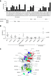
Anti-Fb antibodies recognize hotspots on serine protease and von Willebrand type A domains of FB. (A) Reactivity of autoantibodies against 16 linear peptides of vW type A domain and 16 linear peptides of SP domain of FB was tested by ELISA in plasma samples from 29 patients with APIGN. A total of 27 of 32 peptides were recognized by at least one patient sample positive for anti-FB antibodies. Five hotspot peptides of the vW type A domain and four hotspot peptides of the SP domain recognized by more than four patient samples (indicated by an asterisk) were identified. Location of the amino acids forming the catalytic triads and MIDAS site are indicated below the graph. (B) Reactivity against recombinant FB (wild-type and nine recombinant FBs bearing mutations on hotspot peptide sequences [K312A, N318A, D321A, D378E, E379D P407A, D506A, E509A, and the double mutant D345A-D346A]) in plasma samples from 13 patients with APIGN was tested by ELISA. Results are expressed in OD. Means±2 SD (m+2SD, m-2SD) of binding to the wild type are indicated in dotted lines. Five mutations were associated with a significant decrease of anti-FB antibody binding to recombinant FB (D321A, D378E, E379D, P407A, and D506A), whereas one mutation was associated with a significant increase (E509A). (C) Localization of the hotspots on the FB structure. The five hotspot peptides of the vW domain are indicated by red ribbon, and the four hotspot peptides of SP domain are indicated in green. The hotspot amino acid changes are in dark blue. Amino acids of the MIDAS are indicated in magenta and amino acids of the catalytic triads are indicated in yellow. Cyan indicates amino acids referred to in the literature as being associated with hyperfunctional C3 convertase. MIDAS, magnesium ion–dependent, metal ion–dependent adhesion site.
Figure 5.
Figure 5.
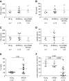
Anti-FB antibodies from children with APIGN enhance activity of fluid-phase and cell-surface C3 convertase but do not stabilize solid-phase C3 convertase. Activation of the fluid-phase complement alternative pathway in the presence of anti-FB antibodies. After incubation of normal human serum (EGTA-magnesium) in the presence of total purified IgG from 13 patients with APIGN, eight patients with C3G positive for anti-C3bBb antibodies, and seven healthy controls, C3a, Bb, C5a, and sC5b9 generation was quantified by ELISA. In the experimental condition, at baseline, median C3a, Bb, C5a, and sC5b9 levels in normal human serum are 6 ng/ml, 18 µg/ml, 9 ng/ml, and 1.8 µg/ml, respectively. After 30 minutes of incubation, median levels obtained with IgG from healthy donors (HD-Ig) are 253.9, 40.6, 159 ng/ml, and 18.6 µg/ml, respectively. (A-B) C3a and Bb releases were significantly increased in patients with APIGN (four of 13 and four of 13, respectively) and in patients with C3G positive for anti-C3bBb antibodies (four of eight and four of eight patients, respectively) compared with IgG from healthy donors (upper mean+2 SD). (C-D) C5a and sC5b9 were significantly increased in patients with APIGN but not in the presence of anti-C3bBb antibodies from patients with C3G. (E-F) Activation of the solid-phase complement alternative pathway in the presence of anti-FB antibodies. The capacity of total purified IgG from patients and controls (healthy donors and patients with C3G positive for anti-C3bBb antibodies) to enhance C3/C5 convertase formation and stabilization activity was tested using hemolytic assays. Results are expressed as the percentage of the maximum lysis of erythrocytes (osmotic lysis with water). (E) To evaluate the capacity of anti-FB antibodies to enhance cell-surface C3 convertase formation, C3 convertase was formed on sensitized sheep erythrocytes bearing C3b in the presence of Ig from patients with APIGN or controls. In the presence of Ig from healthy donors, the median of the maximum lysis is 20%. Lysis was significantly increased in presence of Ig from patients with APIGN. (F) To evaluate the capacity of anti-FB antibodies to stabilize a preformed surface-bound C3 convertase, total purified IgG from patients with APIGN, diluted in gelatin veronal buffer-EDTA, were incubated on sheep erythrocyte bearing preformed C3bBb. IgG from patients with H-C3G positive for anti-C3bBb antibodies were used as positive control and Ig from healthy donors as negative controls. At baseline (T0), the median percentage of maximum lysis is 43% (40%–53%), 79% (43%–100%), and 75% (42%–100%) in the presence of Ig from those with HD-Ig, APIGN, or H-C3G, respectively. At 20 minutes (T20), median percentage of maximum lysis is 13% (15%–19%), 48% (15%–65%) and 65% (31%–100%) in the presence of Ig from those with HD-Ig, APIGN, or H-C3G, respectively. The change in lysis (Δ) between T0 and T20, reflecting dissociation of C3 convertase, is significantly increased in patients with APIGN compared with those with H-C3G. Results are expressed as ΔLysis, representing the difference of lysis between baseline and after 20 minutes of incubation. An increase of ΔLysis suggests a decay of the enzyme.  Ranges are the extrems. HD-Ig, Ig from healthy donors.

Comment in

References

    1. Carapetis JR, Steer AC, Mulholland EK, Weber M: The global burden of group A streptococcal diseases. Lancet Infect Dis 5: 685–694, 2005. - PubMed
    1. Kambham N: Postinfectious glomerulonephritis. Adv Anat Pathol 19: 338–347, 2012. - PubMed
    1. Chapter 9: Infection-related glomerulonephritis. Kidney Int Suppl (2011) 2: 200–208, 2012. - PMC - PubMed
    1. Rodriguez-Iturbe B, Haas M: Post-streptococcal glomerulonephritis. In: Streptococcus pyogenes : Basic Biology to Clinical Manifestations, edited by Ferretti JJ, Stevens DL, Fischetti VA, Oklahoma City, OK, University of Oklahoma Health Sciences Center, 2016. Available at: http://www.ncbi.nlm.nih.gov/books/NBK333429/ - PubMed
    1. Yoshizawa N, Yamakami K, Fujino M, Oda T, Tamura K, Matsumoto K, et al. .: Nephritis-associated plasmin receptor and acute poststreptococcal glomerulonephritis: Characterization of the antigen and associated immune response. J Am Soc Nephrol 15: 1785–1793, 2004. - PubMed

Publication types