

68Ga-PSMA PET/CT Combined with PET/Ultrasound-Guided Prostate Biopsy Can Diagnose Clinically Significant Prostate Cancer in Men with Previous Negative Biopsy Results
- PMID: 32034111
- PMCID: PMC7456174
- DOI: 10.2967/jnumed.119.235333
68Ga-PSMA PET/CT Combined with PET/Ultrasound-Guided Prostate Biopsy Can Diagnose Clinically Significant Prostate Cancer in Men with Previous Negative Biopsy Results
Abstract
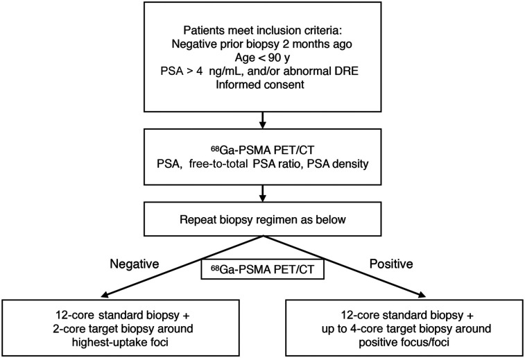
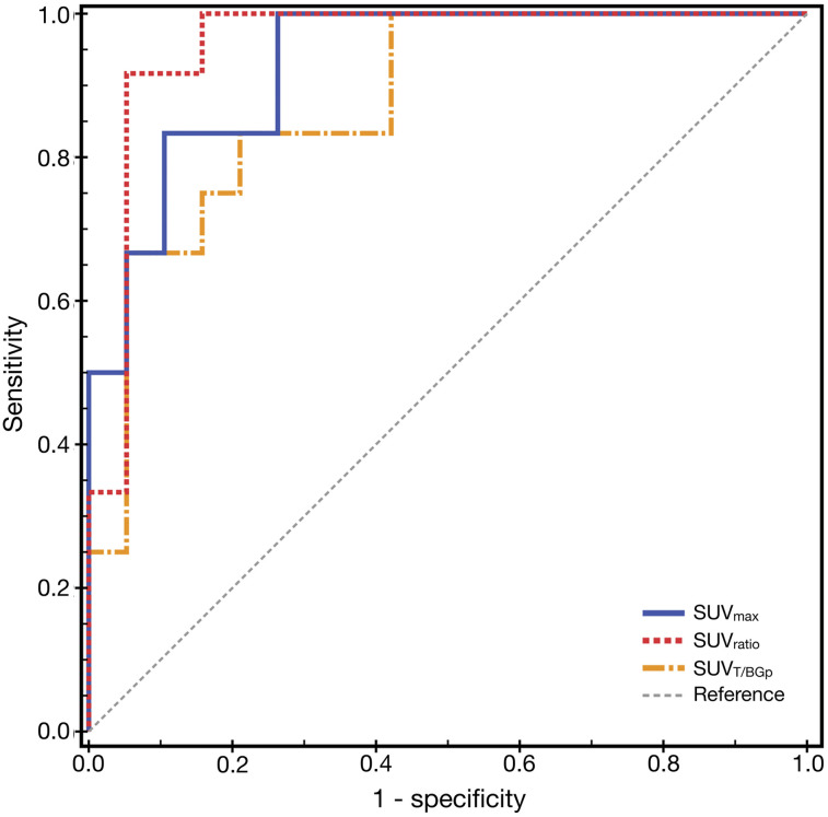
The purpose of this study was to investigate the feasibility and diagnostic efficacy of 68Ga-prostate-specific membrane antigen (PSMA) PET/CT combined with PET/ultrasound-guided biopsy in the diagnosis of prostate cancer (PCa). Methods: In total, 31 patients with a previously negative prostate biopsy but persistent elevated serum prostate-specific antigen (PSA) were imaged with a 68Ga-PSMA PET/CT ligand before undergoing repeat prostate biopsy. On the basis of the proposed Prostate Cancer Molecular Imaging Standardized Evaluation criteria, 68Ga-PSMA PET/CT results were interpreted as negative (molecular-imaging-for-PSMA expression score [miPSMA-ES] of 0-1) or positive (miPSMA-ES of 2-3). All patients underwent standard template systematic biopsy with up to 4 additional PET/ultrasound-guided biopsy cores. The sensitivity, specificity, positive and negative predictive values, and accuracy of 68Ga-PSMA PET/CT were determined. In addition, the correlation between the miPSMA-ES and the detection rate of PCa was also analyzed. Univariate logistic regression models were established using 68Ga-PSMA PET/CT semiquantitative analysis parameters to predict the outcome of repeat prostate biopsy. Results: The median age of patients was 65 y (range, 53-81 y), and the median PSA level was 18.0 ng/mL (range, 5.48-49.77 ng/mL). PCa was detected in 15 of 31 patients (48.4%), and 12 of 31 patients (38.7%) had clinically significant PCa (csPCa). The sensitivity, specificity, positive predictive value, negative predictive value, and accuracy of 68Ga-PSMA PET/CT in the diagnosis of csPCa were 100.0%, 68.4%, 66.7%, 100.0%, and 80.6%, respectively. The detection rate of PCa increased with the increase in miPSMA-ES. The detection rates of csPCa in the miPSMA-ES 0-1, 2, and 3 groups were 0%, 54.5%, and 85.7%, respectively. Semiquantitative analysis of 68Ga-PSMA PET/CT images showed that predictive models based on the SUVmax of prostate lesion, tumor-to-normal-prostate background SUVmax, and tumor-to-normal-liver background SUVmax could effectively predict csPCa; area under the curves were 0.930, 0.877, and 0.956, respectively. Conclusion: This study preliminarily confirmed that 68Ga-PSMA PET/CT imaging, combined with PET/ultrasound-guided prostate biopsy, can effectively detect csPCa. Prebiopsy 68Ga-PSMA PET/CT had predictive value for csPCa in the studied patient population.
Keywords: PET/CT; PSMA; biopsy; prostate cancer.
© 2020 by the Society of Nuclear Medicine and Molecular Imaging.
Figures
References
-
- Ahmed HU, El-Shater Bosaily A, Brown LC, et al. Diagnostic accuracy of multi-parametric MRI and TRUS biopsy in prostate cancer (PROMIS): a paired validating confirmatory study. Lancet. 2017;389:815–822. - PubMed
-
- Wegelin O, van Melick HHE, Hooft L, et al. Comparing three different techniques for magnetic resonance imaging-targeted prostate biopsies: a systematic review of in-bore versus magnetic resonance imaging-transrectal ultrasound fusion versus cognitive registration—is there a preferred technique? Eur Urol. 2017;71:517–531. - PubMed
Publication types
MeSH terms
Substances
LinkOut - more resources
Full Text Sources
Medical
Research Materials
Miscellaneous