. 2020 Feb 7;10(1):2135.
doi: 10.1038/s41598-020-58923-6.

The Golgi Glycoprotein MGAT4D is an Intrinsic Protector of Testicular Germ Cells From Mild Heat Stress

Affiliations

The Golgi Glycoprotein MGAT4D is an Intrinsic Protector of Testicular Germ Cells From Mild Heat Stress

Ayodele Akintayo et al. Sci Rep. .

Abstract

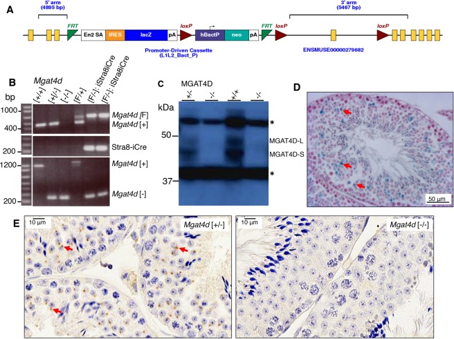
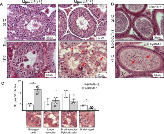
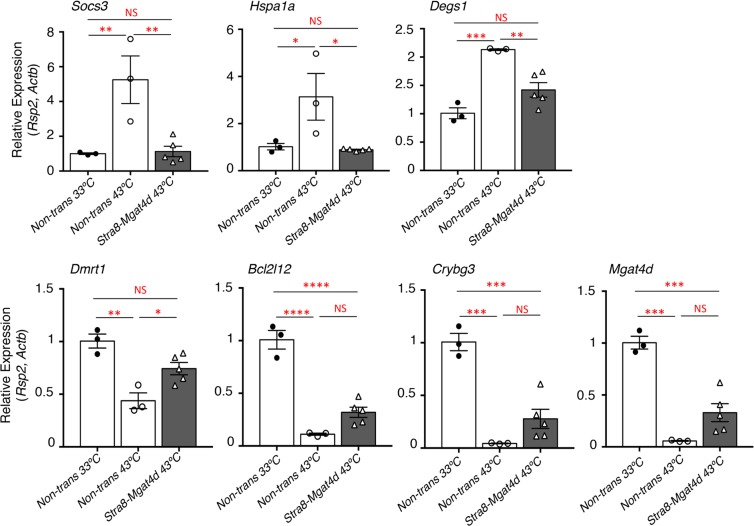
Male germ cells are sensitive to heat stress and testes must be maintained outside the body for optimal fertility. However, no germ cell intrinsic mechanism that protects from heat has been reported. Here, we identify the germ cell specific Golgi glycoprotein MGAT4D as a protector of male germ cells from heat stress. Mgat4d is highly expressed in spermatocytes and spermatids. Unexpectedly, when the Mgat4d gene was inactivated globally or conditionally in spermatogonia, or mis-expressed in spermatogonia, spermatocytes or spermatids, neither spermatogenesis nor fertility were affected. On the other hand, when males were subjected to mild heat stress of the testis (43 °C for 25 min), germ cells with inactivated Mgat4d were markedly more sensitive to the effects of heat stress, and transgenic mice expressing Mgat4d were partially protected from heat stress. Germ cells lacking Mgat4d generally mounted a similar heat shock response to control germ cells, but could not maintain that response. Several pathways activated by heat stress in wild type were induced to a lesser extent in Mgat4d[-/-] heat-stressed germ cells (NFκB response, TNF and TGFβ signaling, Hif1α and Myc genes). Thus, the Golgi glycoprotein MGAT4D is a novel, intrinsic protector of male germ cells from heat stress.


Conflict of interest statement

The authors declare no competing interests.

Figures

Figure 1
Figure 1
Generation of Mgat4d mutant mice. (A) Map of the targeted Mgat4dtm1a(KOMP)Wtsi allele in ES cells obtained from KOMP. Exon 4 is flanked by two loxP sites. LacZ and the neomycin cassettes are flanked by two Frt sites. (B) PCR of genomic DNA from Mgat4d[+/+], Mgat4d[+/−], Mgat4d[−/−] and Mgat4d[F/−]:Stra8-iCre pups to determine genotype. Primers are given in Supplementary Table S1. (C) Western blot analysis of protein extracts of germ cells purified from 28 dpp Mgat4d[+/+], Mgat4d[+/−] and Mgat4d[−/−] mice. Long and short forms of MGAT4D are identified. *is a non-specific band. Full blot in Supplementary Fig. S1. (D) Representative testis section from a mouse carrying the LacZ gene under the control of the Mgat4d promoter after staining for β-galactosidase (blue). Nuclei were stained with eosin. (E) Immunohistochemistry of representative testis sections from Mgat4d[+/−] and Mgat4d[−/−] mice of 28 dpp. The presence of MGAT4D is shown by the brown stain consistent with a Golgi localization (arrows). Nuclei were stained with hematoxylin.
Figure 2
Figure 2
Effects of heat treatment on Mgat4d[−/−] testes. (A) Representative testis sections stained with H&E. Upper panels from mice whose lower body was submerged for 25 min at 33 °C and lower panels from mice treated similarly at 43 °C. Arrows indicate enlarged cells, arrow heads show vacuoles in germ cells. (B) Representative epididymis sections from a Mgat4d[−/−] male treated at 33 °C (upper) or 43 °C (lower) and stained with H&E. Arrows in the 43 °C sample indicate pyknotic cells in the tubule lumen. (C) Quantification of different tubule categories in testis sections from heat-treated (43 °C) Mgat4d[+/−] and Mgat4d[−/−] males. Positive tubules were counted as those with at least one cell of radius ≥10 μm; large vacuoles were tubules with at least one vacuole ≥10 μm; small vacuoles, pyknotic cells were tubules with at least one vacuole of radius <10 μm or tubules containing pyknotic cells; undamaged tubules were tubules with no apparent damage. Mice were from an aged cohort (592–596 days) of Mgat4d[+/−](n = 2) and Mgat4d[−/−](n = 5) mice. Thirty (30) tubules were counted in one section per mouse. Student’s t test (two-tailed, unpaired) **p < 0.01; *p < 0.05.
Figure 3
Figure 3
Apoptosis of germ cells in heat-treated testes. Representative testis sections from 33 °C- or 43 °C-treated aged FVB males were subjected to the TUNEL “Apoptag” assay for in situ detection of DNA strand breaks. (A) Section from a 33 °C-treated Mgat4d[−/−] male. (B) Section from a 43 °C-treated Mgat4d[+/−] male. (C) Section from a 43 °C-treated Mgat4d[−/−] male. DNA breaks stained brown (red arrows). (D) quantification of apoptotic signal in ≥100 tubules using FIJI software. Student’s t test (unpaired, two-tailed) *p < 0.05.
Figure 4
Figure 4
Generation and characterization of Mgat4d transgenic mice. (A) Schematic representation of constructs used to generate transgenic (Tg) mice. Expression of Mgat4d-L-Myc was driven by promoters (Stra8, Ldhc and Prm1) specific for different germ cell types. Lower diagram, position of primers used for qRT-PCR amplification. “LongFw” and “LongRev” to amplify the 5′ region of Mgat4d-L; “TrFw” and “TrRev” to amplify the transgene junction. (B) qRT-PCR of Mgat4d-L 5′ primer transcripts relative to Actb and Rps2. Testis RNA was isolated from males of 28 dpp. (C) Representative H&E stained testis sections from 120 dpp control and Mgat4d-L-Myc transgenic males.
Figure 5
Figure 5
Effects of heat stress in Mgat4d transgenic testis. Quantification of heat-induced damage in testes of Mgat4d[+/+] (n = 5), Stra8-Mgat4d-L-Myc (n = 8), Ldhc-Mgat4d-L-Myc (n = 7), and Prm1-Mgat4d-L-Myc (n = 5) C57BL6/J males. 30 tubules were investigated per mouse in one H&E stained testis section to detect (A) Enlarged germ cells; (B) Large vacuoles; (C) Small vacuoles, pyknotic cells; (D) Undamaged tubules. Differences from control two- tailed, unpaired Student t-test *p < 0.05, **p < 0.01.
Figure 6
Figure 6
Effects of heat treatment on apoptosis in Mgat4d transgenic mice. (A) Representative images of testis sections from 43 °C-treated Mgat4d [+/+] (n = 3) and Mgat4d transgenic mice (n = 4 for each) stained by the Apoptag kit to detect DNA breaks. (B) Quantification of Apoptag signal in ≥100 tubules of 43 °C-treated non-transgenic and transgenic mice. Statistical differences determined by two-tailed unpaired Student’s t-test *p < 0.05, ***p < 0.001.
Figure 7
Figure 7
qRT-PCR of cDNA from testis of 33 °C- or 43 °C-treated control vs Stra8-Mgat4d-L-Myc males of 7 months. Testes were isolated 24 hr after treatment. qRT-PCR was performed in triplicate. Relative gene expression was normalized to Actb and Rps2. Mean ± SEM; statistical analysis by two-tailed, unpaired Student’s t-test.; *p < 0.05, **p < 0.01, ***p < 0.001, ****p < 0.0001.
Figure 8
Figure 8
Microarray analysis of germ cell cDNA. Two month C57BL/6J males were treated at 33 °C or 43 °C for 25 min and germ cells were harvested 8 hr after recovery. (A) Principal component analysis (PCA) for 33 °C-treated Mgat4d[+/+] (n = 4) and Mgat4d[−/−] (n = 3) arrays and 43 °C-treated Mgat4d[+/+] (n = 3) and Mgat4d[−/−] (n = 5) samples. (B) Volcano plots showing the distribution of DEGs based on their expressed log2 fold-change and −log10 p value. Red dots represent genes with a log2 fold-change lower than −0.6 or higher than 0.6 (equivalent to +/−1.5 fold-change) and a p value below the threshold of 0.05 (−log10 (0.05)  =  1.3). (C) Correlation analysis of microarray chip data of wild type (WT) and Mgat4d[−/−] (KO) germ cells at 33 °C and 43 °C.
Figure 9
Figure 9
Validation and Ingenuity Pathway analysis (IPA). (A) qRT-PCR validation of up-regulated genes in control (n = 3) and Mgat4d[−/−] (n = 4) cDNA samples from the same mice used for microarray analyses. (B) qRT-PCR of down-regulated genes in the same samples. Relative expression was determined using Actb and Rps2. Assays were performed in triplicate. Error bars represent mean ± SEM; statistical analysis by two-tailed, unpaired Student’s t-test *p < 0.05, **p < 0.01, ***p < 0.001. (C) Top canonical pathways in IPA significantly overrepresented in heat-treated Mgat4d[−/−] germ cells compared to wild type, normalized to their respective 33 °C counterparts, according to −log p value.
Figure 10
Figure 10
Differential gene interactions between Mgat4d genotype and heat shock conditions. (A) Heat maps showing DEGs either down- or up-regulated specifically in Mgat4d wild type, but not Mgat4d KO cells following heat shock, representative of significantly enriched pathways identified by EnrichR (http://amp.pharm.mssm.edu/Enrichr/). Adjusted p values and odds ratios (OR) for the respective pathways are shown. Full names of the pathways are: Single Gene Perturbations from GEO: Il10 KO mouse GSE25846 sample 3062; ARCHS4 TFs Coexp: HIF1A_human_tf_ARCHS4_coexpression; TRRUST Transcription Factors 2019: NFKB1_ human; Disease Perturbations from GEO up: Infertility due to azoospermia C1321542 mouse GSE3676 sample 151. Color scales represent gene-wise Z-scores. (B). Box plots showing expression of representative genes of the indicated pathways across genotypes and heat shock conditions. These genes were much more up-regulated by heat treatment in WT compared to mutant (KO) germ cells.

References

    1. Huang HH, Stanley P. A testis-specific regulator of complex and hybrid N-glycan synthesis. J. Cell Biol. 2010;190:893–910. doi: 10.1083/jcb.201004102. - DOI - PMC - PubMed
    1. Wu C, et al. BioGPS: an extensible and customizable portal for querying and organizing gene annotation resources. Genome Biol. 2009;10:R130. doi: 10.1186/gb-2009-10-11-r130. - DOI - PMC - PubMed
    1. Huang, H. H. et al. GnT1IP-L specifically inhibits MGAT1 in the Golgi via its luminal domain. Elife 4, 10.7554/eLife.08916 (2015). - PMC - PubMed
    1. Au CE, et al. Expression, sorting, and segregation of Golgi proteins during germ cell differentiation in the testis. Mol. Biol. Cell. 2015;26:4015–4032. doi: 10.1091/mbc.E14-12-1632. - DOI - PMC - PubMed
    1. Batista F, Lu L, Williams SA, Stanley P. Complex N-glycans are essential, but core 1 and 2 mucin O-glycans, O-fucose glycans, and NOTCH1 are dispensable, for mammalian spermatogenesis. Biol. Reprod. 2012;86:179. doi: 10.1095/biolreprod.111.098103. - DOI - PMC - PubMed

Publication types

LinkOut - more resources