

On the localization of the cleavage site in human alpha-2-antiplasmin, involved in the generation of the non-plasminogen binding form
- PMID: 32034861
- PMCID: PMC7317795
- DOI: 10.1111/jth.14761
On the localization of the cleavage site in human alpha-2-antiplasmin, involved in the generation of the non-plasminogen binding form
Abstract
Background: Alpha-2-antiplasmin (α2AP) is the main natural inhibitor of plasmin. The C-terminus of α2AP is crucial for the initial interaction with plasmin(ogen) and the rapid inhibitory mechanism. Approximately 35% of circulating α2AP has lost its C-terminus (non-plasminogen binding α2AP/NPB-α2AP) and thereby its rapid inhibitory capacity. The C-terminal cleavage site of α2AP is still unknown. A commercially available monoclonal antibody against α2AP (TC 3AP) detects intact but not NPB-α2AP, suggesting that the cleavage site is located N-terminally from the epitope of TC 3AP.
Objectives: To determine the epitope of TC 3AP and then to localize the C-terminal cleavage site of α2AP.
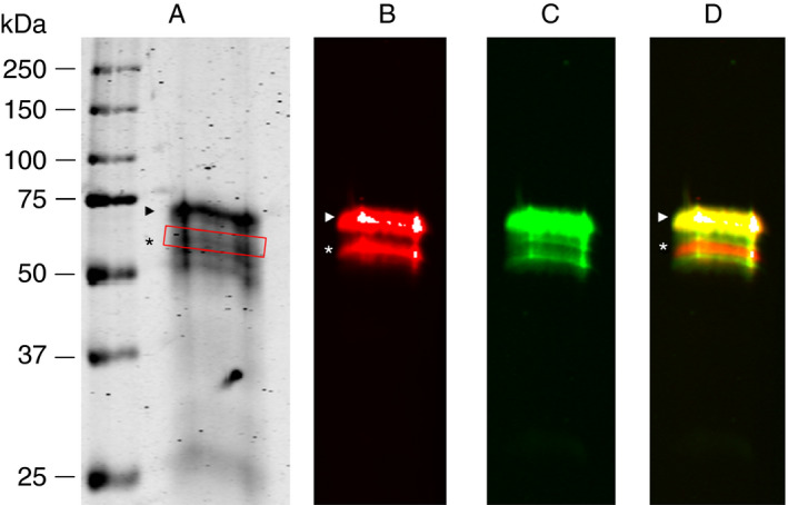
Methods: For epitope mapping of TC 3AP, commercially available plasma purified α2AP was enzymatically digested with Asp-N, Glu-C, or Lys-N. The resulting peptides were immunoprecipitated using TC 3AP-loaded Dynabeads® Protein G. Bound peptides were eluted and analyzed by liquid chromatography-tandem mass spectometry (LC-MS/MS). To localize the C-terminal cleavage site precisely, α2AP (intact and NPB) was purified from plasma and analyzed by LC-MS/MS after enzymatic digestion with Arg-C.
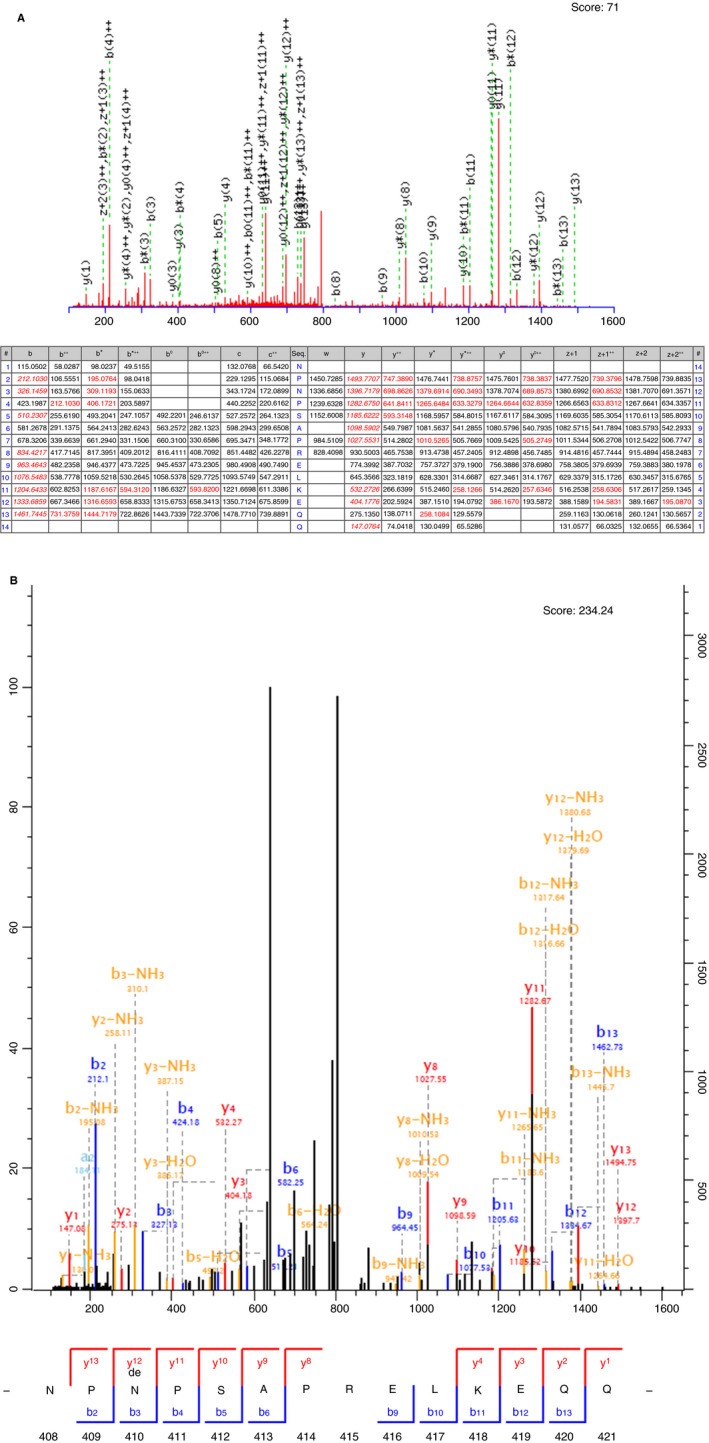
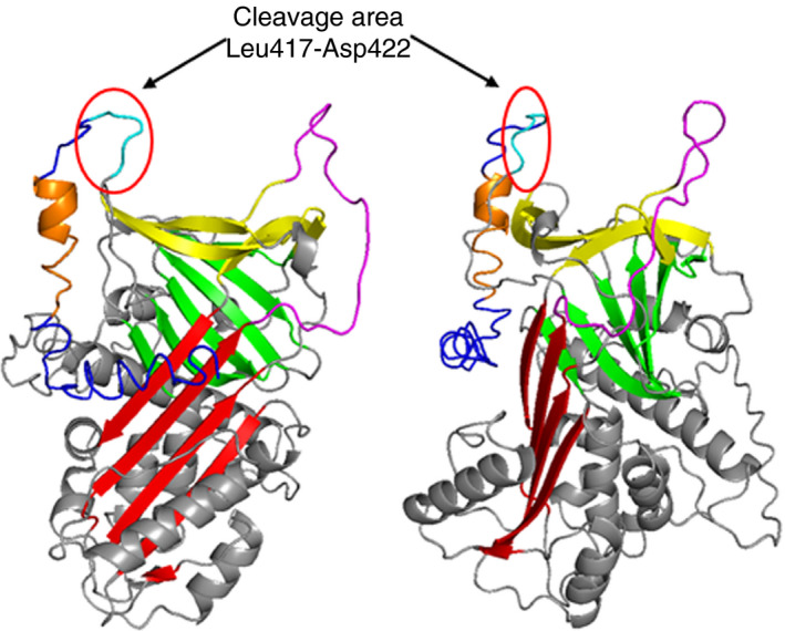
Results: We localized the epitope of TC 3AP between amino acid residues Asp428 and Gly439. LC-MS/MS data from plasma purified α2AP showed that NPB-α2AP results from cleavage at Gln421-Asp422 as preferred site, but also after Leu417, Glu419, Gln420, or Asp422.
Conclusions: The C-terminal cleavage site of human α2AP is located N-terminally from the TC 3AP epitope. Because C-terminal cleavage of α2AP can occur after multiple residues, different proteases may be responsible for the generation of NPB-α2AP.
Keywords: alpha-2-antiplasmin; epitope mapping; mass spectrometry; proteolysis; western blot.
© 2020 The Authors. Journal of Thrombosis and Haemostasis published by Wiley Periodicals, Inc. on behalf of International Society on Thrombosis and Haemostasis.
Conflict of interest statement
The authors state that they have no conflict of interest.
Figures

References
-
- Moroi M, Aoki N. Isolation and characterization of alpha2‐plasmin inhibitor from human plasma. A novel proteinase inhibitor which inhibits activator‐induced clot lysis. J Biol Chem. 1976;251:5956‐5965. - PubMed
-
- Collen D. Identification and some properties of a new fast‐reacting plasmin inhibitor in human plasma. Eur J Biochem. 1976;69:209‐216. - PubMed
-
- Wiman B, Collen D. Purification and characterization of human antiplasmin, the fast‐acting plasmin inhibitor in plasma. Eur J Biochem. 1977;78:19‐26. - PubMed
-
- Holmes WE, Nelles L, Lijnen HR, Collen D. Primary structure of human alpha 2‐antiplasmin, a serine protease inhibitor (serpin). J Biol Chem. 1987;262:1659‐1664. - PubMed
