Review
. 2020 Mar;77(3-4):129-148.
doi: 10.1002/cm.21602. Epub 2020 Feb 14.

Cargo hold and delivery: Ankyrins, spectrins, and their functional patterning of neurons

Affiliations
Review

Cargo hold and delivery: Ankyrins, spectrins, and their functional patterning of neurons

Damaris N Lorenzo. Cytoskeleton (Hoboken). 2020 Mar.

Abstract

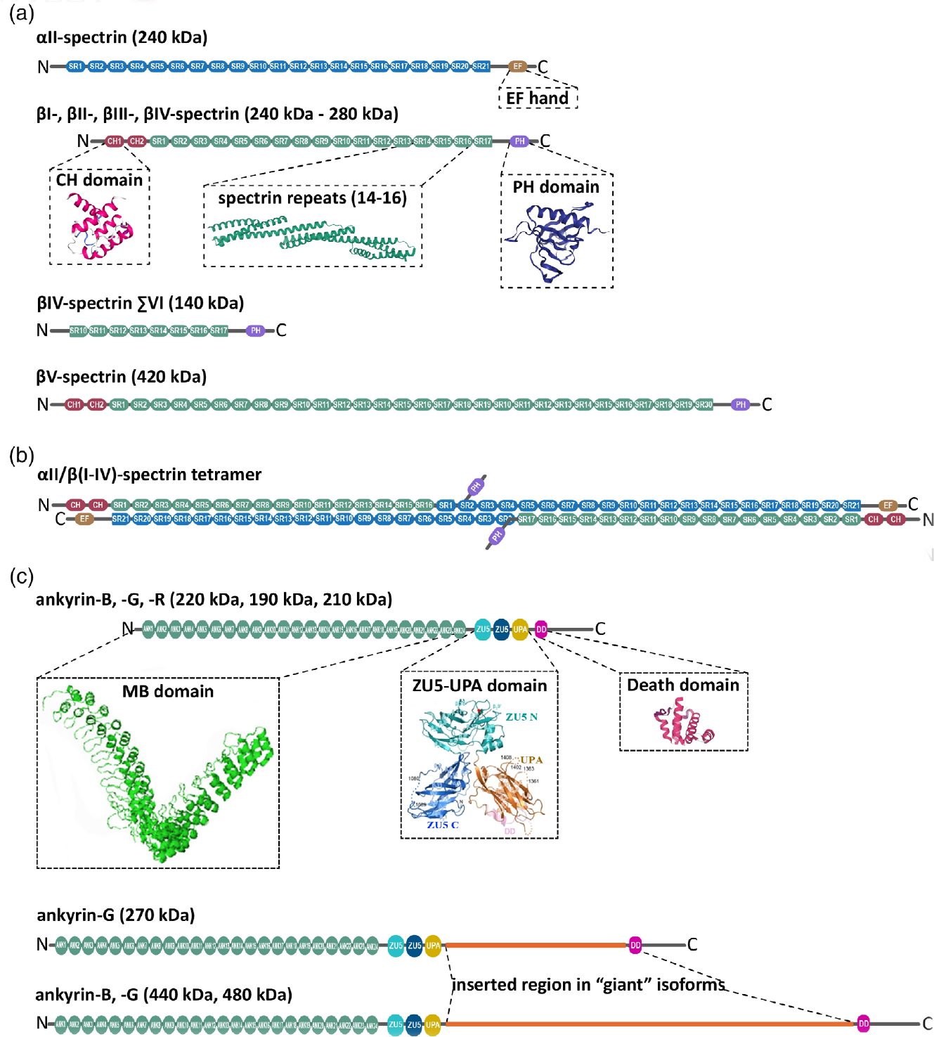
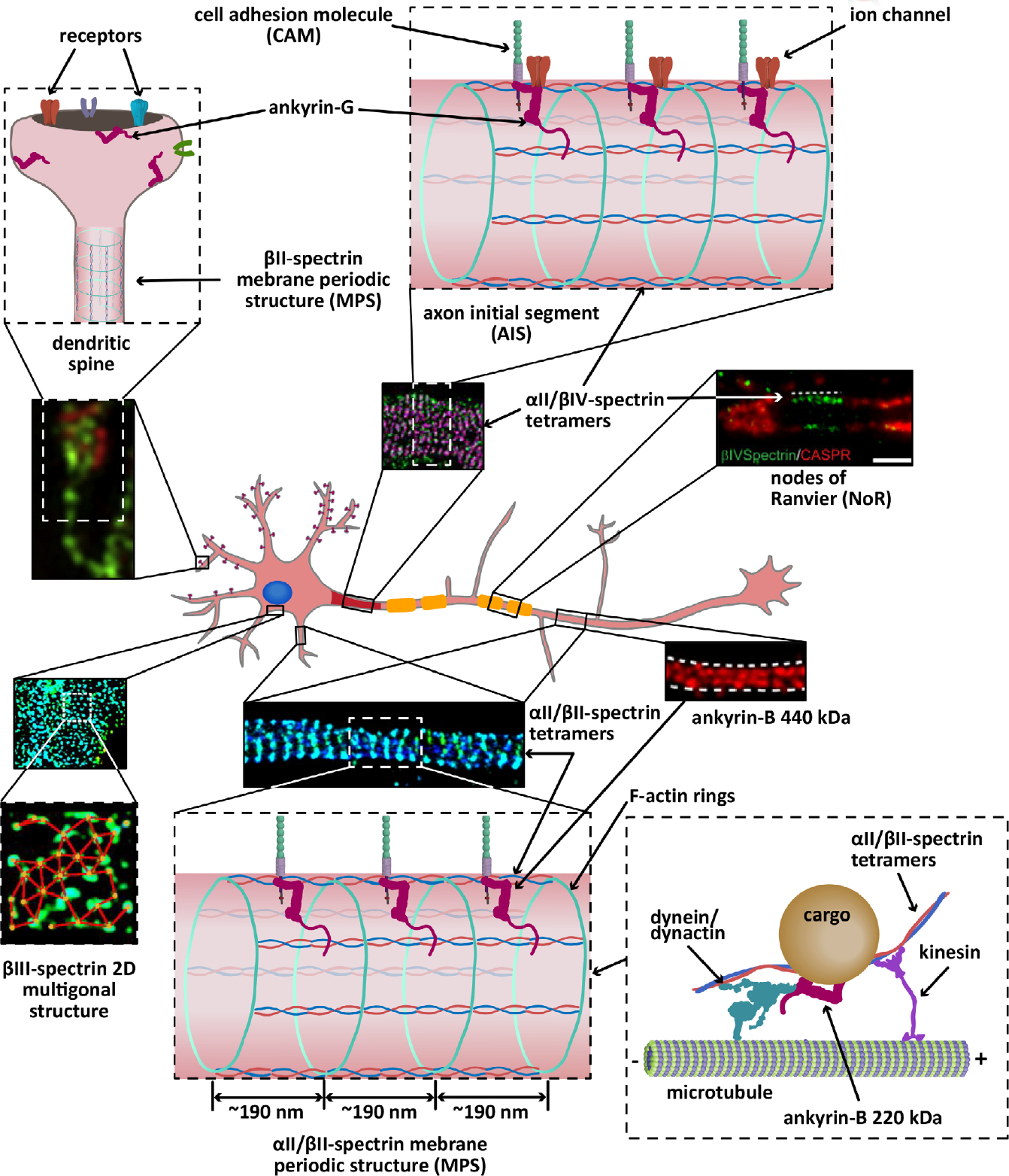
The highly polarized, typically very long, and nonmitotic nature of neurons present them with unique challenges in the maintenance of their homeostasis. This architectural complexity serves a rich and tightly controlled set of functions that enables their fast communication with neighboring cells and endows them with exquisite plasticity. The submembrane neuronal cytoskeleton occupies a pivotal position in orchestrating the structural patterning that determines local and long-range subcellular specialization, membrane dynamics, and a wide range of signaling events. At its center is the partnership between ankyrins and spectrins, which self-assemble with both remarkable long-range regularity and micro- and nanoscale specificity to precisely position and stabilize cell adhesion molecules, membrane transporters, ion channels, and other cytoskeletal proteins. To accomplish these generally conserved, but often functionally divergent and spatially diverse, roles these partners use a combinatorial program of a couple of dozens interacting family members, whose code is not fully unraveled. In a departure from their scaffolding roles, ankyrins and spectrins also enable the delivery of material to the plasma membrane by facilitating intracellular transport. Thus, it is unsurprising that deficits in ankyrins and spectrins underlie several neurodevelopmental, neurodegenerative, and psychiatric disorders. Here, I summarize key aspects of the biology of spectrins and ankyrins in the mammalian neuron and provide a snapshot of the latest advances in decoding their roles in the nervous system.

Keywords: ankyrin; axonal transport; cytoskeleton; plasma membrane; spectrin.


Figures

FIGURE 1
FIGURE 1
Structure domains of neuronal spectrins and ankyrins. (a) αII-spectrin spans 21 modular spectrin repeats (SR, blue), followed by a calcium-binding EF hand domain (light brown) close to the C-terminus. αII-spectrin also contains a Src-homology 3 (SH3) domain in SR9, and a calmodulin-binding loop in SR11. Neuronal spectrins also include canonical βI-, βII-, βIII-, and βIV-spectrins, which are comprised of 17 SR (green; ribbon diagram of the crystal structure of SR14–16 from Grum, MacDonald, & Mondragón, 1999), two in tandem calponin homology domains (CH, red; ribbon diagram of the crystal structure from Djinović-Carugo, Bañuelos, & Saraste, 1997) close to the N-terminus, and a pleckstrin homology domain (PH, purple; ribbon diagram of the crystal structure from Lemmon, Ferguson, & Abrams, 2002). βV-spectrin, the largest known mammalian spectrin, contains 13 additional SR. Alternatively spliced βIV-spectrin ΣVI, which lacks the PH domain and the first 9 SR, is a well-studied noncanonical, neuronal β-spectrin isoform. (b) αII-spectrin uses its N-terminus SR to bind the N-terminus SR of β-spectrins and associate as heterodimers. These heterodimers use antiparallel head-to-head association to form tetrameric units assembles with actin and networks with long-range regularity. (c) Canonical neuronal ankyrins contain a membrane-binding domain comprised of 24 ankyrin repeats (ANK) in the N-terminus (MBD, green; ribbon representation of the crystal structure from Michaely, Tomchick, Machius, & Anderson, 2002), followed by a supermodule that contains two ZU5 domains a UPA domain (teal, blue, orange; ribbon representation of crystal structure from Wang, Yu, Ye, Wei, & Zhang, 2012), which contains the ankyrin-binding site, a death domain (pink; ribbon representation of crystal structure from Wang, Simon, et al., 2019; Wang, Taki, et al., 2019) and a C-terminal unstructured regulatory domain (gray). The giant, neuron-specific ankyrin isoforms have an insertion of a single exon (orange) after the UPA domain
FIGURE 2
FIGURE 2
Cartoon representation of the localization and organization of ankyrins and spectrins in a hippocampal neuron. αII/βII-spectrin tetramers crosslink actin rings to line the axon (STORM image adapted from Zhong et al., 2014), parts of the shafts of dendrites, and a portion of spine necks (not shown), all with an average spacing regularity of ~190 nm. Both αII-spectrin and βII-spectrin also associate with microtubule motors to promote axonal transport of synaptic vesicles. The 440 kDa ankyrin-G isoform, which also shows long range periodicity (STED image adapted from Yang, Walder-Christensen, Kim, et al., 2019; Yang, Walder-Christensen, Lalani, et al., 2019), likely binds the periodic axonal βII-spectrin to stabilize cell adhesion molecules, such as L1CAM. Ankyrin-B 220 kDa (not shown) couples the retrograde transport machinery to organelles to facilitate axonal transport. Like βII-spectrin, βIII-spectrin forms periodic quasi-1D structures in dendritic processes (not shown), in addition to 2D polygonal lattices in the soma (STORM image adapted from Han et al., 2017, with permission). Similarly, αII/βIV-spectrin tetramers form periodic quasi-1D structures in the axon initial segment (AIS) (STORM image adapted from Huang, Zhang, Ho, et al., 2017) and nodes of Ranviers (NoR) (STED image adapted from D’Este, Kamin, Balzarotti, & Hell, 2017, with permission) in myelinated axons, where it binds ankyrin-G to stabilize cell adhesion molecules and ion channels. Ankyrin-R and βI-spectrin concentrate at the nodes to stabilize ion channels (not shown). βI-spectrin is also found in the soma, dendrites, and spines (not shown). Ankryin-G also localizes to dendritic spines (SIM image adapted from Smith et al., 2014, with permission), where it is required for the retention of AMPA receptors

References

    1. Agre P, Orringer EP, & Bennett V (1982). Deficient red-cell spectrin in severe, recessively inherited spherocytosis. The New England Journal of Medicine, 306, 1155–1161. 10.1056/nejm198205133061906 - DOI - PubMed
    1. Alemu EA, Lamark T, Torgersen KM, Birgisdottir AB, Larsen KB, Jain A, et al. (2012). ATG8 family proteins act as scaffolds for assembly of the ULK complex: Sequence requirements for LC3-interacting region (LIR) motifs. Journal of Biological Chemistry, 287, 39275–39290. 10.1074/jbc.M112.378109 - DOI - PMC - PubMed
    1. Amor V, Zhang C, Vainshtein A, Zhang A, Zollinger DR, Eshed-Eisenbach Y, et al. (2017). The paranodal cytoskeleton clusters Na(+) channels at nodes of Ranvier. eLife, 6, e21392 10.7554/eLife.21392 - DOI - PMC - PubMed
    1. An X, Guo X, Sum H, Morrow J, Gratzer W, & Mohandas N (2004). Phosphatidylserine binding sites in erythroid spectrin: Location and implications for membrane stability. Biochemistry, 43, 310–315. - PubMed
    1. Armbrust KR, Wang X, Hathorn TJ, Cramer SW, Chen G, Zu T, et al. (2014). Mutant β-III spectrin causes mGluR1α mislocalization and functional deficits in a mouse model of spinocerebellar ataxia type 5. The Journal of Neuroscience, 34, 9891–9904. 10.1523/JNEUROSCI.0876-14.2014 - DOI - PMC - PubMed

Publication types

MeSH terms

LinkOut - more resources