. 2020 Jul;68(7):1445-1465.
doi: 10.1002/glia.23792. Epub 2020 Feb 8.

Inflammation and matrix metalloproteinase 9 (Mmp-9) regulate photoreceptor regeneration in adult zebrafish

Affiliations

Inflammation and matrix metalloproteinase 9 (Mmp-9) regulate photoreceptor regeneration in adult zebrafish

Nicholas J Silva et al. Glia. 2020 Jul.

Abstract

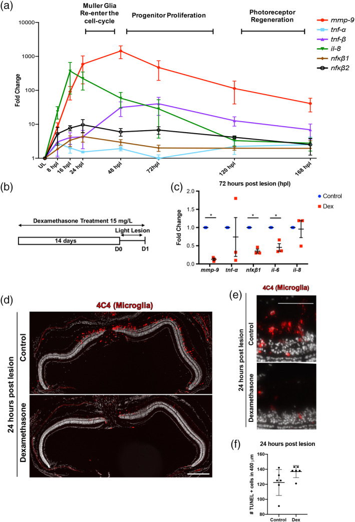
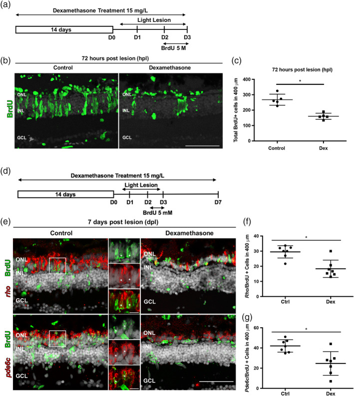
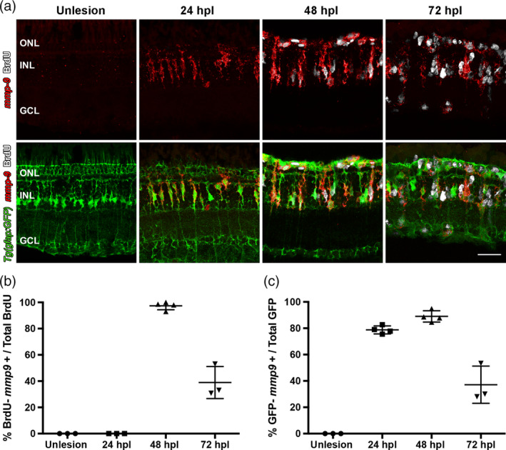
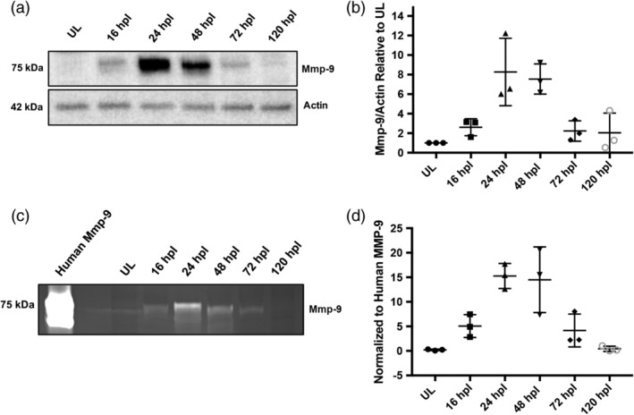
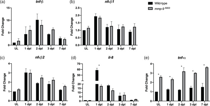
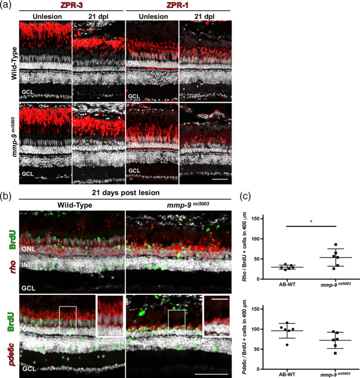
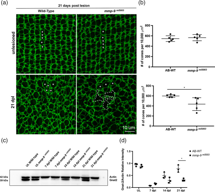
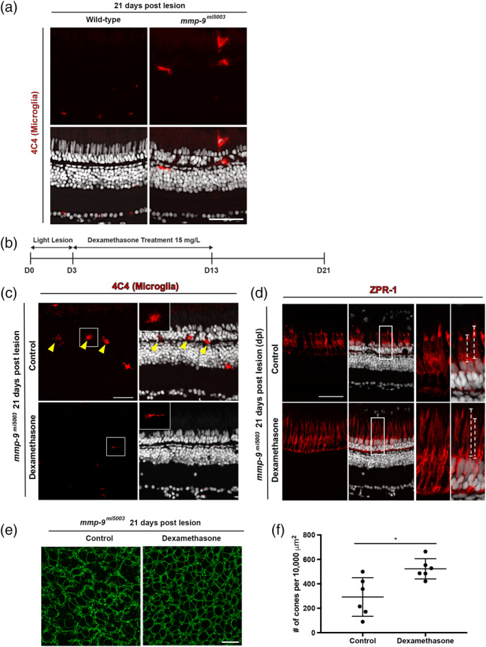
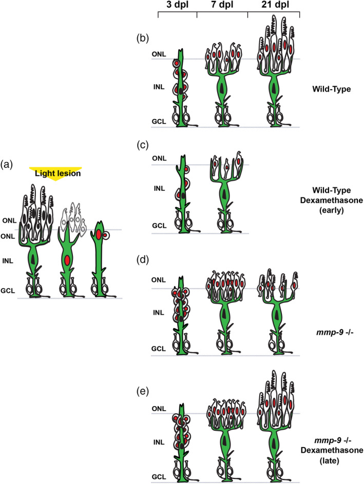
Brain injury activates complex inflammatory signals in dying neurons, surviving neurons, and glia. Here, we establish that inflammation regulates the regeneration of photoreceptors in the zebrafish retina and determine the cellular expression and function of the inflammatory protease, matrix metalloproteinase 9 (Mmp-9), during this regenerative neurogenesis. Following photoreceptor ablation, anti-inflammatory treatment suppresses the number of injury-induced progenitors and regenerated photoreceptors. Upon photoreceptor injury, mmp-9 is induced in Müller glia and Müller glia-derived photoreceptor progenitors. Deleting mmp-9 results in over production of injury-induced progenitors and regenerated photoreceptors, but over time the absence of Mmp-9 compromises the survival of the regenerated cones. At all time-points studied, the levels of tnf-α are significantly elevated in mutant retinas. Anti-inflammatory treatment in mutants rescues the defects in cone survival. These data provide a link between injury-induced inflammation in the vertebrate CNS, Mmp-9 function during neuronal regeneration and the requirement of Mmp-9 for the survival of regenerated cones.

Keywords: Müller glia; cytokines; immunosuppression; microglia; proliferation; stem cell.


Conflict of interest statement

The authors declare no potential conflict of interest.

Figures

Figure 1
Figure 1
Inflammation is induced following photoreceptor ablation. Dexamethasone treatment suppresses microglia activation and cytokine production, but not photoreceptor death. (a) Time‐course for the expression of inflammatory genes, from 8 to 168 hr post lesion (hpl). Unlesioned retinas served as controls. Expression levels are represented as fold change calculated using DDC T method. (b) Experimental paradigm for the immune suppression. (c) qRT‐PCR for the inflammatory genes mmp‐9 (ANOVA F‐ratio = 22.237, p < .0001), tnf‐α (ANOVA F‐ratio = 1.5507, p = .2209), tnf‐β (ANOVA F‐ratio = 6.5120, p = .001), il‐8 (ANOVA F‐ratio = 8.2296, p = .0003), nfκb1 (ANOVA F‐ratio = 2.5207, p = .0596), and nfκb2 (ANOVA F‐ratio = 3.5591, p = .0168) from control and Dex‐treated retinas at 72 hpl. *p ≤ .05. (d) Immunostaining for microglia using the 4C4 antibody in control (top) and Dex‐treated retinas (bottom). (e) High magnification image of 4C4 staining at 1 dpl control (top) and Dex‐treated retinas (bottom). (f) The number of TUNEL positive cells at 1 dpl in control (122.27 ± 17.03 cells; n = 6) and Dex‐treated animals (136.93 ± 8.29 cells; n = 6). Scale bar equals 50 μm [Color figure can be viewed at wileyonlinelibrary.com]
Figure 2
Figure 2
Dexamethasone treatment suppresses proliferation of Müller glia and photoreceptor regeneration. (a) Experimental paradigm of anti‐inflammatory treatment to assay proliferation. (b) BrdU immunostained cells (green) in controls (left) and Dex‐treated animals (right) at 72 hpl. (c) Number of BrdU‐labeled cells in controls (268 ± 36.1 cells; n = 5) and Dex‐treated animals (160.2 ± 20.02 cells; n = 5) at 72 hpl; *p = .0079. (d) Experimental paradigm of anti‐inflammatory treatment. Regenerated photoreceptors were identified as BrdU‐labeled nuclei surrounded by in situ hybridization signal for either rho (rods) or pde6c (cones) at 7 dpl. (e) Double labeled, regenerated photoreceptors using in situ hybridization for rods (rho) and cones (pde6c; red signal) and BrdU (green). The high magnification insets show the colocalization of the two labels (asterisks). (f) Number of regenerated rod photoreceptors in control (29.52 ± 4.1 cells; n = 7) and experimental retinas (18.33 ± 5.71 cells; n = 7); *p = .0047. (g) Number of regenerated cone photoreceptors in control (42 ± 6.1 cells; n = 7) and Dex‐treated animals (25 ± 11.72 cells; n = 7). *p = .0012. Scale bar equals 50 μm. GCL, ganglion cell layer; INL, inner nuclear layer; ONL, outer nuclear layer [Color figure can be viewed at wileyonlinelibrary.com]
Figure 3
Figure 3
Following a photolytic lesion, Müller glia and Müller glia‐derived progenitors express mmp‐9. These experiments were performed using the transgenic line Tg[gfap:EGFP]mi2002, in which eGFP is expressed in Müller glia. (a) Triple labeling using in situ hybridization for mmp‐9 (red) and immunostaining for BrdU for dividing cells (white), combined with GFP immunostaining (green). (b) Number of BrdU+ cells that express mmp‐9 in unlesioned animals and lesioned animals at 24, 48, and 72 hpl. (c) Number of GFP+ Müller glia that also express mmp‐9 expression at 24, 48, and 72 hpl. Scale bar equals 25 μm. GCL, ganglion cell layer; INL, inner nuclear layer; ONL, outer nuclear layer [Color figure can be viewed at wileyonlinelibrary.com]
Figure 4
Figure 4
Mmp‐9 is expressed and catalytically active following photoreceptor death. (a) Western blot of retinal proteins from unlesioned retinas (UL) and lesioned retinas at 16, 24, 48, 72, and 120 hpl, stained with anti‐Mmp‐9 and anti‐Actin (loading control) antibodies. (b) Densitometry of Mmp‐9 protein, plotted relative to unlesioned controls. (ANOVA F‐ratio = 8.377, p = .0013) (c) Zymogram of Mmp‐9 catalytic activity from unlesioned retinas (UL) and lesioned retinas at 16, 24, 48, 72, and 120 hpl. Lanes in zymogram correspond to those in the Western blot. Purified human recombinant protein serves as a positive control. (d) Densitometry of Mmp‐9 catalytic activity, plotted relative to the positive control. (ANOVA F‐ratio = 11.870, p = .0003)
Figure 5
Figure 5
CRISPR mutants lack catalytically‐active Mmp‐9. (a) Genomic structure of mmp‐9 and the gRNA target sequence. (b) Sequence alignment for two indel mutations—8 bp insertion (mmp‐9 mi5004) and 23 bp deletion (mmp‐9 mi5003). Red underline indicates the sequence targeted by the 19 bp gRNA. (c) Western blot of retinal proteins from unlesioned retinas (UL wild‐type; UL mmp‐9 mi5004; UL mmp‐9 mi5003) and lesioned retinas (wild‐type; mmp‐9 mi5004; mmp‐9 mi5003) at 24, and 48 hpl stained with anti‐Mmp‐9 and anti‐Actin (loading control) antibodies. (d) Zymogram of Mmp‐9 catalytic activity. Lanes in zymogram correspond to those in the Western blot. Purified human recombinant protein serves as a positive control [Color figure can be viewed at wileyonlinelibrary.com]
Figure 6
Figure 6
Photolytic lesions in mmp‐9 mutants results in the over production of injury‐induced progenitors and regenerated photoreceptors. (a) BrdU‐labeled cells (green) in wild‐type and mmp‐9 mi5003. (b) Number of BrdU+ cells from wild‐type (109 ± 19.66 cells; n = 6) mutant retinas (142.3 ± 25.72 cells; n = 9) at 72 hpl. *p = .0186. (c) BrdU‐labeled cells (green) in wild‐type and mutant retinas at 168 hpl. (d) Number of BrdU+ cells in the ONL of wild‐type (48 ± 10.24 cells; n = 8) and mutant retinas (71.71 ± 3.7 cells; n = 8). *p = .0001. (e) Double labeled, regenerated photoreceptors using in situ hybridization for rods (rho) and cones (pde6c; red signal) and BrdU (green) at 168hpl. (f) Number of regenerated cone photoreceptors in wild‐type (31.42 ± 9.88 cell; n = 8) and mutant retinas (43.83 ± 10.68 cells; n = 8) at 168 hpl. *p = .0301. (g) Number of regenerated rod photoreceptors in wild‐type (14.88 ± 4.02 cells; n = 8) and mutant retinas (26.04 ± 5.69 cells; n = 8) at 168 hpl. *p = .0005. Scale bars equal 50 μm. GCL, ganglion cell layer; INL, inner nuclear layer; ONL, outer nuclear layer [Color figure can be viewed at wileyonlinelibrary.com]
Figure 7
Figure 7
Absence of Mmp‐9 results in an altered inflammatory response following photoreceptor death. Time‐course of the expression of inflammatory genes, tnf‐β (a; ANOVA F‐ratio = 19.2406, p < .0001,), nfκβ1 (b; ANOVA F‐ratio = 10.4104, p < .0001), nfκβ2 (c; ANOVA F‐ratio = 10.4104, p < .0001), il‐8 (d; ANOVA F‐ratio = 185.42, p < .0001), and tnf‐α (e; ANOVA F‐ratio = 54.53, p < .0001) in Wild‐type (black bars) and Mmp‐9 mutant (gray bars) retinas following photoreceptor death. Fold changes relative to unlesioned retinas of Wild‐type are calculated using DDCt method. ANOVA with post‐hoc Tukey, *p < .0001
Figure 8
Figure 8
Mmp‐9 regulates the survival of regenerated cone photoreceptors. (a) Immunostaining for red‐green double cones with ZPR‐1 and rods with ZPR‐3. Wild‐type are in the top row; mutants are in the bottom row. Unlesioned retinas are left; lesioned retinas at 21 dpl are right. (b) Double labeled, regenerated photoreceptors at 21dpl using in situ hybridization for rods (rho) and cones (pde6c; red signal) and BrdU (green). Insets detail the morphology of regenerated photoreceptors in wild‐type (left) and mutant retinas (right). (c) Number of regenerated cone photoreceptors (bottom) in wild‐type (95.81 ± 18.49 cells; n = 6) and mutant retinas (71.89 ± 20.37 cells; n = 6) at 21 dpl. p = .059. Number of regenerated rod photoreceptors (top) in wild‐type (29.6 ± 4.94 cells; n = 6) and mutant retinas (53.39 ± 21.62 cells; n = 6) at 21 dpl. *p = .0270. Scale bar equals 50 μm. GCL, ganglion cell layer; INL, inner nuclear layer; ONL, outer nuclear layer [Color figure can be viewed at wileyonlinelibrary.com]
Figure 9
Figure 9
Mmp‐9 is required for the survival of regenerated cone photoreceptors. (a) Wholemounts of wild‐type (left) and mutant retinas (right) immunostained for ZO‐1. Unlesioned retinas are top; lesioned retinas at 21dpl are bottom. Asterisks indicate profiles of cones. In mutants, gaps due to missing cones are replaced by the irregular apical processes of Müller glia (dashed lines). (b) Number of unlesioned cones from wild‐type (546.49 ± 55.69; n = 5) and mmp‐9 mi5003 (565.33 ± 27.42; n = 5) *p = .516; below the number of regenerated cones from wild‐type (599.11 ± 27.42 cones; n = 5) and mutant retinas (436.09 ± 128.04 cones; n = 5) at 21 dpl. *p = .0238. (c) Western blot of retinas stained with antibodies against Gnat‐2 and Actin at 7, 14, and 21 dpl. (d) Densitometry of Gnat‐2 labeling in the Western blot. A significant difference in gnat‐2 levels were observed at 21 dpl. *p = .0014. Scale bar equals 10 μm [Color figure can be viewed at wileyonlinelibrary.com]
Figure 10
Figure 10
Late anti‐inflammatory treatment rescues defects of regenerated cones in mutants at 21 dpl. (a) Immunostaining for microglia marker, 4C4 in Wild‐type (0.1 ± 0.3 cells per 400 μm, n = 4) and mutant (4.0 ± 2.2 cells per 400 μm, n = 4, p = .0148) at 21 dpl. (b) Experimental paradigm for the photolytic lesions and Dexamethasone treatment. (c) Immunostaining for 4C4 in control (top) and Dex‐treated retinas (bottom). Inserts illustrate ameboid (top) and ramified (bottom) microglia. (d) Immunostaining with ZPR‐1 for red‐green double cones in control (top) and Dex mutants (bottom) 21 dpl. Insets illustrate differences in the lengths of the cone photoreceptors (dashed lines; control, 22.1 ± 7.2 μm, n = 40 cells; Dex‐treated, 29.7 ± 4.8 μm, n = 69 cells; p < .001) (e) Wholemounts of mutant retinas immunostained for ZO‐1. Control retina is left; Dex‐treated retina is right. (f) Number of regenerated cones in control (292.20 ± 158.11 cones; n = 6) and Dex‐treated retinas (522.79 ± 82.55 cones; n = 6) at 21 dpl. Scale bars equal 50 μm in panel (a), 40 μm in panels (c) and (d), and 10 μm in (e) [Color figure can be viewed at wileyonlinelibrary.com]
Figure 11
Figure 11
Summary diagram of cone regeneration. (a) Müller glia respond to photoreceptor death by expressing mmp‐9, undergoing interkinetic nuclear migration and a single asymmetric cell division that gives rise to a neuronal progenitor. (b) In wild‐type animals, neuronal progenitors form a neurogenic cluster around the Müller glia (3 dpl), migrate to the ONL, and differentiate into cone photoreceptors (7 dpl) that then mature (21 dpl). (c) Anti‐inflammatory treatment results in fewer Müller glia‐derived progenitors and fewer regenerated photoreceptors. (d) In the absence of Mmp‐9, there is overproduction of Müller glia‐derived progenitors and regenerating photoreceptors. However, at 21 dpl, survival of cone photoreceptors is compromised. (e) In the absence of Mmp‐9, anti‐inflammatory treatment rescues the defects of cone photoreceptors. GCL, ganglion cell layer; INL, inner nuclear layer; ONL, outer nuclear layer [Color figure can be viewed at wileyonlinelibrary.com]

References

    1. Amor, S. , Puentes, F. , Baker, D. , & van der Valk, P. (2010). Inflammation in neurodegenerative diseases. Immunology, 129, 154–169. - PMC - PubMed
    1. Ando, K. , Shibata, E. , Hans, S. , Brand, M. , & Kawakami, A. (2017). Osteoblast production by reserved progenitor cells in zebrafish bone regeneration and maintenance. Developmental Cell, 43, 643–650.e3. - PubMed
    1. Bernardos, R. L. , Barthel, L. K. , Meyers, J. R. , & Raymond, P. A. (2007). Late‐stage neuronal progenitors in the retina are radial Müller glia that function as retinal stem cells. The Journal of Neuroscience, 27, 7028–7040. - PMC - PubMed
    1. Bernardos, R. L. , & Raymond, P. A. (2006). GFAP transgenic zebrafish. Gene Expression Patterns, 6, 1007–1013. - PubMed
    1. Bollaerts I, Van Houcke J, Beckers A, Lemmens K, Vanhunsel S, De Groef L, Moons L. 2019. Prior exposure to immunosuppressors sensitizes retinal microglia and accelerates optic nerve regeneration in zebrafish. Mediators of Inflammation. 6135795, 2019, 1, 16. - PMC - PubMed

Publication types

MeSH terms

Substances