. 2020 Feb:139:225-237.
doi: 10.1016/j.yjmcc.2020.02.001. Epub 2020 Feb 5.

Identification and characterization of self-association domains on small ankyrin 1 isoforms

Affiliations

Identification and characterization of self-association domains on small ankyrin 1 isoforms

Janani Subramaniam et al. J Mol Cell Cardiol. 2020 Feb.

Abstract

In striated muscles, the large scaffolding protein obscurin and a small SR-integral membrane protein sAnk1.5 control the retention of longitudinal SR across the sarcomere. How a complex of these proteins facilitates localization of longitudinal SR has yet to be resolved, but we hypothesize that obscurin interacts with a complex of sAnk1.5 proteins. To begin to address this hypothesis, we demonstrate that sAnk1.5 interacts with itself and identify two domains mediating self-association. Specifically, we show by co-precipitation and FLIM-FRET analysis that sAnk1.5 and another small AnkR isoform (sAnk1.6) interact with themselves and each other. We demonstrate that obscurin interacts with a complex of sAnk1.5 proteins and that this complex formation is enhanced by obscurin-binding. Using FLIM-FRET analysis, we show that obscurin interacts with sAnk1.5 alone and with sAnk1.6 in the presence of sAnk1.5. We find that sAnk1.5 self-association is disrupted by mutagenesis of residues Arg64-Arg69, residues previously associated with obscurin-binding. Molecular modeling of two interacting sAnk1.5 monomers facilitated the identification of Gly31-Val36 as an additional site of interaction, which was subsequently corroborated by co-precipitation and FLIM-FRET analysis. In closing, these results support a model in which sAnk1.5 forms large oligomers that interact with obscurin to facilitate the retention of longitudinal SR throughout skeletal and cardiac myocytes.

Keywords: Ankyrin; Obscurin; Sarcoplasmic reticulum; Self-association; sAnk1.5.


Conflict of interest statement

Declaration of Competing Interest The authors declare no competing financial interest.

Figures
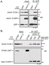
Fig. 1.
Fig. 1.
sAnk1.5 interacts with itself. (A) sAnk1.5-GFP immunoprecipitates sAnk1.5-HA. HA and GFP immunoblots demonstrate expression and precipitation of sAnk1.5-HA by sAnk1.5-GFP but not GFP alone. (B) sAnk1.5-GST precipitates sAnk1.5-HA in stringent wash conditions with increasing salt concentrations from 500 mM to 1 M. HA and GST immunoblots demonstrate expression and precipitation of sAnk1.5-HA by sAnk1.5-GST but not the negative control STIM1-GST.
Fig. 2.
Fig. 2.
sAnk1.6 interacts with itself and with sAnk1.5. (A) Amino acid alignment of sAnk1.5 and 1.6. Transmembrane domain (TMD) and obscurin-binding domains (OBD) are highlighted. HA and GST immunoblots demonstrate expression and precipitation of sAnk1.6-HA by sAnk1.6-GST but not STIM1-GST (B) and of sAnk1.5-HA by sAnk1.6-GST but not STIM1-GST (C).
Fig. 3.
Fig. 3.
sAnk1.5 interacts with sAnk1.5 and obscurin. (A) Schematic of the relative positions of obscurin domains including immunoglobulin (IG), fibronectin type 3 (FN3), calmodulin-binding motif (IQ), Src homology 3 (SH3), guanine nucleotide exchange factor for Rho/Rac/Cdc42-like GTPases (RhoGEF), pleckstrin homology (PH), and ankyrin-binding domains (ABD). (B) HA and GST immunoblots demonstrate expression and precipitation of obscurin-HA by sAnk1.5-GST but not sAnk1.6-GST. (C) Quantification of obscurin-binding by sAnk1.5 and 1.6. (D) HA and GST immunoblots demonstrate expression and precipitation of obscurin-HA and sAnk1.5-HA by sAnk1.5-GST but not STIM1-GST. sAnk1.6-GST only precipitates sAnk1.5-HA but not obscurin-HA.
Fig. 4.
Fig. 4.
FLIM-FRET analysis demonstrating obscurin interaction with sAnk1.5 alone or with sAnk1.6 in the presence of sAnk1.5 in cells. (A) Images from FLIM-FRET analysis (pseudocolored images showing average CFP lifetime: τav) of CFP donor alone (obscurin-CFP) and the positive control (sarcolipin-CFP-YFP). (B) Images of FLIM-FRET analysis of obscurin-CFP (donor) with sAnk1.5-YFP (acceptor) in the presence of sAnk1.5-HA or sAnk1.6-HA. (C) Images of FLIM-FRET analysis of obscurin-CFP (donor) with sAnk1.6-YFP (acceptor) in the presence of sAnk1.5-HA or sAnk1.6-HA. (D) The graph shows mean fluorescence lifetime of CFP ± SEM (n ≥ 80, from three independent experiments). Statistical analysis was performed by unpaired Student’s t-test (*** p < .001). (E) HA and GFP immunoblots demonstrate expression of obscurin-CFP, sAnk1.5-YFP, sAnk1.6-YFP, sAnk1.5-HA and sAnk1.6-HA.
Fig. 5.
Fig. 5.
sAnk1.5 proteins independently self-associate, but obscurin-binding enhances complex formation. (A) Alignment of obscurin amino acid sequences of wild-type and ankyrin-binding domain mutants (ΔABD1, ΔABD2, ΔABD1&2). (B) HA and GST immunoblots demonstrate expression and precipitation of wild-type (wt) and mutant (ΔABD1, ΔABD2) obscurin-HA and sAnk1.5-HA by sAnk1.5-GST. (C) Quantification of sAnk1.5-GST precipitation of sAnk1.5-HA and obscurin-HA.
Fig. 6.
Fig. 6.
Identification of ankyrin-binding site in sAnk1.5. (A) Amino acid alignment of wild-type and mutant (ΔABD2) sAnk1.5. (B) HA and GST immunoblots demonstrate expression and precipitation of wild-type but not ΔABD2 sAnk1.5-HA by sAnk1.5-GST. (C) HA and GST immunoblots demonstrate expression and precipitation of obscurin-HA and sAnk1.5-HA by wild-type sAnk1.5-GST. In contrast, mutant sAnk1.5-GST (ΔABD2) precipitates far less sAnk1.5-HA but maintains its interaction with obscurin-HA. (D) YFP, HA, and GST immunoblots demonstrate expression and precipitation of sAnk1.5-HA and wild-type or mutant sAnk1.5-YFP by obscurin-GST. The decrease in mutant sAnk1.5-YFP precipitated corresponds with an increase in sAnk1.5-HA precipitated by obscurin-GST. (E) Quantification of increased sAnk1.5-HA precipitated and decreased mutant sAnk1.5-YFP precipitated by obscurin compared to obscurin precipitation of wild-type sAnk1.5-YFP and -HA (represented as % change in binding).
Fig. 7.
Fig. 7.
Identification of ankyrin-binding site in sAnk1.6. (A) Amino acid alignment of wild-type and mutant (ΔABD2) sAnk1.6. (B) HA and GST immunoblots demonstrate expression and precipitation of obscurin-HA and sAnk1.6-HA by wild-type sAnk1.6-GST. In contrast, mutant sAnk1.6-GST (ΔABD2) does not precipitate sAnk1.6-HA or obscurin-HA. (C) HA and GST immunoblots demonstrate expression and precipitation of obscurin-HA and sAnk1.5-HA by wild-type sAnk1.6-GST. In contrast, mutant sAnk1.6-GST (ΔABD2) precipitates far less sAnk1.5-HA and does not interact with obscurin-HA.
Fig. 8.
Fig. 8.
Identification of a second ankyrin-binding site in sAnk1.5. (A) Cartoon representation of sAnk1.5 dimer formation with one labeled in green and the other labeled in silver. Predicted sites of interaction are highlighted in red. (B) Magnification of interaction site with gold residues labeling the predicted ankyrin-binding domain (ABD1) and the blue residues labeling ankyrin-binding domain 2 (ABD2). (C) Alignment of sAnk1.5 amino acid sequences of wild-type and ankyrin-binding domain 1 mutant (ΔABD1). (D) HA and GST immunoblots demonstrate expression and precipitation of wild-type sAnk1.5-HA by sAnk1.5-GST. In contrast, sAnk1.5-GST precipitates less mutant sAnk1.5-HA (ΔABD1). (E) HA and GFP immunoblots demonstrate expression and precipitation of wild-type and mutant sAnk1.5-HA by obscurin-GFP. (For interpretation of the references to colour in this figure legend, the reader is referred to the web version of this article.)
Fig. 9.
Fig. 9.
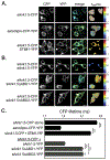
FLIM-FRET analysis demonstrating sAnk1.5 self-association in cells. (A) Images from confocal microscopy (CFP, YFP, merge) and FLIM-FRET analysis (pseudocolored images showing average CFP lifetime: τav) of CFP donor alone (sAnk1.5-CFP), positive control (sarcolipin-CFP-YFP), and negative control (sAnk1.5-CFP and STIM1-YFP). (B) Images from confocal microscopy and FLIM-FRET analysis of wild-type sAnk1.5-CFP (donor) with acceptors: wild-type sAnk1.5-YFP or mutant sAnk1.5-YFP (either ΔABD1 or ΔABD2). Scale bar represents 10 μm. (C) The graph shows mean fluorescence lifetime of CFP ± SEM (n ≥ 65, from three independent experiments). Statistical analysis was performed by unpaired Student’s t-test (*** p < .001).
Fig. 10.
Fig. 10.
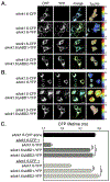
FLIM-FRET analysis demonstrating sAnk1.6 interaction with itself or with sAnk1.5 in cells. (A) Images from confocal microscopy (CFP, YFP, merge) and FLIM-FRET analysis (pseudocolored images showing average CFP lifetime: τav) of wild-type sAnk1.6-CFP (donor) alone or with acceptors: wild-type sAnk1.6-YFP or mutant sAnk1.6-YFP (either ΔABD1 or ΔABD2). (B) Images from confocal microscopy and FLIM-FRET analysis of wild-type sAnk1.5-CFP (donor) with acceptors: wild-type sAnk1.6-YFP or mutant sAnk1.6-YFP (either ΔABD1 or ΔABD2). Scale bar represents 10 μm. (C) The graph shows mean fluorescence lifetime of CFP ± SEM (n ≥ 70, from three independent experiments). Statistical analysis was performed by unpaired Student’s t-test (*** p < .001).

References

    1. Young P, Ehler E, Gautel M, Obscurin, a giant sarcomeric rho guanine nucleotide exchange factor protein involved in sarcomere assembly, J. Cell Biol 154 (2001) 123–136. - PMC - PubMed
    1. Borisov AB, Sutter SB, Kontrogianni-Konstantopoulos A, Bloch RJ, Westfall MV, Russell MW, Essential role of obscurin in cardiac myofibrillogenesis and hypertrophic response: evidence from small interfering RNA-mediated gene silencing, Histochem. Cell Biol 125 (2006) 227–238. - PubMed
    1. Kontrogianni-Konstantopoulos A, Catino DH, Strong JC, Sutter S, Borisov AB, Pumplin DW, Russell MW, Bloch RJ, Obscurin modulates the assembly and organization of sarcomeres and the sarcoplasmic reticulum, FASEB J. 20 (2006) 2102–2111. - PubMed
    1. Raeker MO, Su F, Geisler SB, Borisov AB, Kontrogianni-Konstantopoulos A, Lyons SE, Russell MW, Obscurin is required for the lateral alignment of striated myofibrils in zebrafish, Dev. Dyn 235 (2006) 2018–2029. - PubMed
    1. Borisov AB, Martynova MG, Russell MW, Early incorporation of obscurin into nascent sarcomeres: implication for myofibril assembly during cardiac myogenesis, Histochem. Cell Biol 129 (2008) 463–478. - PMC - PubMed

Publication types