

Cerebral functional activity and connectivity changes in anti-N-methyl-D-aspartate receptor encephalitis: A resting-state fMRI study
- PMID: 32036276
- PMCID: PMC7013171
- DOI: 10.1016/j.nicl.2020.102189
Cerebral functional activity and connectivity changes in anti-N-methyl-D-aspartate receptor encephalitis: A resting-state fMRI study
Abstract
Background: Anti-N-methyl-D-aspartate receptor (NMDAR) encephalitis showing severe neuropsychiatric symptoms is the most common type of autoimmune encephalitis. However, the corresponding standard clinical magnetic resonance imaging (MRI) presents normal or atypical in the majority of patients with anti-NMDAR encephalitis. Here, this study aimed to investigate the alterations in brain functional activity in patients with anti-NMDAR encephalitis and whether these alterations contributed to cognition and mood disorders.
Methods: Seventeen patients with anti-NMDAR encephalitis and eighteen gender, age and education-matched healthy controls were recruited. All participants underwent neuropsychological tests (including Montreal Cognitive Assessment (MoCA), Hamilton Anxiety Scale (HAMA), and Hamilton Depression Scale (HAMD24)) and resting-state functional MRI. MRI data was firstly analyzed by amplitude of low-frequency fluctuation (ALFF), and brain regions with altered ALFF between groups were selected as regions of interest for the further functional connectivity (FC) analysis. Correlation analyses were performed to investigate the associations between brain dysfunction and neuropsychological performance.
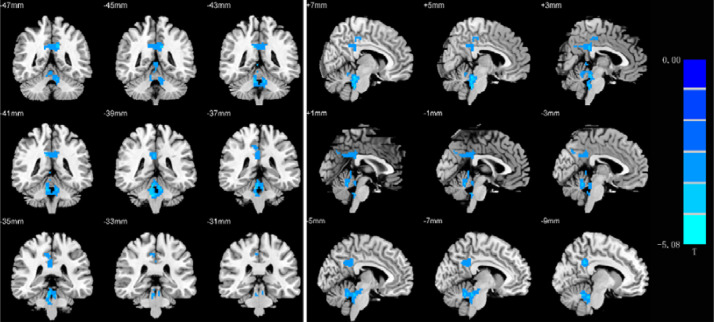
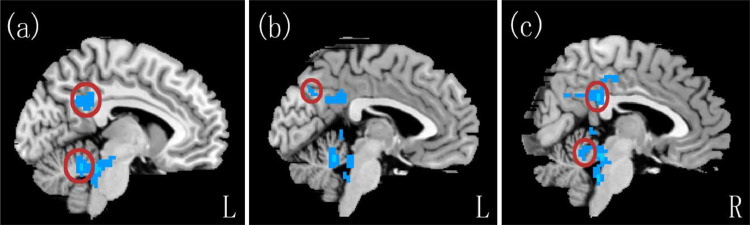
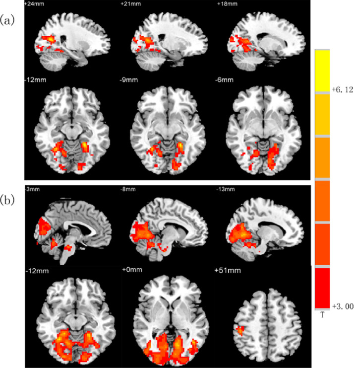
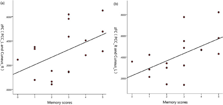
Results: Relative to the healthy controls, patients with anti-NMDAR encephalitis performed inferiorly in the MoCA score, and showed anxiety and depression disorders with higher HAMA and HAMD24 scores (all p < 0.05). In the brain functional activity analysis, the patients showed decreased ALFF values in the bilateral posterior cingulate gyrus, left precuneus, and bilateral cerebellum (false- discovery- rate corrected, p < 0.05). Furthermore, relative to the control group, the patients showed significantly increased FC between the left posterior cingulate cortex (PCC) and the bilateral lingual gyrus, right calcarine, right cuneus, also between the right PCC and the right fusiform gyrus, bilateral lingual gyrus, left calcarine, left cuneus, and right posterior central gyrus (false- discovery- rate corrected, p < 0.05). FC strength between the left posterior cingulate gyrus and right cuneus, and between the right posterior cingulate gyrus and left cuneus were both positively correlated with MoCA memory scores (r = 0.485, p = 0.048; r = 0.550, p = 0.022).
Conclusion: The present study highlight that decreased spontaneous neural activities and abnormal FC exhibited in the patients with anti-NMDAR encephalitis, which may participate in the process of cognition and emotion deficits. These results may help to elucidate the clinical radiological contradictions in anti-NMDAR encephalitis and contribute to deeper understanding of the pathophysiological mechanism of the disease.
Keywords: Amplitude of low-frequency fluctuation; Anti-N-methyl-D-aspartate receptor encephalitis; Functional connectivity; MoCA.
Copyright © 2020 The Authors. Published by Elsevier Inc. All rights reserved.
Conflict of interest statement
Declaration of Competing Interest All other authors declare no competing interests.
Figures
References
-
- Bacchi S., Franke K., Wewegama D., Needham E., Patel S., Menon D. Magnetic resonance imaging and positron emission tomography in anti-NMDA receptor encephalitis: a systematic review. J. Clin. Neurosci. 2018;52:54–59. - PubMed
-
- Buckner R.L., Andrews-Hanna J.R., Schacter D.L. The brain’s default network: anatomy, function, and relevance to disease. Ann. N. Y. Acad. Sci. 2008;1124:1–38. - PubMed
Publication types
MeSH terms
LinkOut - more resources
Full Text Sources
Medical
