

Centella asiatica (L.)-Neurodifferentiated Mesenchymal Stem Cells Promote the Regeneration of Peripheral Nerve
- PMID: 32036567
- PMCID: PMC7105542
- DOI: 10.1007/s13770-019-00235-6
Centella asiatica (L.)-Neurodifferentiated Mesenchymal Stem Cells Promote the Regeneration of Peripheral Nerve
Abstract
Background: Centella asiatica (L.) is a plant with neuroprotective and neuroregenerative properties; however, its effects on the neurodifferentiation of mesenchymal stem cells (MSCs) and on peripheral nerve injury are poorly explored. This study aimed to investigate the effects of C. asiatica (L.)-neurodifferentiated MSCs on the regeneration of peripheral nerve in a critical-size defect animal model.
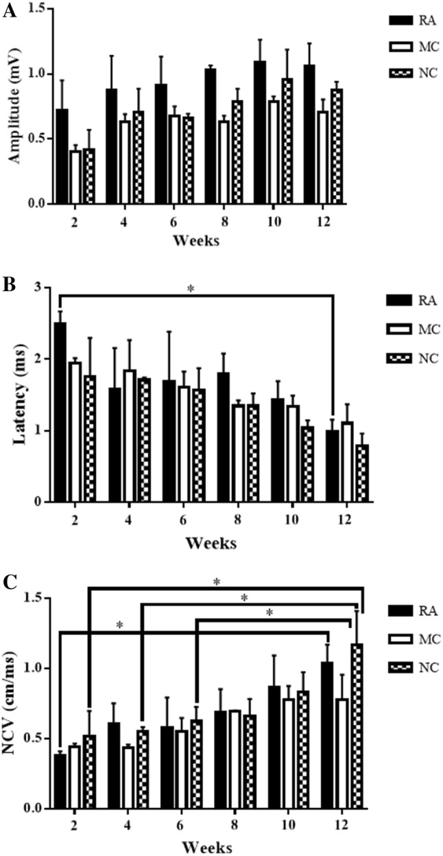
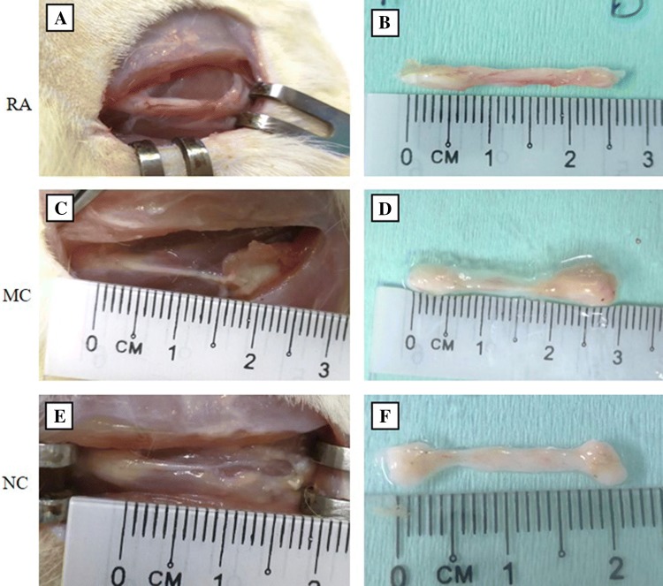
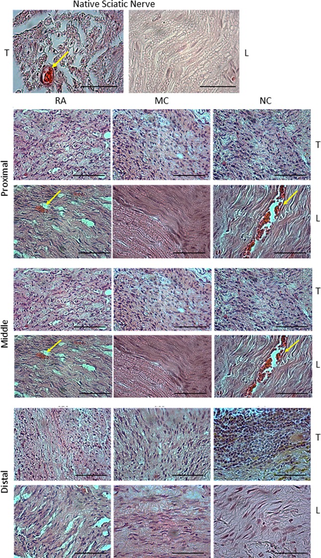
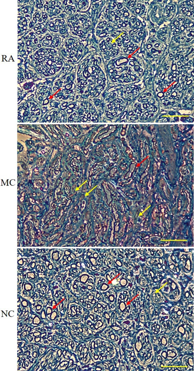
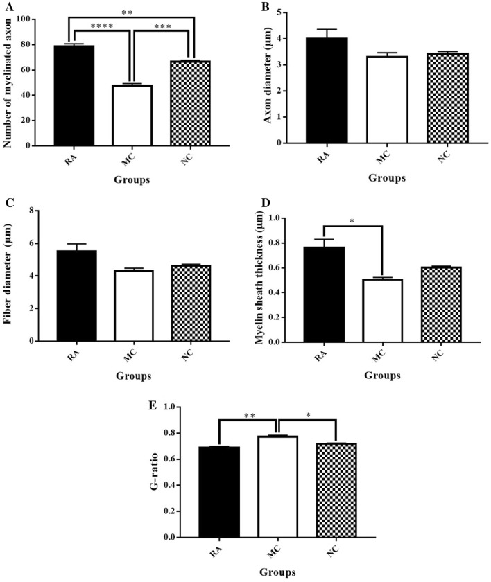
Methods: Nerve conduit was developed using decellularised artery seeded with C. asiatica-neurodifferentiated MSCs (ndMSCs). A 1.5 cm sciatic nerve injury in Sprague-Dawley rat was bridged with reversed autograft (RA) (n = 3, the gold standard treatment), MSC-seeded conduit (MC) (n = 4) or ndMSC-seeded conduit (NC) (n = 4). Pinch test and nerve conduction study were performed every 2 weeks for a total of 12 weeks. At the 12th week, the conduits were examined by histology and transmission electron microscopy.
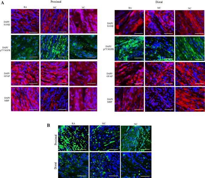
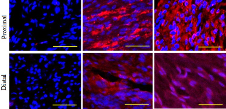
Results: NC implantation improved the rats' sensory sensitivity in a similar manner to RA. At the 12th week, nerve conduction velocity was the highest in NC compared with that of RA and MC. Axonal regeneration was enhanced in NC and RA as shown by the expression of myelin basic protein (MBP). The average number of myelinated axons was significantly higher in NC than in MC but significantly lower than in RA. The myelin sheath thickness was higher in NC than in MC but lower than in RA.
Conclusion: NC showed promising effects on nerve regeneration and functional restoration similar to those of RA. These findings revealed the neuroregenerative properties of C. asiatica and its potential as an alternative strategy for the treatment of critical size nerve defect.
Keywords: Centella; Differentiation; Mesenchymal stem cells; Nerve regeneration.
Conflict of interest statement
The authors declare no conflict of interest.
Figures

Similar articles
-
3D-Printed Poly (Lactic-Co-Glycolic Acid) and Graphene Oxide Nerve Guidance Conduit with Mesenchymal Stem Cells for Effective Axon Regeneration in a Rat Sciatic Nerve Defect Model.Int J Nanomedicine. 2025 Mar 13;20:3201-3217. doi: 10.2147/IJN.S501241. eCollection 2025. Int J Nanomedicine. 2025. PMID: 40098718 Free PMC article.
-
Doxorubicin-Immersed Skeletal Muscle Grafts Promote Peripheral Nerve Regeneration Across a 10-mm Defect in the Rat Sciatic Nerve.J Reconstr Microsurg. 2020 Jan;36(1):41-52. doi: 10.1055/s-0039-1694740. Epub 2019 Aug 13. J Reconstr Microsurg. 2020. PMID: 31408891
-
Centella asiatica accelerates nerve regeneration upon oral administration and contains multiple active fractions increasing neurite elongation in-vitro.J Pharm Pharmacol. 2005 Sep;57(9):1221-9. doi: 10.1211/jpp.57.9.0018. J Pharm Pharmacol. 2005. PMID: 16105244
-
A comprehensive review on therapeutic application of mesenchymal stem cells in neuroregeneration.Life Sci. 2023 Aug 15;327:121785. doi: 10.1016/j.lfs.2023.121785. Epub 2023 May 15. Life Sci. 2023. PMID: 37196856 Review.
-
Potential of Fibrin Glue and Mesenchymal Stem Cells (MSCs) to Regenerate Nerve Injuries: A Systematic Review.Cells. 2022 Jan 10;11(2):221. doi: 10.3390/cells11020221. Cells. 2022. PMID: 35053336 Free PMC article.
Cited by
-
Prospects of Using Chitosan-Based Biopolymers in the Treatment of Peripheral Nerve Injuries.Int J Mol Sci. 2023 Aug 19;24(16):12956. doi: 10.3390/ijms241612956. Int J Mol Sci. 2023. PMID: 37629137 Free PMC article. Review.
-
Stem Cells and Tissue Engineering-Based Therapeutic Interventions: Promising Strategies to Improve Peripheral Nerve Regeneration.Cell Mol Neurobiol. 2023 Mar;43(2):433-454. doi: 10.1007/s10571-022-01199-3. Epub 2022 Feb 2. Cell Mol Neurobiol. 2023. PMID: 35107689 Free PMC article. Review.
-
Plants' Impact on the Human Brain-Exploring the Neuroprotective and Neurotoxic Potential of Plants.Pharmaceuticals (Basel). 2024 Oct 7;17(10):1339. doi: 10.3390/ph17101339. Pharmaceuticals (Basel). 2024. PMID: 39458980 Free PMC article. Review.
-
Effect of Pre-Induced Mesenchymal Stem Cell-Coated Cellulose/Collagen Nanofibrous Nerve Conduit on Regeneration of Transected Facial Nerve.Int J Mol Sci. 2022 Jul 11;23(14):7638. doi: 10.3390/ijms23147638. Int J Mol Sci. 2022. PMID: 35886987 Free PMC article.
-
The Application of Stem Cells and Exosomes in Promoting Nerve Conduits for Peripheral Nerve Repair.Biomater Res. 2025 Apr 14;29:0160. doi: 10.34133/bmr.0160. eCollection 2025. Biomater Res. 2025. PMID: 40231207 Free PMC article. Review.
References
Publication types
MeSH terms
Substances
Grants and funding
LinkOut - more resources
Full Text Sources
Medical
Miscellaneous