. 2020 Apr;39(2):178-189.
doi: 10.14366/usg.19043. Epub 2019 Oct 16.

Role of ultrasound in the evaluation of first-trimester pregnancies in the acute setting

Affiliations

Role of ultrasound in the evaluation of first-trimester pregnancies in the acute setting

Venkatesh A Murugan et al. Ultrasonography. 2020 Apr.

Abstract

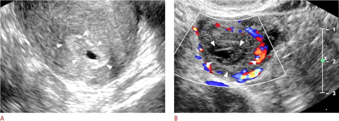
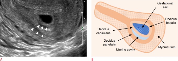
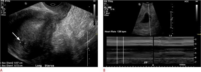
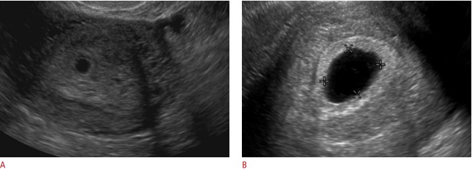
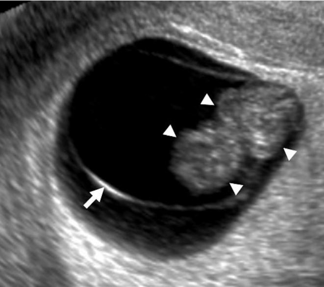
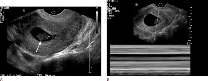
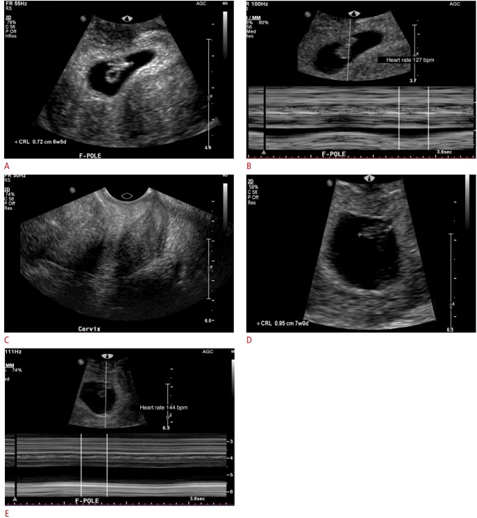
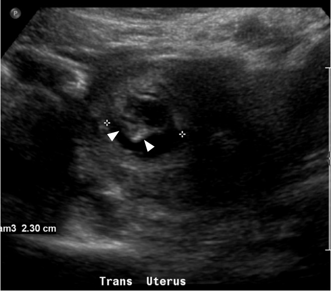
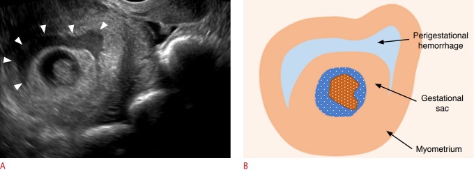
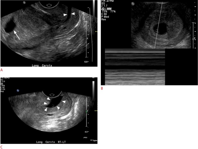
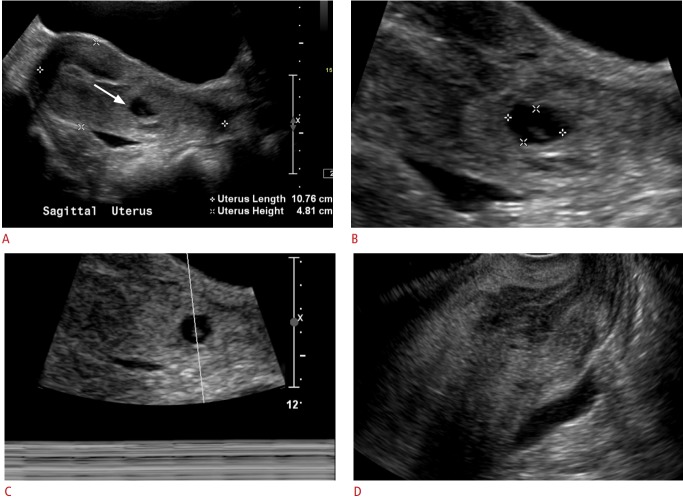
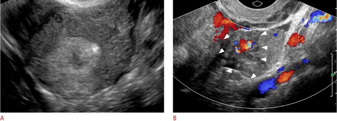
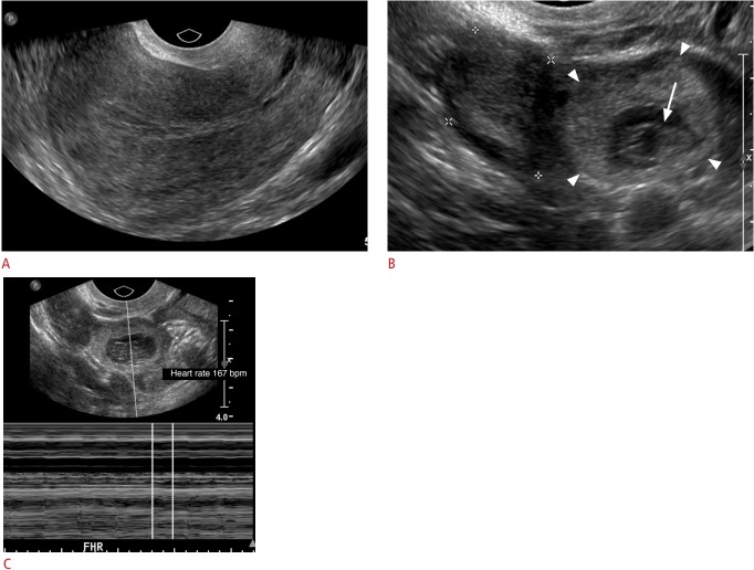
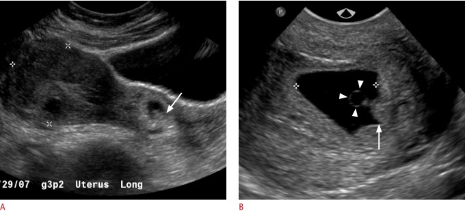
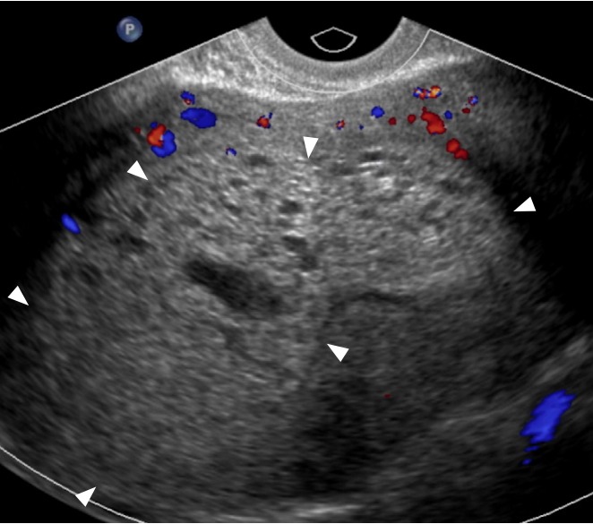
In patients presenting for an evaluation of pregnancy in the first trimester, transvaginal ultrasound is the modality of choice for establishing the presence of an intrauterine pregnancy; evaluating pregnancy viability, gestational age, and multiplicity; detecting pregnancy-related complications; and diagnosing ectopic pregnancy. In this pictorial review article, the sonographic appearance of a normal intrauterine gestation and the most common complications of pregnancy in the first trimester in the acute setting are discussed.

Keywords: Abnormal gestation; Abortion; Ectopic pregnancy; Transvaginal ultrasound; Viability.


Conflict of interest statement

No potential conflict of interest relevant to this article was reported.

Figures

Fig. 1.
Fig. 1.. Normal early gestational sac and corpus luteum.
A. Transvaginal ultrasonography (TVUS) demonstrates an anechoic structure with peripheral echogenic tissue (arrowheads) representing a gestational sac in the uterine cavity of a woman with a positive urine pregnancy test. B. TVUS shows a circumscribed heterogenous structure with peripheral vascularity in the right ovary compatible with a corpus luteum (arrowheads).
Fig. 2.
Fig. 2.. The double decidual sac sign.
A. Transvaginal ultrasonography demonstrates two concentric echogenic rings (arrowheads) with intervening trace hypoechoic material, known as the double decidual sac sign. B. Graphical representation of the double decidual sign is shown.
Fig. 3.
Fig. 3.. A 19-year-old woman with vaginal bleeding and a positive beta-human chorionic gonadotropin test.
A. Initial transvaginal ultrasonography (TVUS) shows a vague hypoechoic collection measuring 7 mm in the uterine fundus (arrow). The morphology was not typical for an intrauterine pregnancy. B. Subsequent TVUS 2 weeks later demonstrates an intrauterine gestational sac with an embryo with heart rate of 126 bpm.
Fig. 4.
Fig. 4.. Transvaginal ultrasonography in a pregnant woman presenting with abdominal pain and cramping.
A. Initial ultrasonography shows a gestational sac without a yolk sac or embryo. B. Follow-up ultrasonography 2 weeks later shows a gestational sac measuring greater than 25 mm in diameter without evidence of a yolk sac or embryo. These findings are diagnostic of early pregnancy loss.
Fig. 5.
Fig. 5.. Transvaginal ultrasonography in a patient with a previously confirmed intrauterine pregnancy (IUP) and vaginal bleeding, showing an IUP with a fetal pole (arrowheads).
A curvilinear echogenic membrane is noted around the embryo, corresponding to the amniotic membrane (arrow).
Fig. 6.
Fig. 6.. Early pregnancy failure.
A. Transvaginal ultrasonography shows an intrauterine pregnancy with an embryo (arrow) with a crown-rump length of 1.1 cm, corresponding to a gestational age of 7 weeks, 2 days. B. No fetal heart rate was identified, compatible with intrauterine embryonic demise.
Fig. 7.
Fig. 7.. Transvaginal ultrasonography in a pregnant woman presenting with abdominal pain, cramping, and vaginal bleeding.
A-E. M-mode images show progressive increase in heart rate with advancing gestational age. A. Intrauterine gestation with an embryo and yolk sac (crown-rump length of 7 mm --> gestational age [GA] of 6 weeks and 5 days) is shown. B. Fetal heart rate is 127 bpm. C. The cervix is closed. D. Two days later, pelvic ultrasonography demonstrates interval growth of the embryo, with a crown-rump length of 1 cm, corresponding to a GA of 7 weeks. E. The heart rate increases progressively with advancing gestational age.
Fig. 8.
Fig. 8.. Early pregnancy with findings suspicious for pregnancy failure.
Transabdominal ultrasonography in a 34-year-old woman with a positive beta-human chorionic gonadotropin test and vaginal bleeding demonstrates an intrauterine gestation with a mean sac diameter of 23 mm and a yolk sac diameter of 19 mm. No definite fetal pole was identified. Instead, an amorphous embryonic structure (arrowheads) was identified. These findings are suspicious for, but not diagnostic of pregnancy failure.
Fig. 9.
Fig. 9.. Subchorionic hemorrhage.
A. Transvaginal ultrasonography in a pregnant woman shows a gestational sac with an embryo and a heterogeneous subchorionic collection (arrowheads) encircling approximately 180° of the gestational sac. B. Graphic depiction of the findings in A is shown.
Fig. 10.
Fig. 10.. Inevitable abortion: transvaginal ultrasonography (TVUS) in a 41-year-old woman with a known intrauterine pregnancy presenting with abdominal pain and vaginal spotting.
A. Initial TVUS shows an intrauterine gestation (arrow), with an open cervix (arrowheads). B. No heart rate was identified. C. Followup ultrasonography obtained the next day shows the gestational sac in the cervical canal (arrowheads), compatible with inevitable abortion.
Fig. 11.
Fig. 11.. Completed abortion: transabdominal and transvaginal ultrasonography obtained in a patient with a confirmed intrauterine gestation.
A, B. Gestational sac containing a fetal pole was identified in the cervix (arrow); the abortion was in progress. C. No fetal heart rate was identified. D. The patient passed a few clots and transvaginal images were obtained. The previously seen gestational sac in the cervix was no longer seen, compatible with a completed abortion.
Fig. 12.
Fig. 12.. Adnexal ectopic pregnancy: transvaginal ultrasonography in a woman with a positive beta-human chorionic gonadotropin test.
A. No intrauterine gestational sac was identified. The right ovary and adnexa were normal. B. A left adnexal heterogenous vascular mass (arrowheads), was suspicious for an adnexal ectopic pregnancy, which was confirmed intraoperatively.
Fig. 13.
Fig. 13.. Adnexal ectopic pregnancy: transvaginal images in a woman with vaginal bleeding, abdominal pain, and a positive beta-human chorionic gonadotropin test.
A. No intrauterine gestational sac was identified. The left ovary and adnexa were normal. B, C. Sonograms demonstrate a right adnexal mass containing a gestational sac (arrowheads) and a fetal pole (arrow), with a heart rate of 167 bpm, compatible with a right adnexal ectopic gestation.
Fig. 14.
Fig. 14.. Cervical ectopic pregnancy.
A. Transabdominal ultrasonography in a woman with a positive beta-human chorionic gonadotropin test, shows a gestational sac containing a fetal pole in the cervix (arrow). B. Transvaginal ultrasonography shows a gestational sac containing a yolk sac (arrowheads) and fetal pole (arrow).
Fig. 15.
Fig. 15.. Gestational trophoblastic disease: complete mole.
Transvaginal ultrasonography in a 35-year-old woman presenting with an elevated serum beta-human chorionic gonadotropin (β -hCG) level (>383,000 mIU/mL) and vaginal bleeding, shows an echogenic, heterogenous mass with minimal peripheral vascularity (arrowheads) and numerous cystic spaces. No fetal parts or myometrial invasion was identified. These findings, given the significantly elevated β-hCG value, were diagnostic of complete mole.

References

    1. AIUM-ACR-ACOG-SMFM-SRU practice parameter for the performance of standard diagnostic obstetric ultrasound examinations. J Ultrasound Med. 2018;37:E13–E24. - PubMed
    1. Doubilet PM. Ultrasound evaluation of the first trimester. Radiol Clin North Am. 2014;52:1191–1199. - PubMed
    1. Nyberg DA, Mack LA, Laing FC, Patten RM. Distinguishing normal from abnormal gestational sac growth in early pregnancy. J Ultrasound Med. 1987;6:23–27. - PubMed
    1. Doubilet PM, Benson CB. First, do no harm... To early pregnancies. J Ultrasound Med. 2010;29:685–689. - PubMed
    1. Doubilet PM, Benson CB, Bourne T, Blaivas M, Society of Radiologists in Ultrasound Multispecialty Panel on Early First Trimester Diagnosis of Miscarriage and Exclusion of a Viable Intrauterine Pregnancy. Barnhart KT, et al. Diagnostic criteria for nonviable pregnancy early in the first trimester. N Engl J Med. 2013;369:1443–1451. - PubMed

LinkOut - more resources