Review
. 2020 Jun;177(11):2413-2433.
doi: 10.1111/bph.15005. Epub 2020 Apr 6.

Update of P2Y receptor pharmacology: IUPHAR Review 27

Affiliations
Review

Update of P2Y receptor pharmacology: IUPHAR Review 27

Kenneth A Jacobson et al. Br J Pharmacol. 2020 Jun.

Abstract

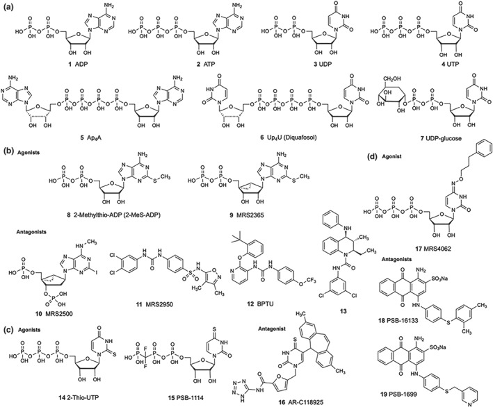
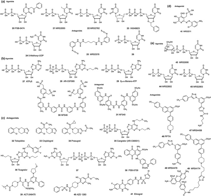
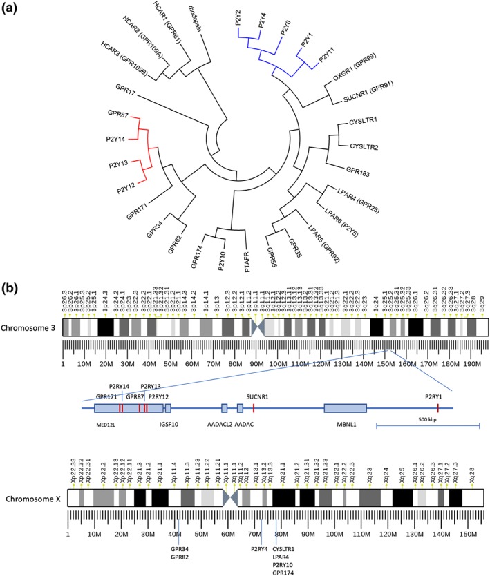
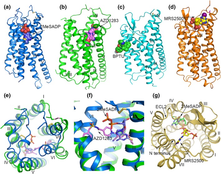
Eight G protein-coupled P2Y receptor subtypes respond to extracellular adenine and uracil mononucleotides and dinucleotides. P2Y receptors belong to the δ group of rhodopsin-like GPCRs and contain two structurally distinct subfamilies: P2Y1 , P2Y2 , P2Y4 , P2Y6 , and P2Y11 (principally Gq protein-coupled P2Y1 -like) and P2Y12-14 (principally Gi protein-coupled P2Y12 -like) receptors. Brain P2Y receptors occur in neurons, glial cells, and vasculature. Endothelial P2Y1 , P2Y2 , P2Y4 , and P2Y6 receptors induce vasodilation, while smooth muscle P2Y2 , P2Y4 , and P2Y6 receptor activation leads to vasoconstriction. Pancreatic P2Y1 and P2Y6 receptors stimulate while P2Y13 receptors inhibits insulin secretion. Antagonists of P2Y12 receptors, and potentially P2Y1 receptors, are anti-thrombotic agents, and a P2Y2 /P2Y4 receptor agonist treats dry eye syndrome in Asia. P2Y receptor agonists are generally pro-inflammatory, and antagonists may eventually treat inflammatory conditions. This article reviews recent developments in P2Y receptor pharmacology (using synthetic agonists and antagonists), structure and biophysical properties (using X-ray crystallography, mutagenesis and modelling), physiological and pathophysiological roles, and present and potentially future therapeutic targeting.


Conflict of interest statement

The authors declare that there are no conflicts of interest.

Figures

FIGURE 1
FIGURE 1
P2Y receptor ligands. (a) Natural agonists of P2Y receptors . (b) P2Y1 receptor agonists and antagonists. (c) P2Y2 receptor agonists and antagonist. (d) P2Y4 receptor agonists and antagonists
FIGURE 2
FIGURE 2
P2Y receptor ligands. (a) P2Y6 receptor agonists and antagonists. (b) P2Y11 receptor agonists and antagonists. (c) P2Y12 receptor agonists and antagonists. (d) P2Y13 receptor antagonist. (e) P2Y14 receptor agonists and antagonists
FIGURE 3
FIGURE 3
(a) Evolutionary relationships of human P2Y1‐ and P2Y12‐like receptors of the δ group of rhodopsin‐like GPCRs (Fredriksson & Schiöth, 2005). P2Y1‐ and P2Y12‐like receptors form separated clusters of receptors indicating that they evolved independently. The evolutionary history was inferred using the neighbour‐joining method by extending a previous analyses (Le Duc et al., 2017). The optimal tree with the sum of branch length = 14.64350117 is shown. The tree is drawn to scale, with branch lengths in the same units as those of the evolutionary distances used to infer the phylogenetic tree. The evolutionary distances were computed using the Poisson correction method and are in the units of the number of amino acid substitutions per site. The analysis involved the amino acid sequences of 30 human receptors which were aligned by using the PAM matrix and default parameters. All positions with less than 95% site coverage were eliminated. That is, fewer than 5% alignment gaps, missing data, and ambiguous bases were allowed at any position. There were a total of 239 positions in the final dataset. Evolutionary analyses were conducted in MEGA7 (Kumar, Stecher, & Tamura, 2016). HCARx, hydroxycarboxylic acid receptors; LPARx, lysophosphatidic acid receptors; OXGR1, 2‐oxoglutarate receptor; SUCNR1, succinate receptor; PTAFR, https://www.guidetopharmacology.org/GRAC/ObjectDisplayForward?objectId=334; P2Yx nucleotide receptors. (b) Genomic clustering of P2Y‐like receptors on chromosome 3 and chromosome X. Several P2Y receptors and other members of the δ group cluster at genomic loci, most prominent clusters at chromosome 3 and chromosome X. The P2Y12 receptor‐like receptors are found in a MED12L gene intron at chromosome 3. Similarly, GPR34 and GPR82 are found in a reverse‐oriented CASK gene intron at Xp11.4. Phylogenetic analysis showed relation of cysteinyl‐LT receptor and lysophosphatidic acid receptors (see (a)). Interestingly, CysLT2 and LPA6 receptors and GPR183 are closely located at chromosome 13 (13q14.2, not shown) whereas CysLT1 receptors genomically cluster with LPA4, P2Y10 receptors, and GPR174 at the X chromosome
FIGURE 4
FIGURE 4
Structures of P2Y12 receptors and P2Y1 receptors. (a) Structure of the P2Y12 receptor‐2MeSADP 8 complex. The P2Y12 receptor is shown in blue cartoon representation. 2MeSADP is shown as blue spheres. The disulphide bonds are shown as yellow sticks. (b) Structure of the P2Y12 receptor‐AZD1283 40 complex. The receptor is coloured green and shown in cartoon representation. AZD1283 is shown as spheres with pink carbons. The disulphide bridge is shown as pink sticks. (c) Structure of the P2Y1 receptor‐BPTU complex. The P2Y1 receptor is coloured cyan and shown as cartoon. The ligand BPTU is shown as spheres with green carbons. The disulphide bonds are shown as yellow sticks. (d) Structure of the P2Y1 receptor‐MRS2500 10 complex. The P2Y1 receptor is shown in orange cartoon representation. MRS2500 is shown as spheres with yellow carbons. The disulphide bonds are show as magenta sticks. (e) Structural comparison of the extracellular region in the P2Y12 receptor‐2MeSADP and P2Y12 receptor‐AZD1283 complexes. In the P2Y12 receptor‐AZD1283 structure, the receptor is shown in green cartoon representation, the ligand AZD1283 is shown as pink sticks. In the P2Y12 receptor‐2MeSADP structure, the receptor is shown in blue cartoon representation and 2MeSADP is shown as blue sticks. The black arrows indicate the movements of helices VI and VII in the 2MeSADP‐bound structure relative to the AZD1283‐bound structure. (f) Comparison of the binding pose of AZD1283 and 2MeSADP in P2Y12 receptors. The colour scheme is the same as that in panel (e). (g) Comparison of the nucleotide binding modes in P2Y1 receptors and P2Y12 receptors. The P2Y1 receptor is shown in yellow cartoon representation. MRS2500 and 2MeSADP are shown as yellow sticks and green carbon respectively

References

    1. Abbas, Z. S. B. , Latif, M. L. , Dovlatova, N. , Fox, S. C. , Heptinstall, S. , Dunn, W. R. , & Ralevic, V. (2018). UDP‐sugars activate P2Y14 receptors to mediate vasoconstriction of the porcine coronary artery. Vascular Pharmacology, 103‐105, 36–46. 10.1016/j.vph.2017.12.063 - DOI - PMC - PubMed
    1. Abbracchio, M. P. , Boeynaems, J. M. , Boyer, J. L. , Burnstock, G. , Ceruti, S. , Fumagalli, M. , … Weisman, G. A. (2019). P2Y receptors (version 2019.4) in the IUPHAR/BPS Guide to Pharmacology Database. IUPHAR/BPS Guide to Pharmacology CITE. 2019(4). Available from: 10.2218/gtopdb/F52/2019.4. - DOI
    1. Abbracchio, M. P. , Burnstock, G. , Boeynaems, J. M. , Barnard, E. A. , Boyer, J. L. , Kennedy, C. , … Weisman, G. A. (2006). International Union of Pharmacology LVIII. Update on the P2Y G protein‐coupled nucleotide receptors: From molecular mechanisms and pathophysiology to therapy. Pharmacological Reviews, 58, 281–341. - PMC - PubMed
    1. Ahn, Y. H. , Lee, J. Y. , Park, H. D. , Kim, T. H. , Park, M. C. , Choi, G. , & Kim, S. (2016). Identification of a new morpholine scaffold as a P2Y12 receptor antagonist. Molecules, 21, E1114. - PMC - PubMed
    1. Alexander, S. P. H. , Christopoulos, A. , Davenport, A. P. , Kelly, E. , Mathie, A. , Peters, J. A. , … Collaborators, C. G. T. P. (2019). The Concise Guide to PHARMACOLOGY 2019/20: G protein‐coupled receptors. British Journal of Pharmacology, 176, S21–S141. 10.1111/bph.14748 - DOI - PMC - PubMed

Publication types

Substances

LinkOut - more resources