. 2020 Aug;528(12):2099-2131.
doi: 10.1002/cne.24879. Epub 2020 Feb 19.

ZEBrA: Zebra finch Expression Brain Atlas-A resource for comparative molecular neuroanatomy and brain evolution studies

Affiliations

ZEBrA: Zebra finch Expression Brain Atlas-A resource for comparative molecular neuroanatomy and brain evolution studies

Peter V Lovell et al. J Comp Neurol. 2020 Aug.

Abstract

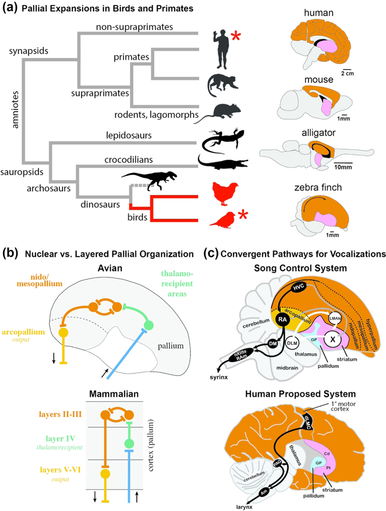
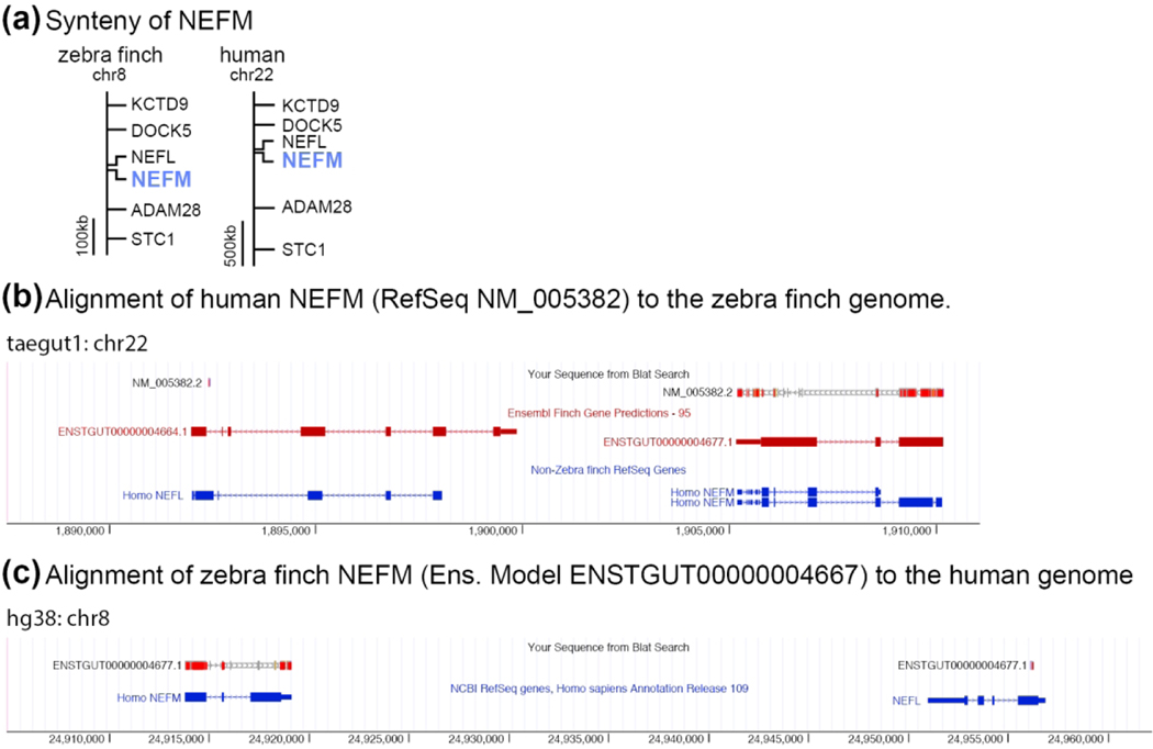
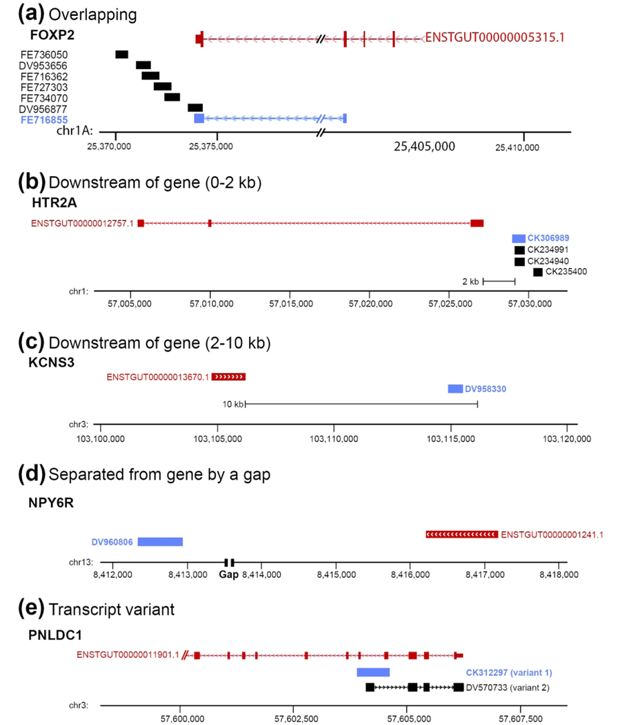
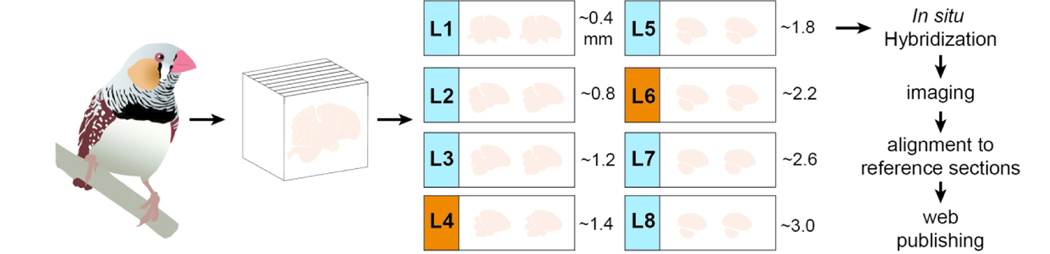
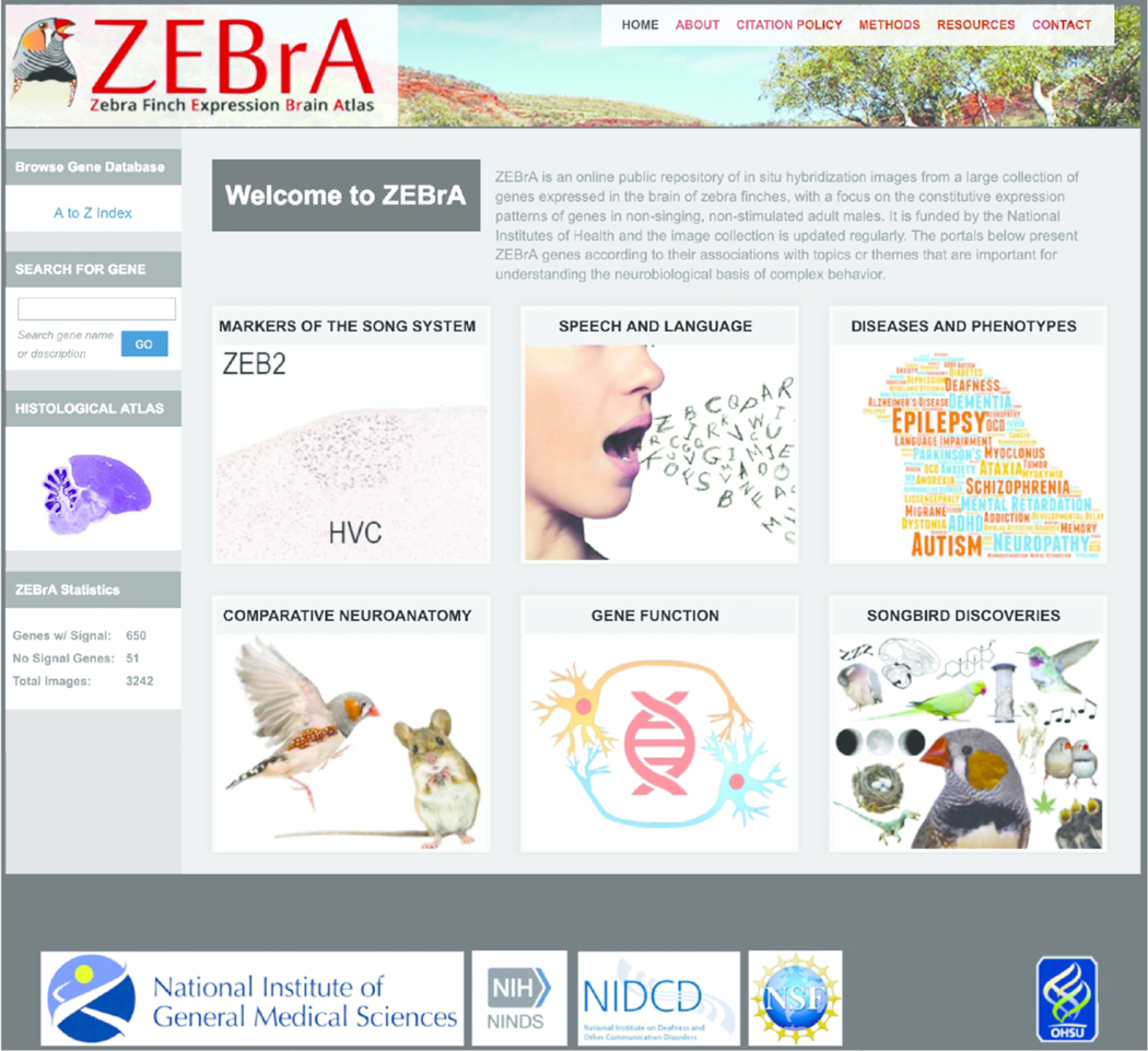
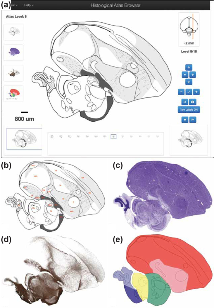
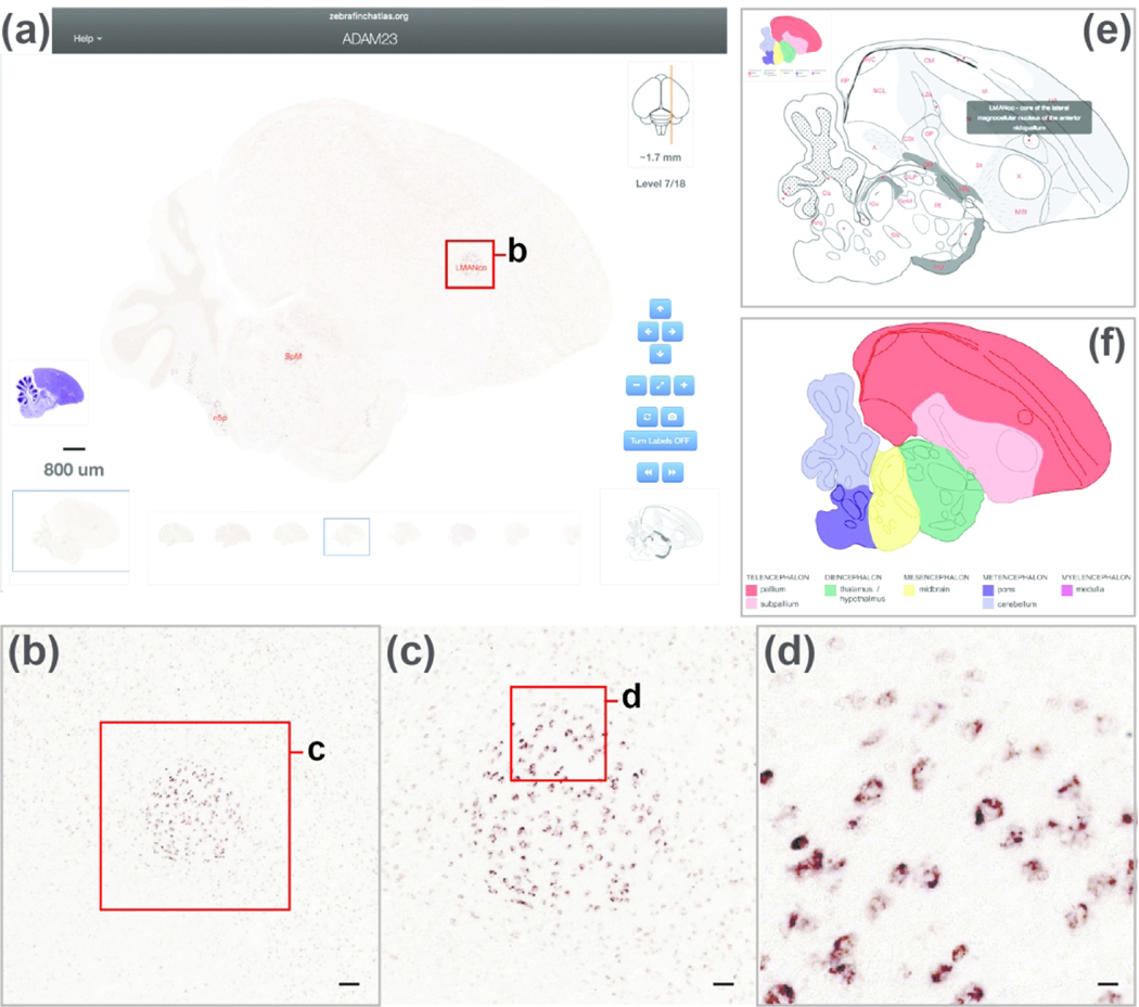
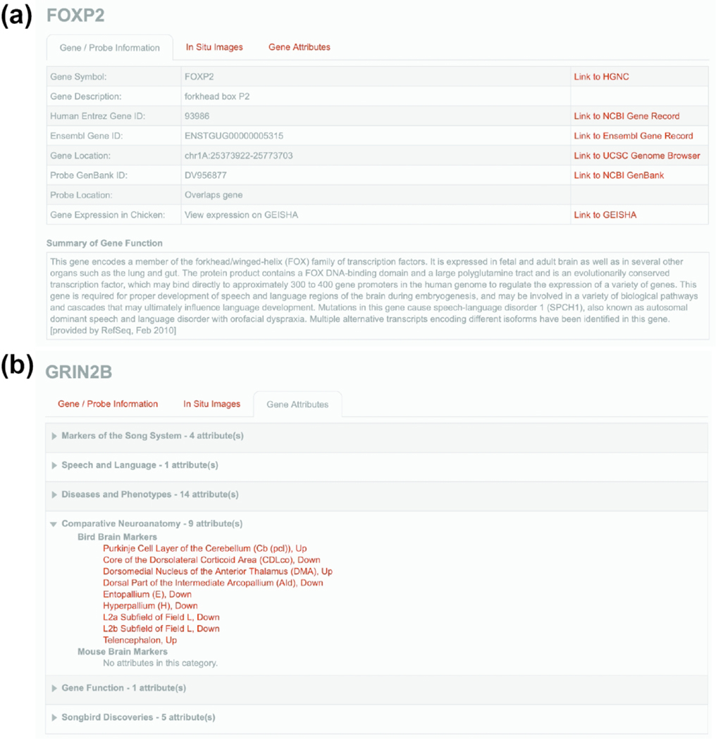
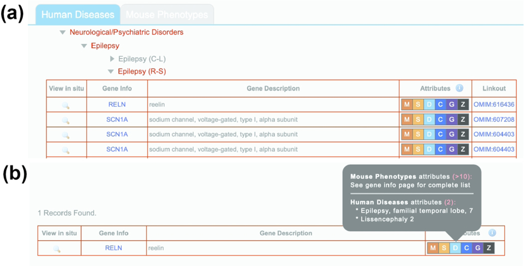
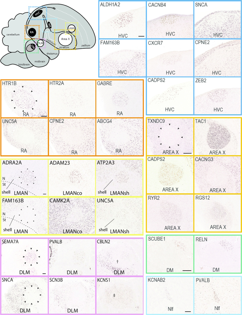
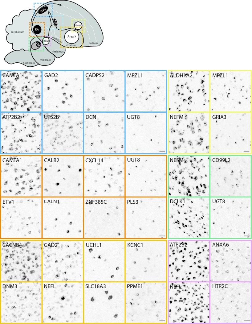
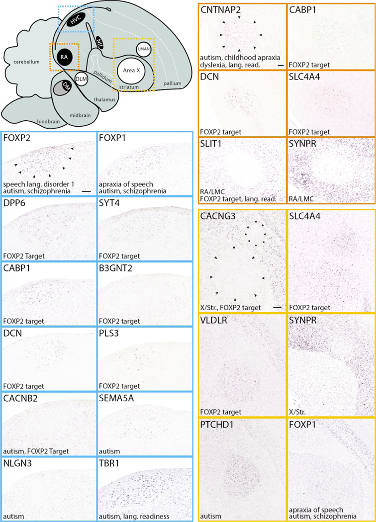
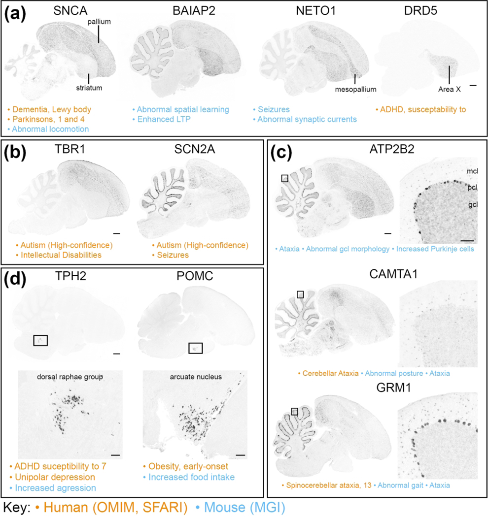
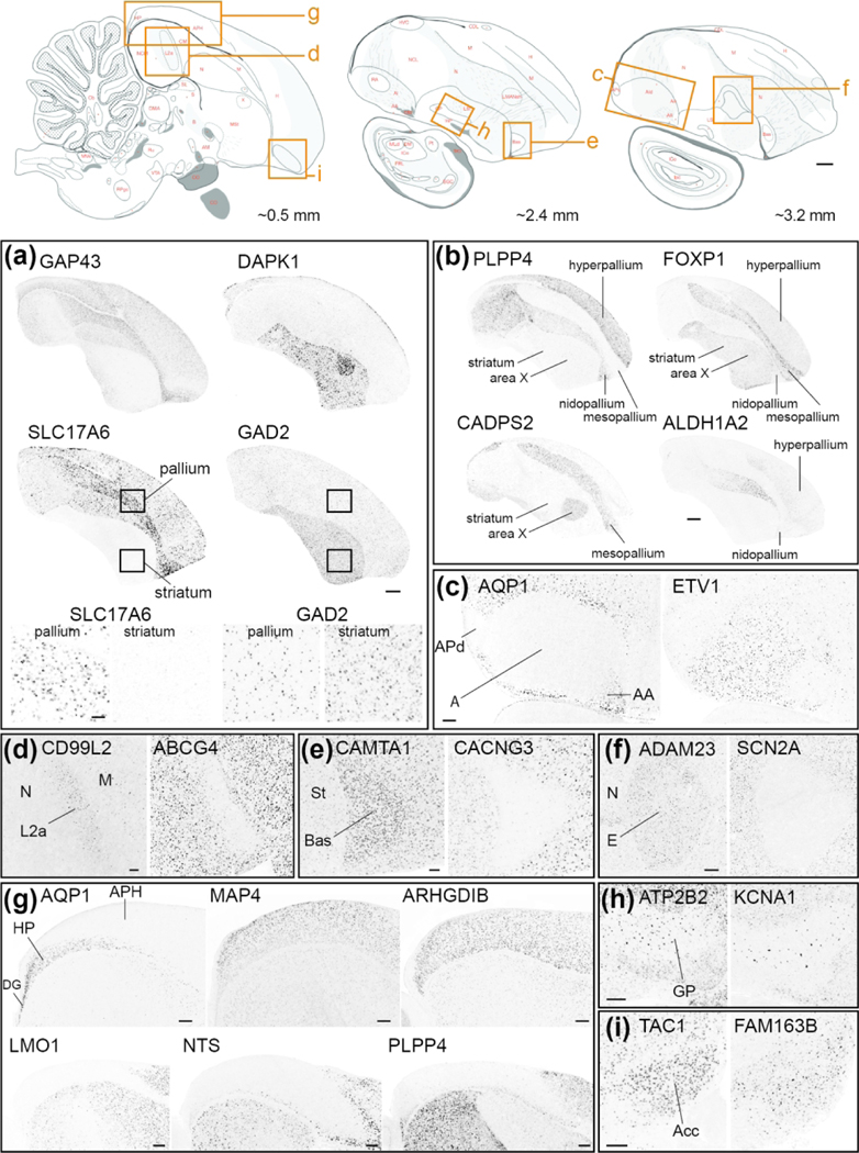
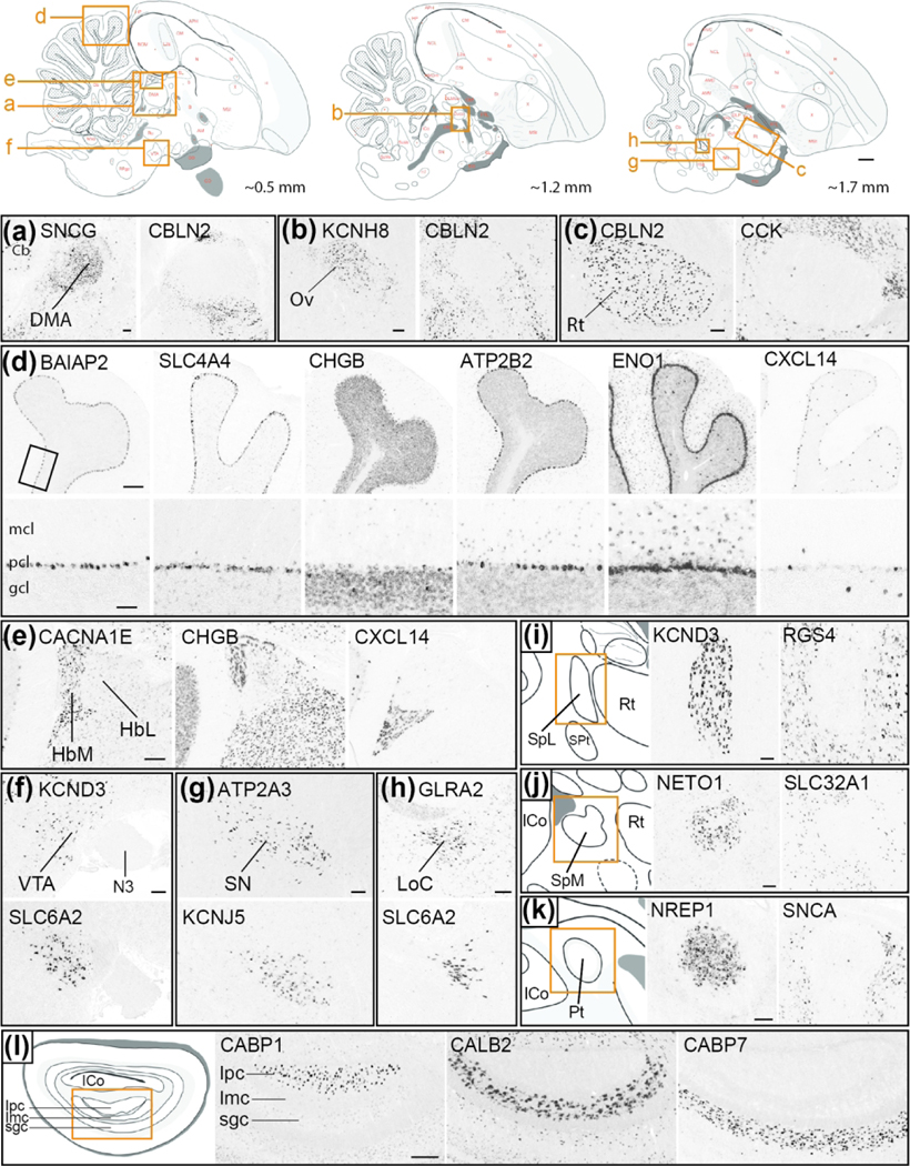
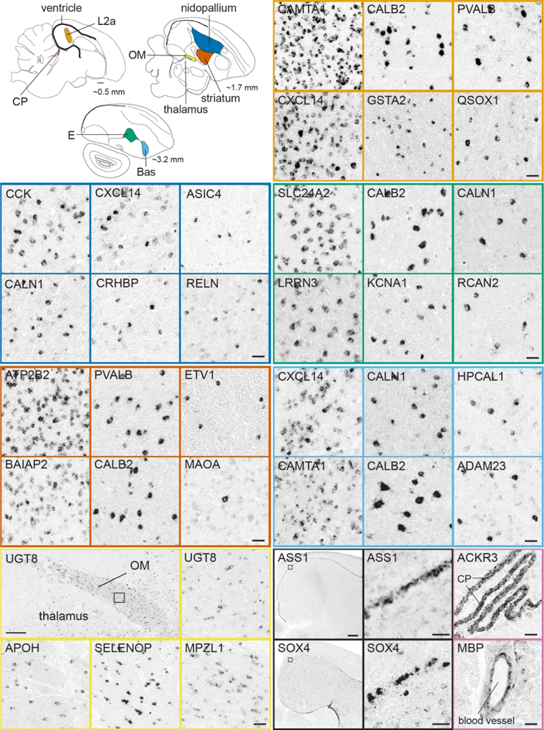
An in-depth understanding of the genetics and evolution of brain function and behavior requires a detailed mapping of gene expression in functional brain circuits across major vertebrate clades. Here we present the Zebra finch Expression Brain Atlas (ZEBrA; www.zebrafinchatlas.org, RRID: SCR_012988), a web-based resource that maps the expression of genes linked to a broad range of functions onto the brain of zebra finches. ZEBrA is a first of its kind gene expression brain atlas for a bird species and a first for any sauropsid. ZEBrA's >3,200 high-resolution digital images of in situ hybridized sections for ~650 genes (as of June 2019) are presented in alignment with an annotated histological atlas and can be browsed down to cellular resolution. An extensive relational database connects expression patterns to information about gene function, mouse expression patterns and phenotypes, and gene involvement in human diseases and communication disorders. By enabling brain-wide gene expression assessments in a bird, ZEBrA provides important substrates for comparative neuroanatomy and molecular brain evolution studies. ZEBrA also provides unique opportunities for linking genetic pathways to vocal learning and motor control circuits, as well as for novel insights into the molecular basis of sex steroids actions, brain dimorphisms, reproductive and social behaviors, sleep function, and adult neurogenesis, among many fundamental themes.

Keywords: brain atlas; brain organization; functional circuits; gene expression; molecular signature; song system; zebra finch.


Figures

Figure 1.
Figure 1.. Comparison of brains and circuit organization across vertebrates.
(a) Simplified phylogeny and brain evolution in amniotes. Left: Schematic diagram depicting phylogenetic relationships across major amniote lineages; branches containing bird lineages are highlighted in blue. Only select major extant groups are depicted; dashed line indicates dinosaur lineage extinction. Red asterisks indicate species/groups that possess the trait of vocal learning. Right: Schematic diagrams of representative vertebrate brains highlighting the relative sizes of pallial (orange) and subpallial (pink) areas in representative synapsids and sauropsids. (b) Schematic generic representation of the ‘nuclear’ vs. ‘layered’ hypothesis of pallial organization in avian (top) and mammalian (bottom) brains (after Karten, 1991). Color coding indicates analogous (and possibly homologous) elements between lineages. (c) Schematic diagrams of specialized pathways for birdsong (top) and speech (bottom). Top: Vocal control circuitry of the adult male zebra finch, depicted in the parasagittal plane; some structures have been omitted for clarity. The system consists of a direct motor pathway for song production (in black) that includes connections from HVC to RA, and from RA to brainstem nuclei involved in vocal-motor and respiratory control (nXIIts, RAm), and an anterior forebrain pathway for song learning (in white) containing a cortico-striato-thalamo-cortical loop with projections from pallial LMAN to striatal Area X, from Area X to thalamic DLM, from DLM to LMAN, and from LMAN back to Area X. Additional connections are shown in grey. Bottom: Proposed vocal control circuitry in humans consisting of a direct motor pathway for speech production (in black), and striatal brain regions (Cd, Pt) that have been proposed to be involved in speech learning (in white), adapted from Pfenning et al. 2014. The descending projections from RA and dLMC to brainstem vocal nuclei are thought to be convergently evolved in songbirds and humans. Abbreviations: Am, nucleus ambiguus; Cd, Caudate nucleus of the striatum; dLMC, dorsal part of the laryngeal motor cortex; DLM, medial part of the dorsolateral nucleus of the anterior thalamus; GP, Globus pallidus; HVC, HVC proper name; LMAN, lateral magnocellular nucleus of the anterior nidopallium; nXIIts, tracheosyringeal portion of the nucleus of the hypoglossal nerve (XII); PAG, periaqueductal grey; Pt, Putamen; RA, robust nucleus of the arcopallium; RAm, nucleus retroambiguus medialis; X, Area X.
Figure 2.
Figure 2.. Gene Orthology Verification.
(a) Schematic depiction showing how conserved synteny establishes the one-to-one homology of zebra finch and human NEFM. (b-c) Screenshots from UCSC’s genome browser showing how NEFM gene predictions from zebra finch (Ensemble model ENSTGUT00000004667) and human (Refseq NM_005382.2) align to the correct loci with the highest reciprocal alignment scores. (b) Human NEFM (NM_005382.2) aligns to a region of the zebra finch genome containing NEFM (ENSTGUT00000004677.1) and NEFL (ENSGUT00000004664.1). The alignment of human NEFM has a higher scoring alignment, and more completely covers (i.e. higher exon coverage) the NEFM locus, than the NEFL locus (BLAT scores = 1085 vs. 25, respectively). (c) Zebra finch NEFM (ENSTGUT00000004677.1) aligns to a region of the human genome that contains NEFM and NEFL. The alignment of zebra finch NEFM model has a higher scoring alignment, and more complete coverage for the NEFM locus than for NEFL locus (BLAT scores = 1154 vs. 65, respectively).
Figure 3.
Figure 3.. In Situ Probe Classification.
(a) Example of overlapping probe type: schematic depiction of genomic region for the zebra finch FOXP2 gene (based on UCSC’s genome browser) shows that clone FE716855 (Genbank ID shaded in red) was selected because it overlaps with coding and non-coding (3’-UTR) portions of the FOXP2 Ensembl Gene model (ENSTGUG00000005315.1). The EST for this clone also aligns unambiguously to this locus. Therefore, the Probe Location in the FOXP2 Gene Info page is listed as ‘Overlaps gene’. We note that the FOXP2 gene and related clones are on the minus strand (indicated by arrowheads in the gene model and selected clone alignments). (b-d) Examples of non-overlapping probes according to clone location relative to the gene. In (d), ‘Gap’ refers to a gap present in the genome assembly (taegut1). (e) Example of apparent spliced variants, where only one variant (CK312297) was analyzed. In all panels, Ensembl models are in red, clones selected for in situs are in blue, and orientation of gene and related clones are indicated by the arrowheads in the model and/or alternative variant clone. For each gene in ZEBrA the chromosomal location of the selected clone is listed under Probe Location in the Gene Info Page
Figure 4.
Figure 4.. In situ pipeline.
Schematic representation of ZEBrA’s in situ hybridization pipeline. Serial cryostat brains sections encompassing most major brain structures are hybridized at close intervals (e.g. L1–8, in blue) for genes that exhibit highly differential patterns, or at select levels (e.g. L4/L6, in vermillion) for non-differential genes, alignment to reference sections from the histological atlas, and ZEBrA web publishing.
Figure 5.
Figure 5.. ZEBrA: The Zebra finch Expression Brain Atlas.
Image of the homepage of the ZEBrA website (www.zebrafinchatlas.org, RRID: SCR_012988), an online public repository of high-resolution in situ hybridization images for a large collection of genes that are expressed in the brain of the adult male zebra finch.
Figure 6.
Figure 6.. The Histological Atlas Browser.
(a) View of the Browser depicting an example of a drawing from one selected level of the Histological Atlas. The full set of parasagittal levels currently available is shown as thumbnails at the bottom of the browser. The thumbnail icons on the left allow users to toggle across each of the four images types. The position of the section being viewed relative to the midline is illustrated by the orange line on the dorsal-view schematic drawing of the brain, in the top right corner of the bowser. The distance (in mm) of the current section from the midline, as well as the section level in the series, are indicated beneath that schematic. The navigation buttons on the lower right provide options for panning (arrows) or zooming (+ and −) an image, resetting the image zoom level (diagonally opposing arrows), toggling across the four image types for a selected level (circular arrows), requesting an image from ZEBrA (camera symbol), turning anatomical labels ON/OFF, and moving to the next or previous image in the series (>> or <<). The Pan function can also be applied by clicking and dragging the main image in any desired direction, and all other navigation functions can be activated by the appropriate keyboard stroke, as described under the “Navigation Help” tab in the upper-left corner of the browser. The size and position of the field of view shown in the browser is indicated by the blue rectangle over the icon on the bottom left, and the image scale is indicated above that icon. (b) View of the same example drawing as in (a), with Turn Labels button activated (ON position). (c-e) Views of the Nissl- and myelin-stained sections, and of the colorized drawing depicting the: pallium (red), sub-pallium (pink), diencephalon (green), midbrain (yellow), pons (dark blue) and cerebellum (light blue).
Figure 7.
Figure 7.. The in Situ Image Browser
(a) View of the Browser depicting an in situ image from an example gene (ADAM23), with Turn Labels button activated. The set of icons at the bottom of the browser indicate the levels of the in situ images available for this gene, and the highlighted icon indicates the level of the enlarged image at the center of the browser. The position of the section being viewed relative to the midline is illustrated by the orange line on the dorsal-view schematic drawing of the brain, in the top right corner of the bowser. The distance (in mm) of the current section from the midline, as well as the section level in the series, are indicated beneath that schematic. The navigation buttons on the lower right provide options for panning (arrows) or zooming (+ and −) an image, resetting the image zoom level (diagonally opposing arrows), toggling between the in situ image and the corresponding drawing from the histological reference atlas (circular arrows; note that the atlas drawing cannot be zoomed from this browser), requesting an image from ZEBrA (camera symbol), turning anatomical labels ON/OFF, and moving to the next or previous image in the series (>> or <<). The Pan function can also be applied by clicking and dragging the main image in any desired direction, and all other navigation functions can be activated by the appropriate keyboard stroke, as described under the “Navigation Help” tab in the upper-left corner of the browser. The size and position of the field of view shown at the center of the browser is indicated by the blue rectangle over the icon on the bottom left, and the image scale is indicated above that icon. The icon above the scale provides access to the histological atlas browser, which opens in another window. (b-d) Enlarged views of the areas indicated by the red boxes in (a-c), corresponding to the core region of song nucleus LMAN (LMANco). Scale bars: (b): 100 μm; (c): 50 μm, (d): 25 μm. (e) View of the reference drawing (with labels ON) associated with the in situ image in (a) depicts specific brain structures present at the level of that in situ image. (f) View of the corresponding colorized drawing depicts the position of general brain subdivisions present at that level.
Figure 8.
Figure 8.. The Gene Info Page.
(a) Example of Gene/Probe Information tab (for FOXP2) presents the official gene symbol and related description (HGNC-based), the NCBI entry for the human ortholog, the zebra finch gene entry from Ensembl, the gene’s chromosomal location in the zebra finch genome (UCSC genome browser-based), the GenBank entry and genome location for the clone used to generate the in situ hybridization probe, and information on expression in chick embryos are depicted. A summary of gene function (from NCBI), and hyperlinks to relevant databases are also presented, when available. (b) Example of Gene Attributes tab providing counts and full lists of attributes for a given gene, in this case GRIN2B.
Figure 9.
Figure 9.. Structure of gene lists.
(a) View of the organization and layout of a typical list of genes associated with a specific ZEBrA portal theme. In this example, a subset of genes associated with the Neurological/Psychiatric Disorder Epilepsy within the Human Diseases sub-portal is shown. Each gene entry includes a magnifying glass icon under the View In Situ column (which provides access to in situ images in the Image browser), and entries for gene name (which provides access to the Gene Info page), Gene Description, Attributes, and Linkouts (which in this case provide access to an entry in OMIM). (b-c) Views of example pop-up menus showing Attributes associated with the gene RELN that are derived from the Disease and Phenotypes (b) and Speech and Language (c) portals. The association of RELN with the OMIM Attribute “Epilepsy, familial temporal lobe, 7” provides the reasoning behind the classification of this gene under the Human Disease theme of Epilepsy.
Figure 10.
Figure 10.. Markers of the Song System (Portal 1).
Upper left: Schematic drawing of a parasagittal view of the adult male zebra finch brain depicts major brain subdivisions and the relative location of major song control nuclei; nuclei of the direct vocal-motor pathway are in black, nuclei of the anterior forebrain pathway are in white, colored dashed rectangles indicate the areas shown in the in situ hybridization images. Other panels: Examples of in situ hybridization images of positive or negative markers (up- or down-regulated expression compared to adjacent areas) of zebra finch song nuclei HVC (blue), RA (vermillion), LMAN (yellow), Area X (orange), DLM (reddish purple), DM (green) and NIf (sky blue). We note that various markers of LMAN specifically label the core (LMANco), shell (LMANsh), or both core and shell regions of LMAN. DM: mediodorsal nucleus of the intercollicular complex; NIf: nucleus interfacialis; N: nidopallium; St: Striatum; †: Unknown region of the dorsal thalamus surrounding nucleus DLM; ‡: Unknown region of the dorsal thalamus likely corresponding to the DLM defined by Wada et al., 2004. For other abbreviations, see Figure 1 legend. Scale bars for LMAN, DLM, and Area X = 200 μm; for all other nuclei = 100 μm.
Figure 11.
Figure 11.. Markers of the Song System: cellular level (Portal 1).
Upper left: Schematic drawing of a parasagittal view of the adult male zebra finch brain depicts major brain subdivisions and the relative location of major song control nuclei; nuclei of the direct vocal-motor pathway are in black, nuclei of the anterior forebrain pathway are in white, colored dashed rectangles indicate the areas shown in the in situ hybridization images. Other panels: Examples of in situ hybridization images of markers that show high density of labeling (left panels for each nucleus) vs sparse labeling (other panels) for song nuclei HVC (blue), RA (vermillion), LMAN (yellow), NIf (sky blue), Area X (orange), and DLM (reddish purple). For abbreviations, see legends of Figures 1 and 11. Scale bars: 50 μm.
Figure 12.
Figure 12.. Speech and Language genes (Portal 2).
Upper left: Schematic drawing of a parasagittal view of the adult male zebra finch brain depicts major brain subdivisions and the relative location of major song control nuclei; nuclei of the direct vocal-motor pathway are in black, nuclei of the anterior forebrain pathway are in white, colored dashed rectangles indicate the areas shown in the in situ hybridization images. Other panels: Examples of in situ hybridization images of genes linked to Speech and Language function that are positive or negative markers (up- or down-regulated expression compared to adjacent areas) of zebra finch song nuclei HVC (blue), RA (vermillion), and LMAN and/or Area X (orange). Gene attributes related to this portal are indicated in the bottom left of each image; song nuclei borders are indicated by small black arrowheads in the first image for each nucleus. For abbreviations, see legends of Figures 1 and 11. Scale bars for HVC and RA = 200 μm; Area X/LMAN = 400 μm.
Figure 13.
Figure 13.. Diseases and Phenotypes genes (Portal 3).
Examples of in situ hybridization images of genes linked to OMIM- or SFARI-based human neurological and/or psychiatric disorders (sub-portal 3A), or to MGI-based mouse neurological and/or behavioral phenotypes, that show differential expression in various avian brain areas (sub-portal 3B). Gene attributes related to this portal are indicated under each image (vermillion annotations are OMIM- or SFARI-based, blue annotations are MGI-based). (a) Broad expression in telencephalic fields, including pallium, pallial subdivisions, and/or striatum, for genes involved in disorders like seizures or neurodegenerative diseases, or phenotypes disrupting broad learning or locomotor functions. (b) Broad pallial and/or cerebellar expression of genes involved in autism. (c) Differential cerebellar expression for genes involved in cerebellar-related disorders and phenotypes, like ataxias and/or abnormal gait. (d) Discrete expression in nuclei in the pons or hypothalamus for genes involved in disruptions of the serotonergic (left panels) or neuroendocrine (right panels) systems. In (c) and (d), the location of the higher magnification panels in the right or bottom panels are indicated by small rectangles in the left or top panels. Abbreviations for cerebellar cortical layers in (b): gcl, granule cell layer; mcl, molecular layer; pcl, Purkinje cell layer. Scale bars: low-power images = 1 mm; high-power images in (c) and (d) = 200 μm.
Figure 14.
Figure 14.. Comparative Neuroanatomy genes: telencephalon (Portal 4).
Top row: Schematic drawings of parasagittal sections of the adult male zebra finch brain depict major brain subdivisions and specific nuclei shown in this figure. Red letters and dots indicate structure annotations as they appear in ZEBrA, vermillion rectangles indicate the position of the images shown in other panels, and numbers at bottom right of each diagram provide position (in mm) relative to the midline. Other panels: Examples of in situ hybridization images of genes that show differential expression in various parts of the telencephalon, taken from the Bird Brain Markers (sub-portal 4A). (a) Markers showing differential enrichment in pallium (GAP43, SLC17A6) vs striatum (DAPK1, GAD2); the top and bottom small squares show the position of the left and right insets respectively, depicting pallial vs. striatal differences in cell labeling densities. (b) Markers showing differential enrichment in specific pallial subdivisions, including negative and positive markers of the mesopallium (PLPP4, CADPS2 and FOXP1) and nidopallium (ALDH1A2), in different combinations with St and Area X. (c) General negative/positive markers of the arcopallium (AQP1/ETV1, the latter also a negative marker of subdomains APd and AA). (d-f) Positive/negative markers of pallial thalamo-recipient zones L2a (CD99L2/ABCG4), nucleus basorostralis (CAMTA1/CACNG3) and entopallium (ADAM23/SCN2A). (g) Markers of the entire hippocampal formation (positive: MAP4, ARGHDIB; negative: LMO1, NTS) or parts thereof (AQP1, PLPP4). (h) Markers of the globus pallidus (ATP2B2, KCNA1). (i) Markers of the striatal nucleus accumbens (TAC1, FAM163B). For simplicity, only telencephalic structures are shown in (a) and (b). Abbreviations: A: arcopallium; Acc, nucleus accumbens; APH, parahippocampal area; Bas, nucleus basorostralis; DG, dentate gyrus; E, entopallium; GP, globus pallidus; H, hyperpallium; HA, apical part of the hyperpallium; Hp, hippocampus proper; L2a, subfield L2a with auditory Field L; M, mesopallium; N, nidopallium; St, striatum. For other abbreviations in top row diagrams, please consult the histological atlas in ZEBrA. Scale bars: top row diagrams = 1 mm; (a): low-power photomicrographs = 1mm, high-power insets = 100 μm; (b) = 1 mm; (c) = 400 μm; (d) and (e) = 200 μm; (f) – I = 400 μm.
Figure 15.
Figure 15.. Comparative Neuroanatomy genes: diencephalon and brainstem (Portal 4).
Top row: Schematic drawings of parasagittal sections of the adult male zebra finch brain depict major brain subdivisions and specific nuclei shown in this figure with the exception of the brain areas shown in panels I-L. Red letters and dots indicate structure annotations as they appear in ZEBrA, vermillion rectangles indicate the position of the images shown in other panels, and numbers at bottom right of each diagram provide position (in mm) relative to the midline. Other panels: Examples of in situ hybridization images of genes that show differential expression in various parts of the diencephalon and brainstem, taken from the Bird Brain Markers (sub-portal 4A). (a-c) Positive/negative markers of thalamic nuclei DMA (SNCG/CBLN2), Ov (KCNH8/CBLN2) and Rt (CBLN2/CCK). (d) Markers of cerebellar cortical layers show distinctly differential expression in pcl (BAIAP2, SLC4A4), gcl (CHGB), pcl/mcl (ATP2B2, ENO1), or in sparse cells in gcl (CXCL14, among other patterns; the bottom panels show high magnification views of interface zone across layers, taken from region depicted by rectangle in top left panel. (e) Markers that delineate the habenula and/or differentiate its medial and lateral subdivisions. (f-h) Markers of VTA (KCND3, SLC6A2), SN (ATP2A3, KCNJ5) and LoC (GLRA2, SLC6A2) in brainstem tegmentum. (i-k) Positive/negative markers of pre-tectal nuclei SpL (KCND1/RGS4), SpM (NETO1/SLC32A1) and Pt (NREP1/SNCA). (l) Markers of ipc (CABP1), imc (CALB2) and sgc (CABP7) in the optic tectum. Abbreviations: Cb, cerebellum; DMA, dorsomedial nucleus of the anterior thalamus; gcl, granule cell layer of Cb; HbL, lateral habenular nucleus; HbM, medial habenular nucleus; Imc, magnocellular part of the isthmic nucleus; Ipc, parvocellular part of the isthmic nucleus; LoC, locus coeruleus; mcl, molecular layer of Cb; N3, root of third cranial nerve; Ov, nucleus ovoidalis; pcl, Purkinje cell layer of Cb; Rt, nucleus rotundus; SN, substantia nigra; sgc, stratum griseum centrale of the optic tectum; SpL, nucleus spiriformis lateralis; SpM, nucleus spiriformis medialis; VTA, ventral tegmental area. For other abbreviations in top row diagrams, please consult the histological atlas in ZEBrA. Scale bars: top row diagrams = 1 mm; (a-c) = 200 μm; (d): low-resolution panels = 200 μm, high-resolution panels = 100 μm; (e-k) = 200 μm; (l) = 400 μm.
Figure 16.
Figure 16.. Comparative Neuroanatomy genes: cellular level (Portal 4).
Upper left: Schematic drawings of parasagittal sections of the adult male zebra finch brain depict the locations (colored regions) of the brain regions sampled in the in situ hybridizations images. Other panels: Examples of in situ hybridization images of genes that show high density of labeling (left panels for most brain areas) vs. sparse cell labeling (other panels) in pallium (dark blue), striatum (red), L2a (orange), Bas (light blue) or E (green), as well as markers of OM (yellow), ventricle (black), and choroid plexus and blood vessel endothelium (pinkish purple). Abbreviations: Bas, nucleus basorostralis; CP, choroid plexus; E, entopallium; L2a, subfield L2a within auditory Field L; OM, occipitomesencephalic tract. Scale bars: Top-row drawings = 1 mm; Low-power photomicrographs of UGT8 = 400 μm; ASS1 and SOX4 = 800 μm; all other panels = 50 μm.

References

    1. Achiro JM, Shen J, & Bottjer SW (2017). Neural activity in cortico-basal ganglia circuits of juvenile songbirds encodes performance during goal-directed learning. Elife, 6. doi:10.7554/eLife.26973 - DOI - PMC - PubMed
    1. Acquaah-Mensah GK, & Taylor RC (2016). Brain in situ hybridization maps as a source for reverse-engineering transcriptional regulatory networks: Alzheimer’s disease insights. Gene, 586(1), 77–86. doi:10.1016/j.gene.2016.03.045 - DOI - PubMed
    1. Adam I, Mendoza E, Kobalz U, Wohlgemuth S, & Scharff C. (2016). FoxP2 directly regulates the reelin receptor VLDLR developmentally and by singing. Mol Cell Neurosci, 74, 96–105. doi:10.1016/j.mcn.2016.04.002 - DOI - PubMed
    1. Alarcon M, Abrahams BS, Stone JL, Duvall JA, Perederiy JV, Bomar JM, . . . Geschwind DH (2008). Linkage, association, and gene-expression analyses identify CNTNAP2 as an autism-susceptibility gene. Am J Hum Genet, 82(1), 150–159. doi:10.1016/j.ajhg.2007.09.005 - DOI - PMC - PubMed
    1. Alvarez-Buylla A, Kirn JR, & Nottebohm F. (1990). Birth of projection neurons in adult avian brain may be related to perceptual or motor learning. Science, 249(4975), 1444–1446. doi:10.1126/science.1698312 - DOI - PubMed

Publication types