. 2020 Mar 12;63(5):2588-2619.
doi: 10.1021/acs.jmedchem.9b01526. Epub 2020 Feb 24.

Synthetic Lethality in Pancreatic Cancer: Discovery of a New RAD51-BRCA2 Small Molecule Disruptor That Inhibits Homologous Recombination and Synergizes with Olaparib

Affiliations

Synthetic Lethality in Pancreatic Cancer: Discovery of a New RAD51-BRCA2 Small Molecule Disruptor That Inhibits Homologous Recombination and Synergizes with Olaparib

Greta Bagnolini et al. J Med Chem. .

Abstract

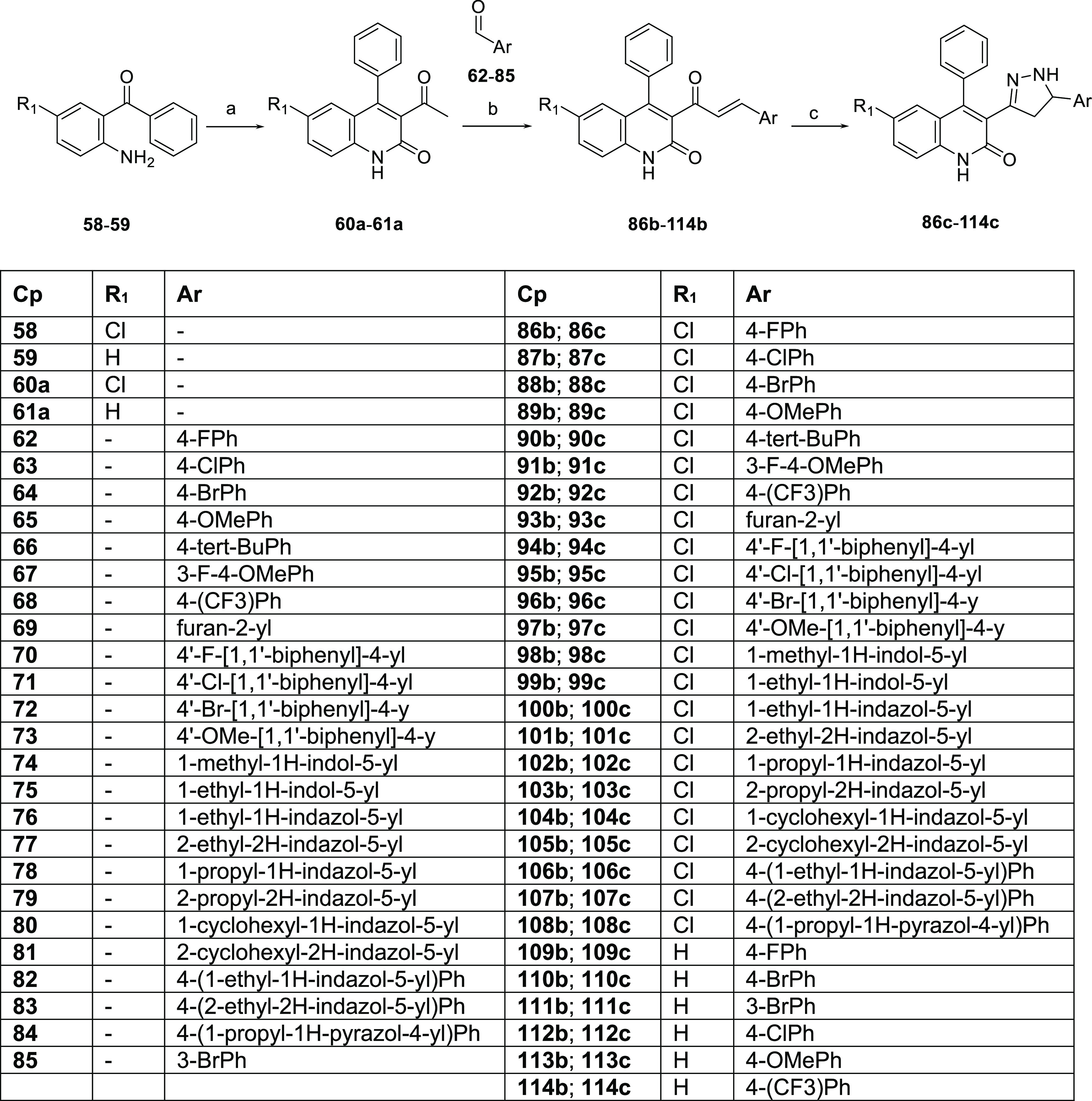
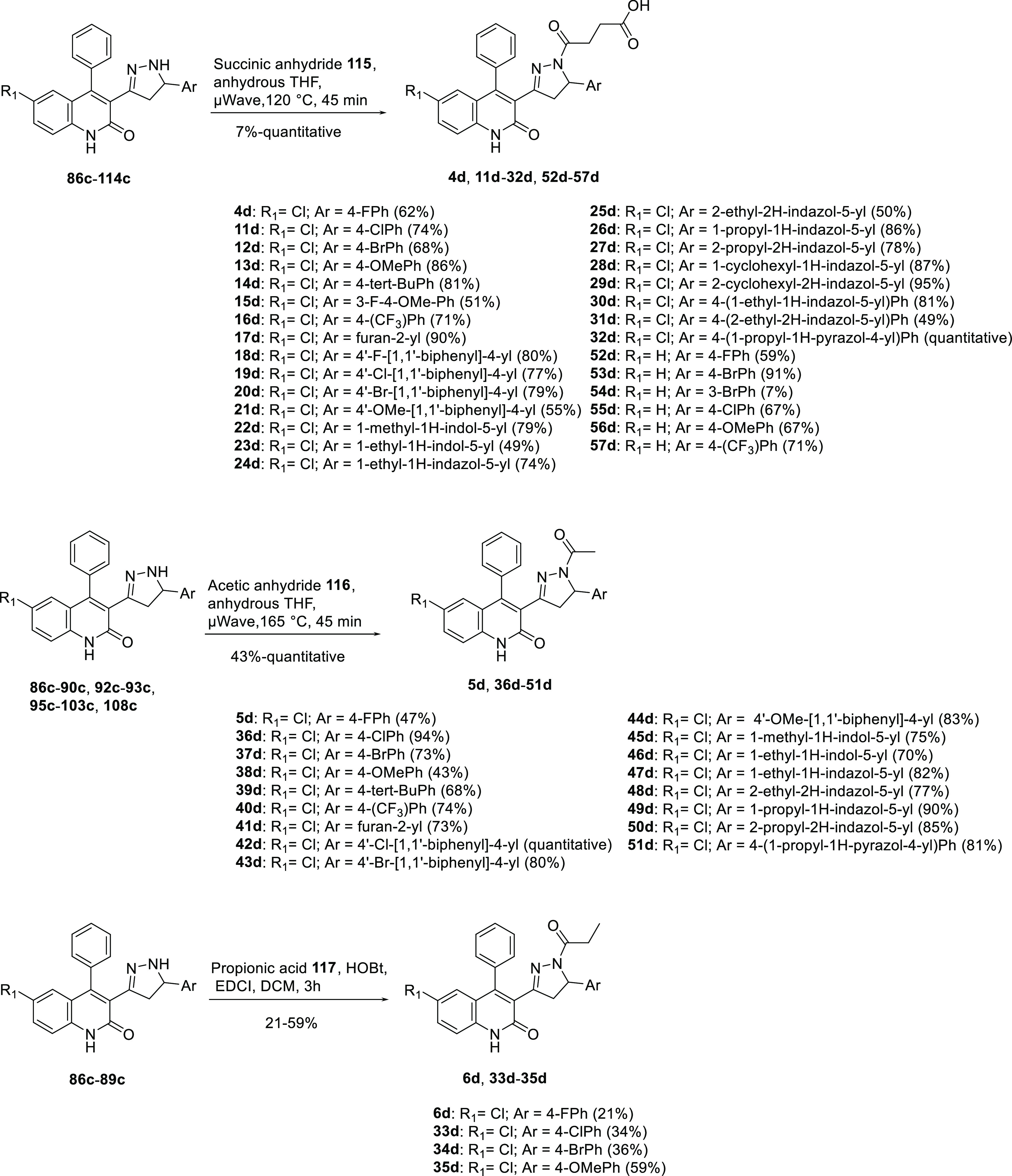
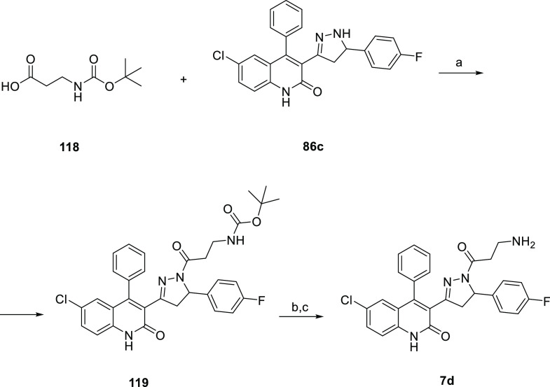
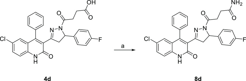
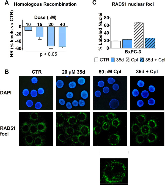
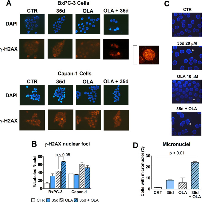
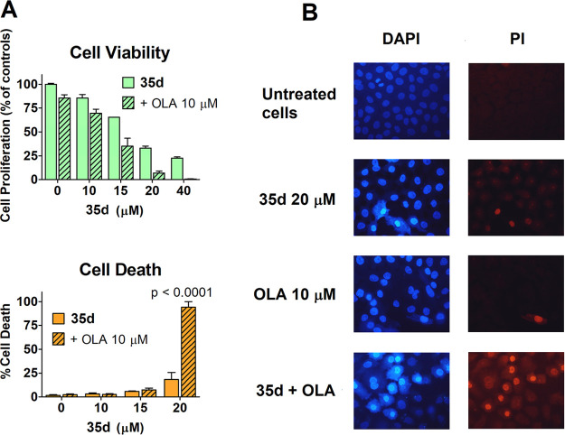
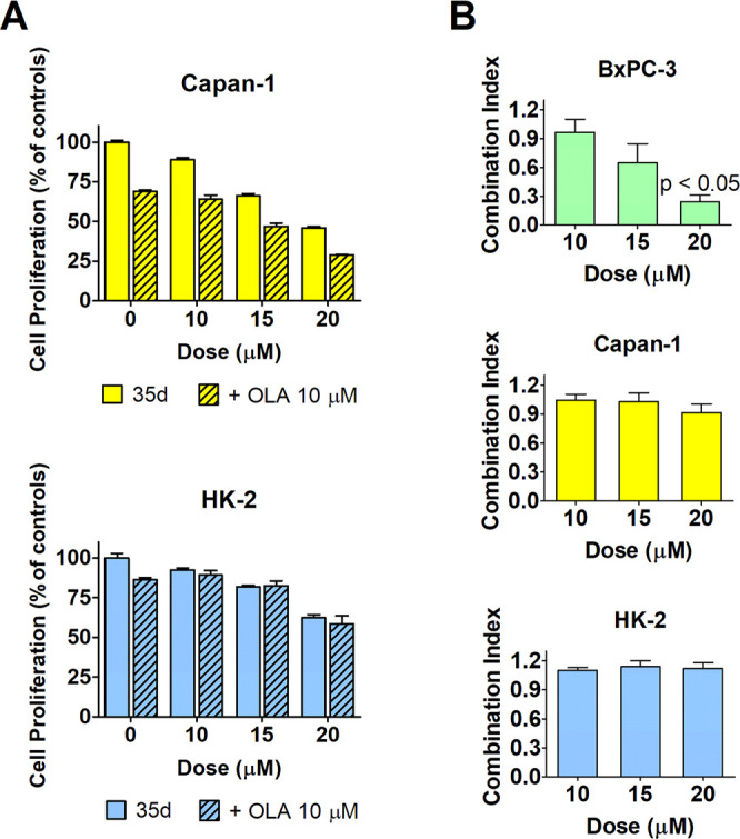
Synthetic lethality is an innovative framework for discovering novel anticancer drug candidates. One example is the use of PARP inhibitors (PARPi) in oncology patients with BRCA mutations. Here, we exploit a new paradigm based on the possibility of triggering synthetic lethality using only small organic molecules (dubbed "fully small-molecule-induced synthetic lethality"). We exploited this paradigm to target pancreatic cancer, one of the major unmet needs in oncology. We discovered a dihydroquinolone pyrazoline-based molecule (35d) that disrupts the RAD51-BRCA2 protein-protein interaction, thus mimicking the effect of BRCA2 mutation. 35d inhibits the homologous recombination in a human pancreatic adenocarcinoma cell line. In addition, it synergizes with olaparib (a PARPi) to trigger synthetic lethality. This strategy aims to widen the use of PARPi in BRCA-competent and olaparib-resistant cancers, making fully small-molecule-induced synthetic lethality an innovative approach toward unmet oncological needs.


Conflict of interest statement

The authors declare the following competing financial interest(s): One patent application protecting the class of compounds disclosed in this article has been filed by the following authors: Greta Bagnolini, Domenico Milano, Marcella Manerba, Jose Antonio Ortega, Francesca De Franco, Roberto Pellicciari, Saverio Minucci, Giuseppina Di Stefano, Marinella Roberti, and Andrea Cavalli.

Figures

Figure 1
Figure 1
Structures of the previously identified triazoles 13.
Figure 2
Figure 2
(A) RAD51-BRCA2 BRC repeat complex (PDB code 1N0W). RAD51 is represented as a surface, BRC4 as a cartoon. The two hot spots of the interaction between the proteins (Phe1524 and Phe1546) are highlighted in sticks. (B) Zone II magnification showing the interacting residues of BRC4 (yellow) and RAD51 (white).
Figure 3
Figure 3
Both enantiomers of compound 4d docked into the LFDE binding site (site II) of RAD51 (PDB code 1N0W).
Figure 4
Figure 4
Overview of the optimization strategy of 4d for SAR exploration.
Scheme 1
Scheme 1. Synthesis of Dihydroquinolone Pyrazoline Intermediates 86c114c
Reagents and conditions: (a) ethyl acetoacetate, DMF, 120 °C μWave or reflux, 60a quantitative, 61a 53%; (b) KOH, EtOH/H2O 4:3 v/v (0.05 M), 0 °C to rt, 53%, quantitative; (c) hydrazine monohydrate, EtOH, 110 °C μWave, 45 min, 64%, quantitative.
Scheme 2
Scheme 2. Synthesis of Final Dihydroquinolone Pyrazolines 4d57d
Scheme 3
Scheme 3. Synthesis of Compound 7d
Reagents and conditions: (a) HOBt, EDCI, DCM, overnight, yield 25%; (b) HCl 4 M in dioxane, rt, 15 min; (c) NaOH 0.5 M in EtOAc, rt, 15 min, yield 50%.
Scheme 4
Scheme 4. Synthesis of Compound 8d
Reagents and conditions: (a) HATU (1.5 equiv), EDC (1.5 equiv), DCM, DMF, ammonium chloride (5.0 equiv), DIPEA (4.0 equiv), rt, 26 h, yield 46%.
Scheme 5
Scheme 5. Synthesis of Compound 9d
Reagents and conditions: (a) HOBt (1.1 equiv), EDC (1.1 equiv), TEA (2.2. equiv) DCM, rt, 16 h, yield 42%.
Scheme 6
Scheme 6. Synthesis of Compound 10d
Reagents and conditions: (a) TEA (5.0 equiv), anhydrous DCM, methanesulfonyl chloride 122 (2.0 equiv), rt, 48 h, yield 82%; (b) MeOH/THF (1:1 v/v), 2 M LiOH, rt, 16 h, yield 82%; (c) HOBt (1.1 equiv), EDC (1.1 equiv), TEA (2.2 equiv), DCM, rt, 16 h, yield 15%.
Figure 5
Figure 5
MST analysis of His-hRAD51-35d binding. Titration curve of (RED-tris-NTA 2nd Generation)-His-hRAD51 (80 nM) with increasing concentrations of 35d. Sigmoidal fitting curve was obtained using the Affinity Analysis software of NanoTemper Technologies. MST data are the average of three replicates.
Figure 6
Figure 6
(A) Effect on HR caused by 35d administered to BxPC3 cells during plasmid transfection (5 h). HR was evaluated by real-time PCR, as described in the Experimental Section. Data were statistically analyzed using the column statistics of Prism 5 software, which applies the inferences analysis and the one-sample t test. The observed inhibitory effect was significantly different from 0 (the level of untreated cultures) for all tested doses, with p < 0.05. (B, C) Immunofluorescence detection of RAD51 in BxPC-3 nuclei after a treatment with 50 μM cisplatin (Cpl) given separately or in combination with 20 μM 35d. Experimental details are reported in the Experimental Section. (B) Representative pictures showing DAPI-stained cell nuclei and the corresponding immune-labeling of RAD51 localization. In untreated cells (CTR), RAD51 labeling is clearly evident in cytoplasm and does not appear in cell nuclei. In the pictures of Cpl-exposed cells, nuclear localization of the protein is clearly evident in 3 out of the 6 shown nuclei. A higher magnification detail was included for this sample. (C) The bar graph shows the percentage of RAD51-labeled nuclei counted by two independent observers who analyzed the treated cultures. Data were statistically evaluated by applying the one-way ANOVA, which indicated a significantly increased nuclear RAD51 labeling caused by Cpl (p < 0.05) and no statistically significant difference between cells treated with 35d and those exposed to 35d + Cpl.
Figure 7
Figure 7
(A, B) Evaluation of DNA damage through immune detection of nuclear γ-H2AX foci in BxPC-3 and Capan-1 cells exposed for 48 h to olaparib (10 μM) or 35d (20 μM), given alone or in combination. (A) Representative pictures showing DAPI-stained cell nuclei and the corresponding immune-labeling of γ-H2AX. In BxPC-3 cells, coadministration of 35d and olaparib produced increased γ-H2AX labeling. A higher magnification detail was included for this sample. As expected, Capan-1 cells showed a constitutive γ-H2AX labeling that was highly increased by olaparib but was unaffected by 35d coadministration. (B) The bar graph shows the percentage of γ-H2AX -labeled nuclei counted by two independent observers who analyzed the treated cultures. Data obtained in bxPC-3 cells were statistically evaluated by applying the one-way ANOVA, which indicated a statistically significant difference between the cultures treated with olaparib and those exposed to olaparib + 35d. (C, D) Evaluation of micronuclei generation in BxPC3 cells treated (72 h) with 35d and olaparib, given alone or in combination. (C) Representative pictures showing DAPI-stained cell nuclei. White asterisks indicate the presence of micronuclei. (D) The percentage of cells bearing micronuclei was estimated by two independent observers, by analyzing 100–250 cells for each treatment sample. The obtained results were statistically analyzed by applying the one-way ANOVA, which indicated a p value of <0.01.
Figure 8
Figure 8
(A) BxPC3 cell viability and death measured after 72 h exposure to 35d and 10 μM olaparib, given alone or in combination. Data were analyzed by two-way ANOVA using the two treatments (35d and olaparib) as variables. In the cell viability experiment, Bonferroni post-test indicated a statistically significant difference produced by olaparib coadministration in all BxPC3 cultures treated with the different 35d doses, with p values ranging from 0.01 (10 μM 35d) to 0.0001 (15 and 20 μM 35d). The same analysis was applied to the data from the cell death experiment and indicated that no statistically significant increase in cell death was produced by olaparib when coadministered with 10–15 μM 35d. In cultures exposed to olaparib + 20 μM 35d the evidence for cell death was markedly increased and statistically significant, with p < 0.0001. (B) After the 72 h treatment, BxPC3 cells were stained with vital dyes. As shown in the microscope pictures, the only culture displaying sharp evidence of cell death was that exposed to the combination of olaparib/20 μM 35d, as demonstrated by PI nuclear staining.
Figure 9
Figure 9
(A) Antiproliferative effect caused by 35d and its association with olaparib, measured in Capan-1 cells (which do not operate RAD51-BRCA2-dependent HR) and in normal immortalized human renal cells (HK-2). The same procedures described for BxPC3 cells were used here. (B) Combination indexes of the olaparib/35d association measured in the three used cell lines, calculated according to the method previously described., For BxPC-3 cells, the data reported in Figure 8A (cell proliferation) were used. Data were analyzed using the column statistics and the one-sample t test of Prism 5 software. For BxPC-3 cells, this test showed a statistically significant difference from 0.8 for the combination of olaparib and 20 μΜ 35d. Values of <0.8 indicate synergism between the two compounds.

References

    1. Bridges C. B. The origin and variations in sexual and sex limited characters. Am. Nat. 1922, 56, 51–63. 10.1086/279847. - DOI
    1. Dobzhansky T. Genetics of natural populations. Xiii. Recombination and variability in populations of Drosophila pseudoobscura. Genetics 1946, 31, 269–290. - PMC - PubMed
    1. Lucchesi J. C. Synthetic lethality and semi-lethality among functionally related mutants of Drosophila melanfgaster. Genetics 1968, 59, 37–44. - PMC - PubMed
    1. Kaiser C. A.; Schekman R. Distinct sets of SEC genes govern transport vesicle formation and fusion early in the secretory pathway. Cell 1990, 61, 723–733. 10.1016/0092-8674(90)90483-U. - DOI - PubMed
    1. Hennessy K. M.; Lee A.; Chen E.; Botstein D. A group of interacting yeast DNA replication genes. Genes Dev. 1991, 5, 958–969. 10.1101/gad.5.6.958. - DOI - PubMed

Publication types

MeSH terms