Review
. 2020 Jan 21:10:1568.
doi: 10.3389/fphar.2019.01568. eCollection 2019.

Role of Endothelial Dysfunction in Cardiovascular Diseases: The Link Between Inflammation and Hydrogen Sulfide

Affiliations
Review

Role of Endothelial Dysfunction in Cardiovascular Diseases: The Link Between Inflammation and Hydrogen Sulfide

Hai-Jian Sun et al. Front Pharmacol. .

Abstract

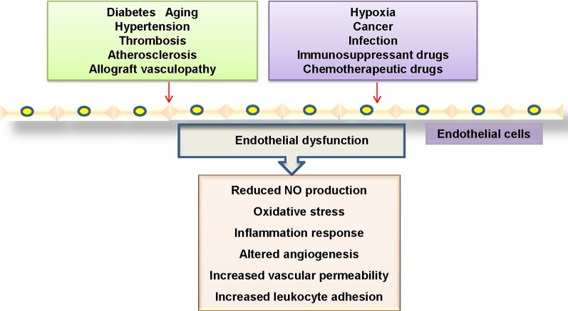
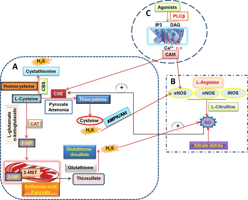
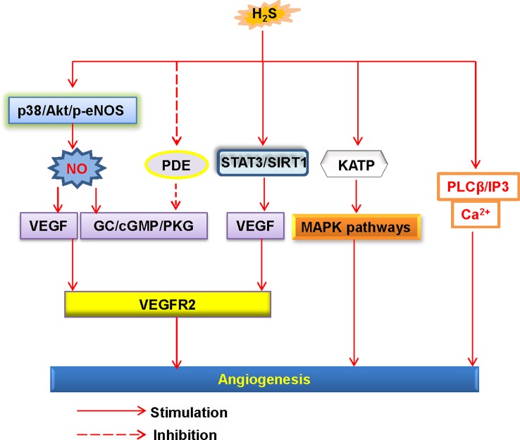
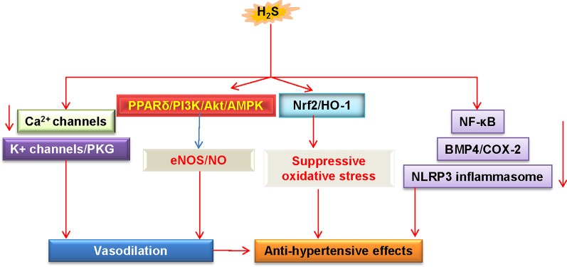
Endothelial cells are important constituents of blood vessels that play critical roles in cardiovascular homeostasis by regulating blood fluidity and fibrinolysis, vascular tone, angiogenesis, monocyte/leukocyte adhesion, and platelet aggregation. The normal vascular endothelium is taken as a gatekeeper of cardiovascular health, whereas abnormality of vascular endothelium is a major contributor to a plethora of cardiovascular ailments, such as atherosclerosis, aging, hypertension, obesity, and diabetes. Endothelial dysfunction is characterized by imbalanced vasodilation and vasoconstriction, elevated reactive oxygen species (ROS), and proinflammatory factors, as well as deficiency of nitric oxide (NO) bioavailability. The occurrence of endothelial dysfunction disrupts the endothelial barrier permeability that is a part of inflammatory response in the development of cardiovascular diseases. As such, abrogation of endothelial cell activation/inflammation is of clinical relevance. Recently, hydrogen sulfide (H2S), an entry as a gasotransmitter, exerts diverse biological effects through acting on various targeted signaling pathways. Within the cardiovascular system, the formation of H2S is detected in smooth muscle cells, vascular endothelial cells, and cardiomyocytes. Disrupted H2S bioavailability is postulated to be a new indicator for endothelial cell inflammation and its associated endothelial dysfunction. In this review, we will summarize recent advances about the roles of H2S in endothelial cell homeostasis, especially under pathological conditions, and discuss its putative therapeutic applications in endothelial inflammation-associated cardiovascular disorders.

Keywords: cardiovascular disease; endothelial cell; gasotransmitters; hydrogen sulfide; inflammation.


Figures

Figure 1
Figure 1
Mechanisms linked to endothelial dysfunction. Several key mechanisms that promote endothelial dysfunction.
Figure 2
Figure 2
H2S and NO biosynthetic pathways in blood vessels. (A) L-cysteine is the substrate for the formation of H2S through three H2S-producing enzymes, L-cysteine is catalyzed by CSE to produce pyruvate, ammonia, and thiocysteine, the latter is then decomposed to cysteine and H2S. The endogenous H2S production by CBS is related with the condensation of homocysteine with L-cysteine, followed by the formation of cystathionine and H2S. Direct reaction of L-cysteine and α-ketoglutarate by CAT yields the release of 3-MP and L-glutamate, 3-Mercaptopyruvate is transported into the mitochondria where it is catalyzed to sulfurous acid, pyruvate and thiosulfate by 3-MST. In the presence of reduced glutathione, the thiosulfate is reduced to glutathione disulfide and H2S. It is well accepted that H2S can increase eNOS activity and thereby subsequent NO production directly or through AMPK/Akt signaling pathway. (B) NO is produced in all tissues by NOS-dependent (L-arginine-NO pathway) and -independent (nitrate-nitrite-NO pathway) pathways. A recently discovered pathway for NO generation is the serial reduction of the inorganic anions nitrate and nitrite. With the assistance of three isoforms of NOS including nNOS, eNOS, and iNOS, L-arginine is oxidized into L-citrulline with NO. NO is found ro increase CSE activity and expression and then stimulate H2S production. (C) In endothelial cells, vasoconstrictor agonists stimulate the release of Ca2+ and cause formation of calcium-calmodulin (CaM) via the PLCβ/IP3/DAG pathway. Then, CaM can simultaneously activate eNOS and CSE that yield NO and H2S, respectively. H2S, hydrogen sulfide; NO, nitric oxide; 3-MP, 3-mercaptopyruvate; CAT, cysteine aminotransferase; CSE, cystathionine γ-lyase; CBS, cystathionine β-synthase; 3-MST, 3-mercaptopyruvate sulfurtransferase; CaM, calcium-calmodulin; PLCβ, phospholipase Cβ; IP3, inositol-3-phosphate (IP3); DAG, diacylglycerol (DAG); eNOS, endothelial NO synthase; iNOS, inducible NO synthase; nNOS, neuronal NO synthase.
Figure 3
Figure 3
Schematic illustration of the underlying mechanisms of H2S-induced angiogenesis. H2S, hydrogen sulfide; NO, nitric oxide; Akt, protein kinase B; p38, p38 mitogen-activated protein kinases; eNOS, endothelial NO synthase; VEGF, vascular endothelial growth factor; VEGFR2, vascular endothelial growth factor receptor 2; PDE, phosphodiesterase; GC, guanylate cyclase; cGMP, cyclic guanosine monophosphate; PKG, protein kinase G; SIRT1, sirtuin 1; KATP, ATP-sensitive K+ channels; MAPK, mitogen-activated protein kinase; STAT3, signal transducer and activator of transcription 3; PLCβ, phospholipase Cβ; IP3, inositol-3-phosphate (IP3).
Figure 4
Figure 4
Schematic illustration of underlying mechanisms in which H2S protects against hypertension. H2S lowers high blood pressure via vasodilatation by activation of vascular KATP channels and inhibition of Ca2+ influx. The PPARδ/PI3K/Akt/AMPK signaling pathway participates in H2S-induced NO production. These above events cause vasodilation. H2S inhibits ROS production via Nrf-2/HO-1 related redox sensitive signaling pathways. In addition, H2S treatment blunts increases in systolic blood pressure by inhibiting inflammation-related signaling pathways. H2S, hydrogen sulfide; NO, nitric oxide; Akt, protein kinase B; eNOS, endothelial NO synthase; PKG, protein kinase G; PI3K, phosphoinositide 3-kinase; PPARδ, peroxisome proliferators-activated receptor δ; AMPK, adenosine 5’-monophosphate (AMP)-activated protein kinase; Nrf2, nuclear factor erythroid 2-related factor 2; HO-1, heme oxygenase 1; NF-κB, nuclear factor-kappa B; BMP4, bone morphogenetic protein 4; COX-2, cyclooxygenase-2; NLRP3, nucleotide-binding oligomerization domain, leucine rich repeat, and pyrin domain-containing protein 3.

References

    1. Ahmad F. U., Sattar M. A., Rathore H. A., Tan Y. C., Akhtar S., Jin O. H., et al. (2014). Hydrogen sulphide and tempol treatments improve the blood pressure and renal excretory responses in spontaneously hypertensive rats. Ren. Fail 36, 598–605. 10.3109/0886022X.2014.882218 - DOI - PubMed
    1. Ali M. Y., Ping C. Y., Mok Y. Y., Ling L., Whiteman M., Bhatia M., et al. (2006). Regulation of vascular nitric oxide in vitro and in vivo; a new role for endogenous hydrogen sulphide? Br. J. Pharmacol. 149, 625–634. 10.1038/sj.bjp.0706906 - DOI - PMC - PubMed
    1. Allende-Vigo M. Z. (2010). Pathophysiologic mechanisms linking adipose tissue and cardiometabolic risk. Endocr. Pract. 16, 692–698. 10.4158/EP09340.RA - DOI - PubMed
    1. Al-Magableh M. R., Kemp-Harper B. K., Hart J. L. (2015). Hydrogen sulfide treatment reduces blood pressure and oxidative stress in angiotensin II-induced hypertensive mice. Hypertens. Res. 38, 13–20. 10.1038/hr.2014.125 - DOI - PubMed
    1. Altaany Z., Moccia F., Munaron L., Mancardi D., Wang R. (2014). Hydrogen sulfide and endothelial dysfunction: relationship with nitric oxide. Curr. Med. Chem. 21, 3646–3661. 10.1097/SHK.0000000000000599 - DOI - PubMed