Review
. 2020 Jan 24:10:1572.
doi: 10.3389/fphar.2019.01572. eCollection 2019.

An Update on the Structure of hERG

Affiliations
Review

An Update on the Structure of hERG

Andrew Butler et al. Front Pharmacol. .

Abstract

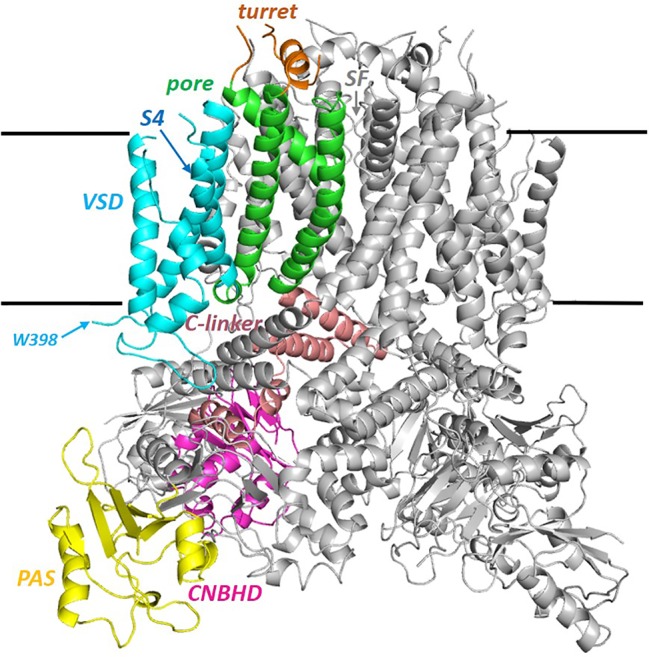
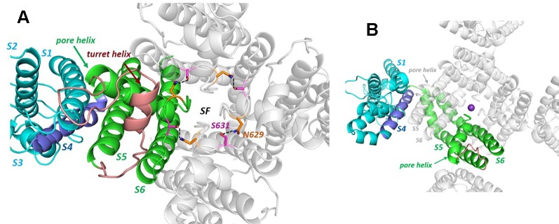
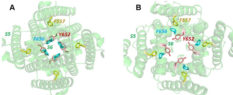
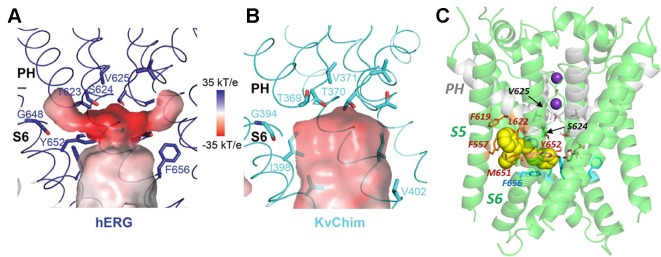
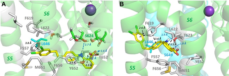
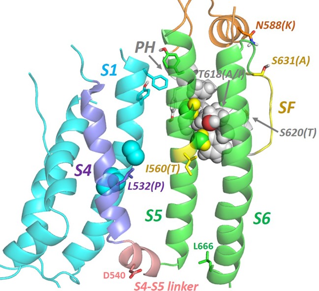
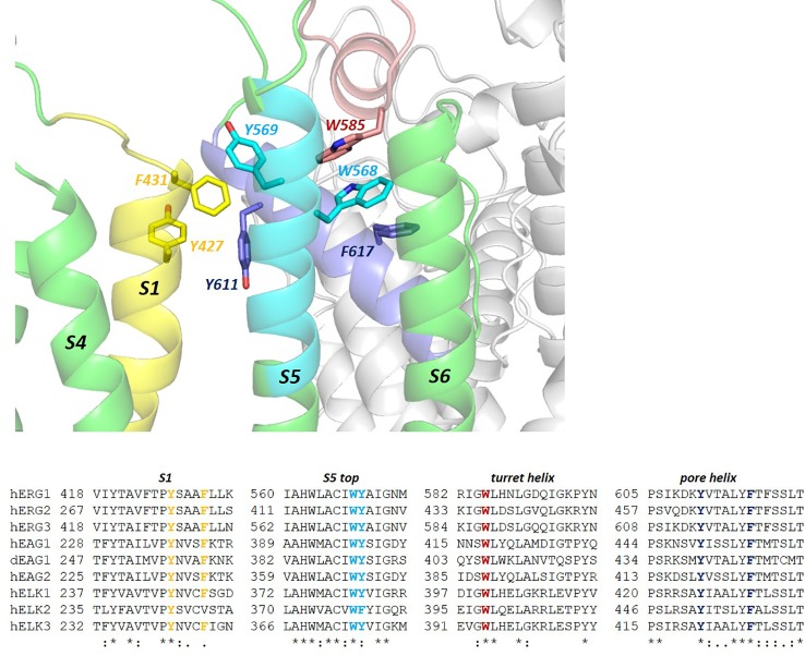
The human voltage-sensitive K+ channel hERG plays a fundamental role in cardiac action potential repolarization, effectively controlling the QT interval of the electrocardiogram. Inherited loss- or gain-of-function mutations in hERG can result in dangerous "long" (LQTS) or "short" QT syndromes (SQTS), respectively, and the anomalous susceptibility of hERG to block by a diverse range of drugs underlies an acquired LQTS. A recent open channel cryo-EM structure of hERG should greatly advance understanding of the molecular basis of hERG channelopathies and drug-induced LQTS. Here we describe an update of recent research that addresses the nature of the particular gated state of hERG captured in the new structure, and the insight afforded by the structure into the molecular basis for high affinity drug block of hERG, the binding of hERG activators and the molecular basis of hERG's peculiar gating properties. Interpretation of the pharmacology of natural SQTS mutants in the context of the structure is a promising approach to understanding the molecular basis of hERG inactivation, and the structure suggests how voltage-dependent changes in the membrane domain may be transmitted to an extracellular "turret" to effect inactivation through aromatic side chain motifs that are conserved throughout the KCNH family of channels.

Keywords: C-type inactivation; KCNH; channelopathy; cryo-EM structure; drug block; hERG; long QT syndrome; short QT syndrome.


Figures

Figure 1
Figure 1
Side view of the full hERG cryo-EM structure (PDB: 5VA2; hERGT) with one of the four subunits colored by domain. PAS (the N-terminal Per-ARNT-Sim) domain; the cytoplasmic C-linker links the S6 helix of the pore domain (green) to the CNBHD (Cyclic Nucleotide Binding Homology Domain). VSD (Voltage Sensor Domain). W398 is the first residue of the membrane domain of hERG for which atom density is defined. Sequence breaks in some extra extracellular loops are regions lacking atom density in the cryo-EM structure. The horizontal black lines mark the approximate limits of the non-polar part of the bilayer membrane.
Figure 2
Figure 2
(A) The hERG cryo-EM structure viewed from the extracellular side of the membrane illustrating the non-domain-swapped subunit organization in which the voltage sensor domain (VSD) is packed against the pore domain of the same subunit. The long turret sequence containing a turret helix, that links the top of S5 with the N-terminal end of the pore helix, is colored brown (some atom density in the non-helical region of the turret is missing in the cryo-EM structure and is modelled into the structure shown). Polar side chains of N629 and S631 form a hydrogen-bonded ring that links subunits around the top of the selectivity filter. (B) Equivalent view of the Kv1.2/2.1 K+ channel chimera structure [KvChim; PDB: 2R9R (Long et al., 2007)] illustrating domain-swapping and a very short “turret” sequence (brown). Although the VSDs are packed against pore domains of adjacent subunits in domain-swapped channels, the relative intersubunit juxtaposition of S5 and the pore helix with the VSD is similar to the intrasubunit juxtaposition of VSD and S5 and pore helix in hERG (and rEAG). The purple sphere is a K+ ion in the S1 position of the selectivity filter.
Figure 3
Figure 3
(A) Bottom up view of the pore domain of a hERG homology model built on the structure of the highly homologous rEAG structure [PDB: 5K7L (Whicher and MacKinnon, 2016)] which has an activated voltage sensor but a closed pore. In this model the side chains of key amino acids for hERG channel block, Y652 and F656, are oriented towards the K+ permeation pathway at the centre of the pore. (B) In the open pore hERG cryo-EM structure, the F656 side chains are oriented away from the pore center towards the F557 side chain on the S5 helix.
Figure 4
Figure 4
Comparison of the hERG pore cavity (A) with that of the Kv1.2/2.1 channel chimera (KvChim) (B). The hERG cavity is smaller than the equivalent KvChim cavity and has hydrophobic “pockets” that project from the central cavity below the bottom of the selectivity filter and underneath the pore helix (PH). The pore helix negative dipole charges focus a strong negative electrostatic potential below the selectivity filter which contributes to the binding energy for positively-charged hERG pore blockers. (C) The hERG blocker “Cavalli-2” [(Cavalli et al., 2012); yellow space filling representation] can be docked partially within a hydrophobic pocket although readjustment of F656 side chains is required for interaction of blocker with more than one F656 side chain [see text; adapted from (Helliwell et al., 2018)]. Panels (A and B) from (Wang and MacKinnon, 2017) with permission.
Figure 5
Figure 5
(A) PD-118057 (yellow sticks) can be docked into the hERG structure in configurations that orient the benzyl carboxyl group to interact with K+ ions as they traverse the hERG conductance pathway as suggested in (Schewe et al., 2019). However in these configurations PD-118057 does not make favourable interactions with F619, L622 (and L646 on the adjacent subunit), identified as key binding determinants for this activator (Perry et al., 2009). Also PD-118057 would be expected to interact with Y652 and F557 in these states whereas mutagenesis of Y652 and F557 has minimal effect on activator binding (Perry et al., 2009). (B) Docked states consistent with mutagenesis (Perry et al., 2009) (aromatic stacking and van der Waals interactions with F619, L622 and adjacent L646 side chain) can be found deeper within the hydrophobic pockets below the pore helix, but these states are not compatible with interaction of the PD-118057 carboxylate with K+ ions in the pore as suggested in (Schewe et al., 2019). To orient the viewer, PD-118057 occupies a hydrophobic pocket similar to that shown in Figure 4C for Cavalli-2 binding, with the dichlorophenyl group (chlorine atoms green) of PD-118057 close to the membrane in panel B. Docking was performed with GOLD version 5.6; Cambridge Crystallographic Data Centre, Cambridge, UK as described previously (Dempsey et al., 2014; Helliwell et al., 2018).
Figure 6
Figure 6
Natural mutations (in brackets) in hERG that perturb inactivation gating are found throughout the membrane domain indicating a network of helix interactions that transmits conformational changes to the selectivity filter (SF) resulting from mutation or voltage sensor (VS) activation. In the VS-activated state captured in the cryo-EM structure, residues on S4 whose mutation perturbs inactivation [e.g. L532P; (Hassel et al., 2008; Zhang et al., 2011)] interact closely with side chains of the S1 helix. The extracytoplasmic ends of the S1, S5 and pore helices interact via a cluster of aromatic side chains (see also Figure 7). S5 and pore helix side chains interdigitate (‘knobs into holes' packing) indicating a strong conformational coupling of these helices. The locations of D540 and L666 that move close together upon membrane repolarization are shown [adapted from (Butler et al., 2019)].
Figure 7
Figure 7
A “tryptophan clamp” connects the hERG turret helix with the top of the S5 helix. This motif that includes an interaction with F617 on the hERG pore helix is conserved throughout the KCNH family of channels that includes ERG, EAG and ELK variants, despite considerable sequence diversity in the turret helix itself (G584-I593 in hERG1). Helix interactions involving a cluster of aromatic side chains at the extracytoplasmic ends of S1, S5 and the pore helix may serve to anchor the VS domain against the pore as observed in other Kv channels (Lee et al., 2009); dEAG1 is drosophila EAG1; h, human.

References

    1. Akdis D., Saguner A. M., Medeiros-Domingo A., Schaller A., Balmer C., Steffel J., et al. (2018). Multiple clinical profiles of families with the short QT syndrome. Europace 20, f113–f121. 10.1093/europace/eux186 - DOI - PubMed
    1. Anderson C. L., Kuzmicki C. E., Childs R. R., Hintz C. J., Delisle B. P., January C. T. (2014). Large-scale mutational analysis of Kv11.1 reveals molecular insights into type 2 long QT syndrome. Nat. Commun. 5, 5535. 10.1038/ncomms6535 - DOI - PMC - PubMed
    1. Armstrong C. M., Hollingworth S. (2018). A perspective on Na and K channel inactivation. J. Gen. Physiol. 150, 7–18. 10.1085/jgp.201711835 - DOI - PMC - PubMed
    1. Barchad-Avitzur O., Priest M. F., Dekel N., Bezanilla F., Parnas H., Ben-Chaim Y. (2016). A novel voltage sensor in the orthosteric ninding site of the M2 Muscarinic Receptor. Biophys. J. 111, 1396–1408. 10.1016/j.bpj.2016.08.035 - DOI - PMC - PubMed
    1. Brugada R., Hong K., Dumaine R., Cordeiro J., Gaita F., Borggrefe M., et al. (2004). Sudden death associated with short-QT syndrome linked to mutations in HERG. Circulation 109, 30–35. 10.1161/01.CIR.0000109482.92774.3A - DOI - PubMed

LinkOut - more resources