Review
. 2020 Jan 22:10:1583.
doi: 10.3389/fphys.2019.01583. eCollection 2019.

Vascular Integrity and Signaling Determining Brain Development, Network Excitability, and Epileptogenesis

Affiliations
Review

Vascular Integrity and Signaling Determining Brain Development, Network Excitability, and Epileptogenesis

Jugajyoti Baruah et al. Front Physiol. .

Abstract

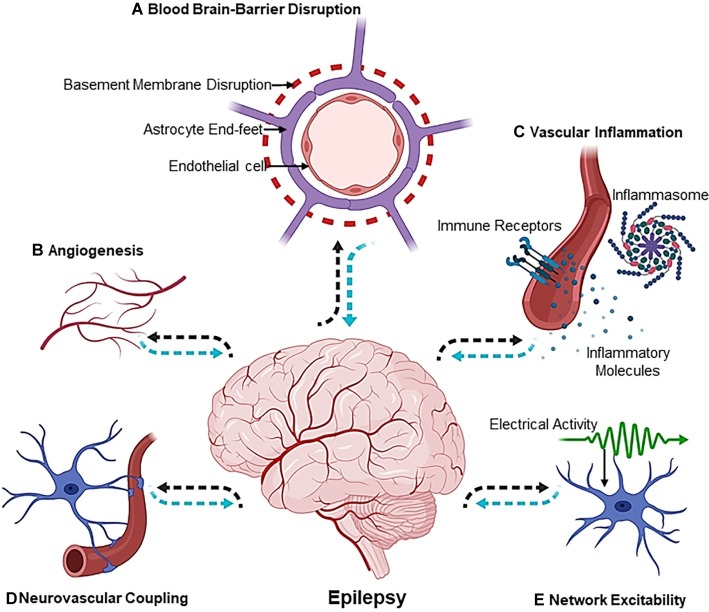
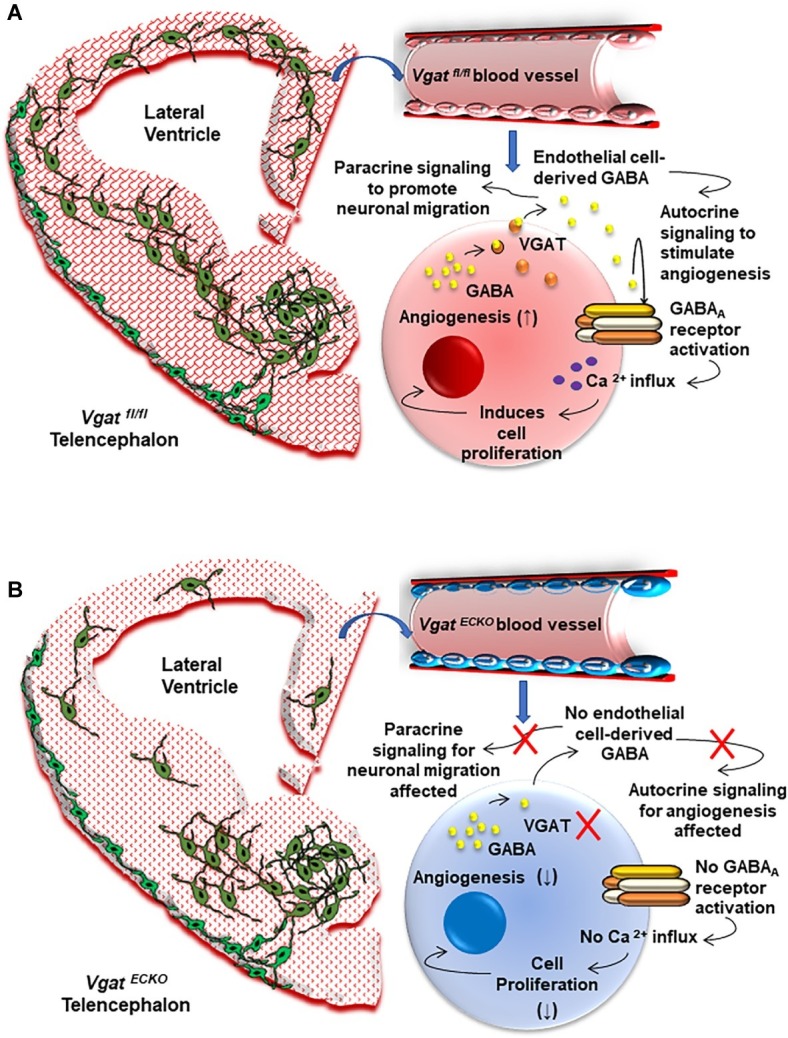
Our understanding of the etiological mechanisms leading up to epilepsy has undergone radical changes over time due to more insights into the complexity of the disease. The traditional hypothesis emphasized network hyperexcitability and an imbalance of inhibition and excitation, eventually leading to seizures. In this context, the contribution of the vascular system, and particularly the interactions between blood vessels and neuronal tissue, came into focus only recently. Thus, one highly exciting causative or contributing factor of epileptogenesis is the disruption of the blood-brain barrier (BBB) in the context of not only posttraumatic epilepsy, but also other etiologies. This hypothesis is now recognized as a synergistic mechanism that can give rise to epilepsy, and BBB repair for restoration of cerebrovascular integrity is considered a therapeutic alternative. Endothelial cells lining the inner surface of blood vessels are an integral component of the BBB system. Sealed by tight junctions, they are crucial in maintaining homeostatic activities of the brain, as well as acting as an interface in the neurovascular unit. Additional potential vascular mechanisms such as inflammation, altered neurovascular coupling, or changes in blood flow that can modulate neuronal circuit activity have been implicated in epilepsy. Our own work has shown how intrinsic defects within endothelial cells from the earliest developmental time points, which preclude neuronal changes, can lead to vascular abnormalities and autonomously support the development of hyperexcitability and epileptiform activity. In this article, we review the importance of vascular integrity and signaling for network excitability and epilepsy by highlighting complementary basic and clinical research studies and by outlining possible novel therapeutic strategies.

Keywords: blood-brain barrier; development; epileptogenesis; hyperexcitability; inflammation; vascular endothelia.


Figures

Figure 1
Figure 1
The Vascular Landscape in Epilepsy. The mechanisms involved in the etiology of epileptogenesis are multiphasic [(A) blood-brain-barrier disruption, (B) angiogenesis, (C) vascular inflammation, (D) neurovascular coupling and, lastly, (E) network excitability] and exist at the crossroads of the neurovascular network. Dotted black arrows indicate the sequence of events leading up to epileptogenesis, whereas dotted blue arrows show the sequence of events that affect the neuron or vascular interface in an epileptic brain. Illustration was created using Biorender.com.
Figure 2
Figure 2
The Vascular Origin of Epilepsy. (A) Schema depicting the Vgat fl/fl or control embryonic telencephalon at day 15 that has normal periventricular vascular network (red lattice pattern) and a normal endothelial GABA signaling pathway that promotes long distance tangential migration of GABAergic interneurons (green) from the ventral telencephalon. A single vessel has been magnified to illustrate the positive feedback GABA signaling pathway that exists in normal telencephalic endothelial cells (red). Endothelial GABA activates GABAA receptors, thereby triggering Ca2+ influx and endothelial cell proliferation. Vesicular GABA transporter (VGAT) is the primary mechanism for GABA release from telencephalic endothelial cells at embryonic day 15 (Li et al., 2018). Endothelial GABA release thus has autocrine roles in stimulating angiogenesis (↑) and paracrine roles in promoting long distance GABAergic neuronal migration in the embryonic telencephalon. (B) Schema depicting altered vascular profiles in Vgat ECKO telencephalon (red dotted pattern) in which there is complete loss of endothelial GABA secretion that causes GABAergic interneurons to stall in the ventral telencephalon. This has significant consequences for GABAergic neuronal tangential migration, resulting in neuronal reductions and abnormal cortical distribution in Vgat ECKO telencephalon. Magnification of a single vessel to depict abnormal Vgat ECKO endothelial cells (blue) and how lack of GABA release from these endothelial cells affects the positive feedback of GABA-GABAA receptor signaling, which in turn significantly affects angiogenesis-related gene expression (↓). Lack of endothelial GABA release also affects paracrine signaling and impairs long-distance migration of GABAergic interneurons.

References

    1. Abbott N. J., Friedman A. (2012). Overview and introduction: the blood-brain barrier in health and disease. Epilepsia 53(Suppl. 6), 1–6. 10.1111/j.1528-1167.2012.03696.x - DOI - PMC - PubMed
    1. Abkur T. M., Kearney H., Hennessy M. J. (2018). Refractory epilepsy following natalizumab associated PML. Mult. Scler. Relat. Disord. 20, 1–2. 10.1016/j.msard.2017.12.004, PMID: - DOI - PubMed
    1. Alonso-Nanclares L., DeFelipe J. (2014). Alterations of the microvascular network in the sclerotic hippocampus of patients with temporal lobe epilepsy. Epilepsy Behav. 38, 48–52. 10.1016/j.yebeh.2013.12.009, PMID: - DOI - PubMed
    1. Alvarez Dolado M., Broccoli V. (2011). GABAergic neuronal precursor grafting: implications in brain regeneration and plasticity. Neural Plast. 2011:384216. 10.1155/2011/384216, PMID: - DOI - PMC - PubMed
    1. Anton E. S., Kreidberg J. A., Rakic P. (1999). Distinct functions of α3 and αV integrin receptors in neuronal migration and laminar organization of the cerebral cortex. Neuron 22, 277–289. 10.1016/S0896-6273(00)81089-2, PMID: - DOI - PubMed

LinkOut - more resources