. 2020 Jan 22:10:2986.
doi: 10.3389/fmicb.2019.02986. eCollection 2019.

Degradation of Recalcitrant Polyurethane and Xenobiotic Additives by a Selected Landfill Microbial Community and Its Biodegradative Potential Revealed by Proximity Ligation-Based Metagenomic Analysis

Affiliations

Degradation of Recalcitrant Polyurethane and Xenobiotic Additives by a Selected Landfill Microbial Community and Its Biodegradative Potential Revealed by Proximity Ligation-Based Metagenomic Analysis

Itzel Gaytán et al. Front Microbiol. .

Abstract

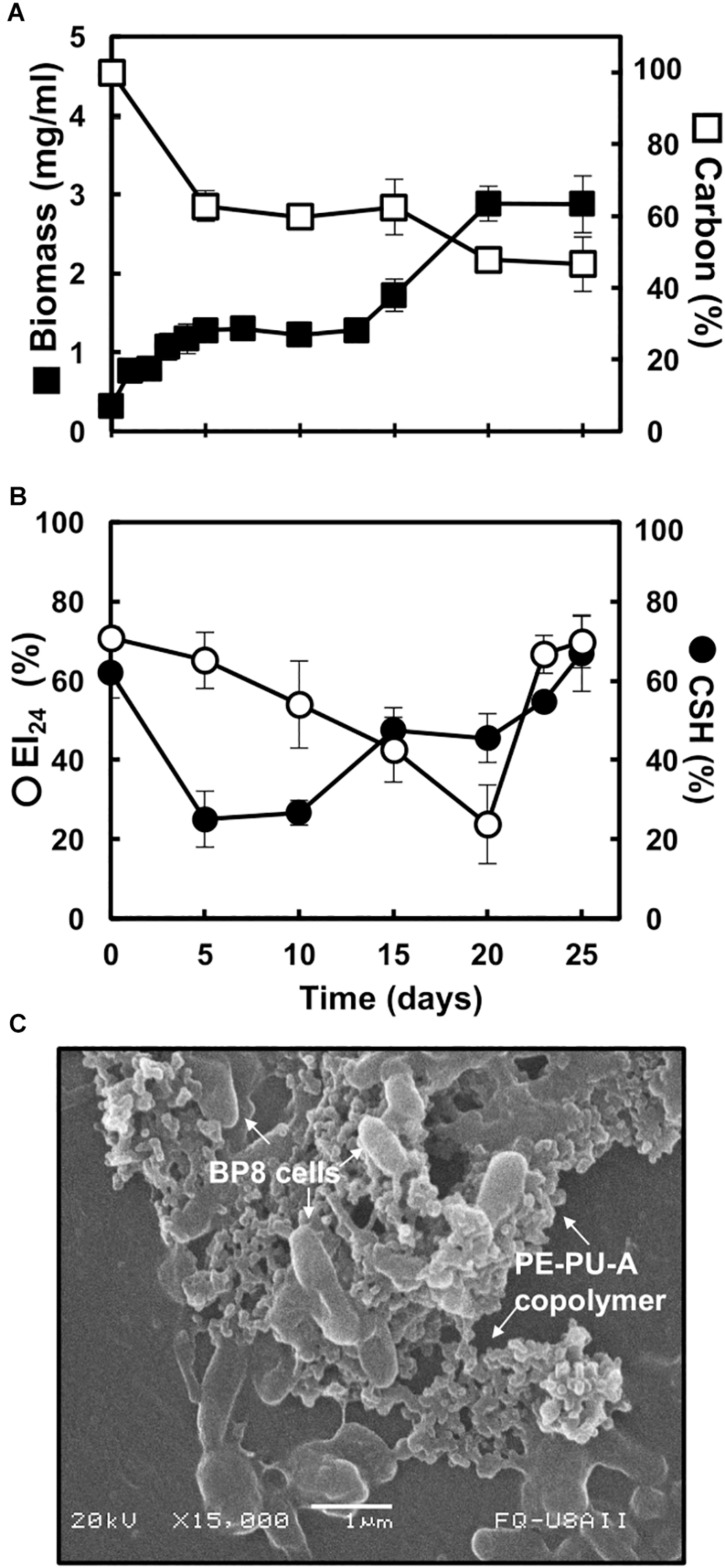
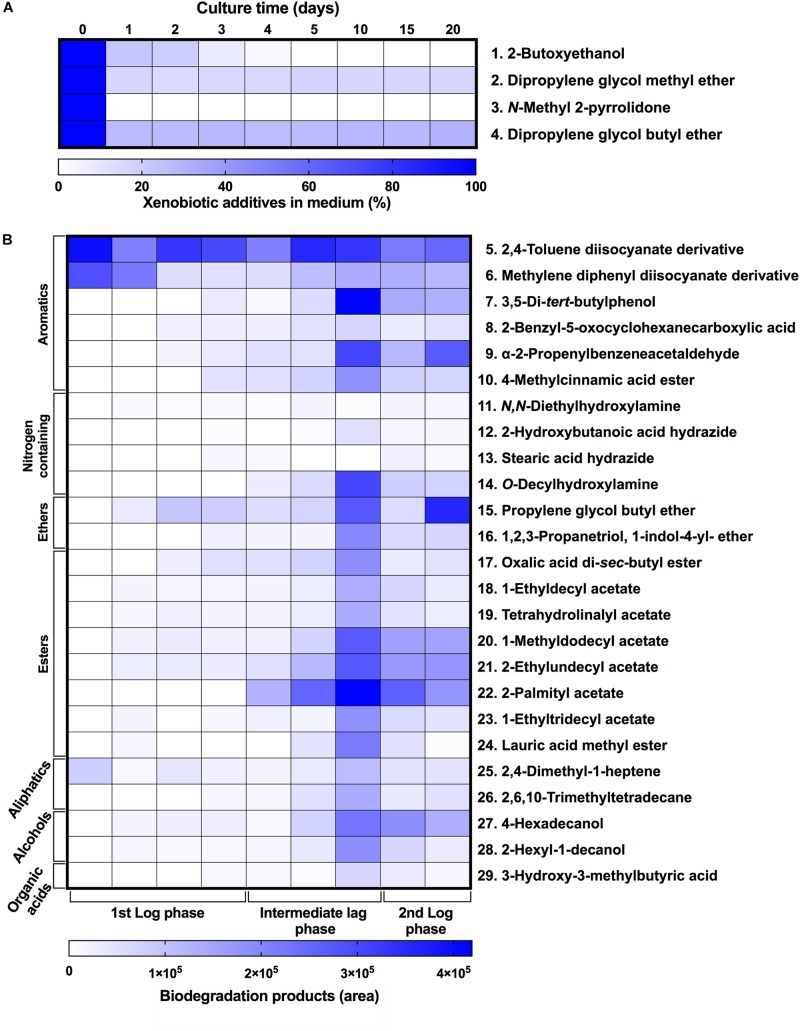
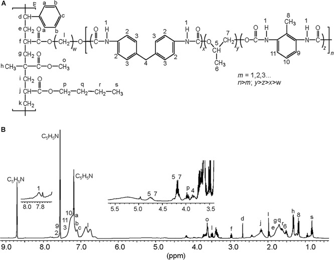
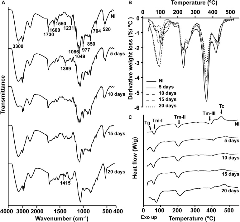
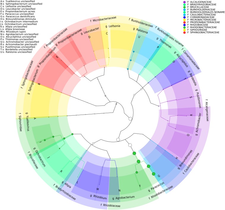
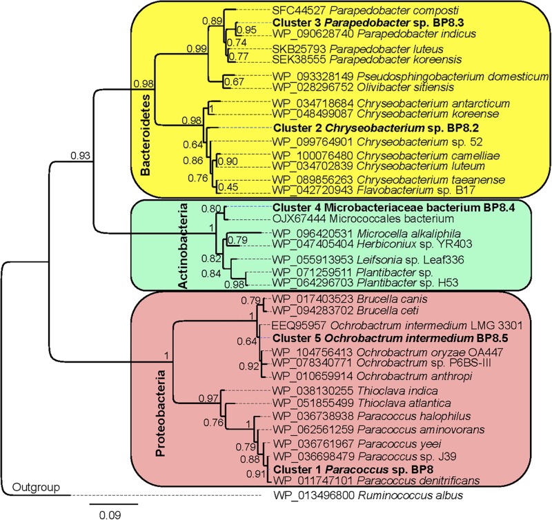
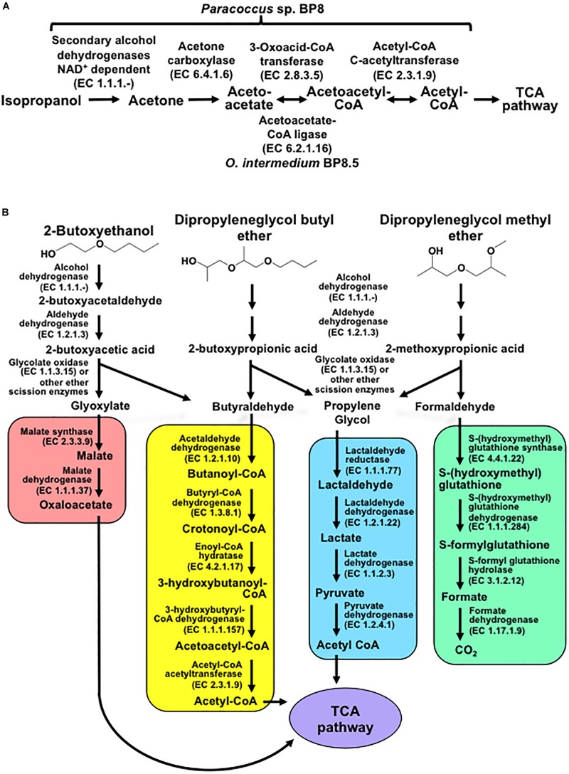
Polyurethanes (PU) are the sixth most produced plastics with around 18-million tons in 2016, but since they are not recyclable, they are burned or landfilled, generating damage to human health and ecosystems. To elucidate the mechanisms that landfill microbial communities perform to attack recalcitrant PU plastics, we studied the degradative activity of a mixed microbial culture, selected from a municipal landfill by its capability to grow in a water PU dispersion (WPUD) as the only carbon source, as a model for the BP8 landfill microbial community. The WPUD contains a polyether-polyurethane-acrylate (PE-PU-A) copolymer and xenobiotic additives (N-methylpyrrolidone, isopropanol and glycol ethers). To identify the changes that the BP8 microbial community culture generates to the WPUD additives and copolymer, we performed chemical and physical analyses of the biodegradation process during 25 days of cultivation. These analyses included Nuclear magnetic resonance, Fourier transform infrared spectroscopy, Thermogravimetry, Differential scanning calorimetry, Gel permeation chromatography, and Gas chromatography coupled to mass spectrometry techniques. Moreover, for revealing the BP8 community structure and its genetically encoded potential biodegradative capability we also performed a proximity ligation-based metagenomic analysis. The additives present in the WPUD were consumed early whereas the copolymer was cleaved throughout the 25-days of incubation. The analysis of the biodegradation process and the identified biodegradation products showed that BP8 cleaves esters, C-C, and the recalcitrant aromatic urethanes and ether groups by hydrolytic and oxidative mechanisms, both in the soft and the hard segments of the copolymer. The proximity ligation-based metagenomic analysis allowed the reconstruction of five genomes, three of them from novel species. In the metagenome, genes encoding known enzymes, and putative enzymes and metabolic pathways accounting for the biodegradative activity of the BP8 community over the additives and PE-PU-A copolymer were identified. This is the first study revealing the genetically encoded potential biodegradative capability of a microbial community selected from a landfill, that thrives within a WPUD system and shows potential for bioremediation of polyurethane- and xenobiotic additives-contamitated sites.

Keywords: Hi-C proximity-ligation; biodegradation; biodegradative potential; community structure; metagenomics; microbial community; polyether-polyurethane-acrylate; xenobiotic additives.


Figures

FIGURE 1
FIGURE 1
Characteristics of the BP8 community growing in MM-PolyLack. (A) Growth and carbon consumption, (B) emulsification index (EI24) and cell surface hydrophobicity (CSH) at different cultivation times. (C) SEM micrograph of BP8 cells attached to the PE-PU-A copolymer at 10 days of cultivation. Bars represent standard deviation. n = 3.
FIGURE 2
FIGURE 2
Xenobiotic additives consumed (A) and PE-PU-A biodegradation products generated (B) by the BP8 community. Cell-free supernatants were extracted at different cultivation times with chloroform:methanol and analyzed by GC-MS. (A) Additives were quantified using standard curves for each compound and (B) biodegradation products by analyzing their areas in independent chromatograms. n = 3. Compounds with mass spectra similarity values over 700 were considered the same compounds of the Library hits. The numbers in the compounds correspond to signals in the chromatograms of Supplementary Figure S1.
FIGURE 3
FIGURE 3
Proposed chemical structure for the PE-PU-A copolymer present in PolyLack®. (A) This structure was proposed based on the (B) 1H-NMR analysis of dried PolyLack®, the information included in the manufacturer technical manual (SayerLack. Poly Lack Aqua Brillante UB-0800), the GC-MS analysis (Figure 2), and the most frequent acrylates used in the synthesis of these types of copolymers (Pardini and Amalvy, 2008; Maurya et al., 2018). Synthesis of PE-PU-A copolymers starts by the polycondensation of polyols [polypropylene glycol (PPG)] (y moiety) and diisocyanates (TDI and MDI) (x and z moieties) followed by end capping with acrylates’ mixture (m moiety). From the most frequently used acrylates we selected methyl methacrylate, butyl acrylate, hydroxy acrylate and styrene as representatives in this structure. In the 1H-NMR spectrum, chemical shifts are provided in parts per million from SiMe4 as internal reference. Signal 1 is assigned to carbamate groups (NH-COO); signals a, b, c, 2, 3, 9-11 are assigned to the aromatic protons; signals 4 and 8 correspond to the protons of methylene (CH2) and methyl (CH3) groups in MDI and TDI, respectively; signals 5–7 correspond to PPG; signals l correspond to the hydroxyl proton (CH2-O) and methylene groups (CH2) in the chain of hydroxy-acrylate; signals f, j, o and p correspond to the acrylic groups (CH-COO, CH2-COO or CH3-COO), signals d (CH), e, g, i, k, q, r (CH2), h and s (CH3) are assigned to methylene and methyl groups in the acrylate mixtures.
FIGURE 4
FIGURE 4
Physical and chemical analyses of the aromatic PE-PU-A copolymer after incubation with the BP8 community. (A) FTIR spectra. (B) DTG analysis. Thermal degradation stages correspond to the following functional groups: (I) Low molecular weight compounds, (II) Urethane, (III) Ester, (IV) Ether. (C) DSC analysis. Glass transition temperature (Tg) represents the relative amount of soft and hard segments; melting temperatures, Tm-I, Tm-II, and Tm-III are associated with hard domains, and crystallization temperature (Tc) represents heat-directed crystallization of copolymer chains. NI, non-inoculated.
FIGURE 5
FIGURE 5
Taxonomic cladogram of BP8 community microbial diversity profiled with MetaPhlAn. Circles size is proportional to the taxon relative abundance. The most abundant taxa were Paracoccus genus (83%) and Ochrobactrum genus (8.7%). Families are color-labeled and predicted species diversity is indicated by capital letters (Asnicar et al., 2015).
FIGURE 6
FIGURE 6
Maximum likelihood phylogeny for taxonomic delimitation of the deconvoluted genomes from the BP8 metagenome. The analysis was performed with three phylogenetic markers: ribosomal protein L3, ribosomal protein S5 and DNA gyrase A subunit, which generated similar results. The analysis for ribosomal protein L3 is presented. Branch support values are indicated in the corresponding nodes. Bar indicates the number of expected substitutions per site under the WAG + G model. A sequence of Ruminococcus albus (Firmicutes) was used as outgroup. Key genome clusters are highlighted in bold and different Phyla are indicated at the left. Sequences for L3 ribosomal proteins of the deconvoluted genomes are accessible in the NCBI GenBank under accession numbers RQP07704.1, RQP15098.1, RQP16503.1, RQP08603.1, and RQP16393.1 for clusters 1–5, respectively.
FIGURE 7
FIGURE 7
Potential degradation pathways for isopropanol (A) and glycol ethers (B) encoded in the BP8 metagenome. (A) Paracoccus sp. BP8 genome encodes ADH enzymes that can oxidize IP to acetone, but genes encoding enzymes for the oxidative metabolism of acetone were not found. Instead, genes encoding the reductive transformation of acetone to acetyl CoA, acetone carboxylase, 3-oxoacid-CoA transferase and acetyl-CoA C-acetyltransferase, were identified. In O. intermedium BP8.5 genome, acetoacetate-CoA ligase, which performs the same reaction that 3-oxoacid-CoA transferase, is encoded. All the enzymes for the TCA pathway are encoded in the BP8 metagenome (see Table 3). (B) Subsequent oxidations of glycol ethers’ hydroxy terminal groups by PEG-DH and PEG-ALDH will produce 2-butoxyacetic acid from 2-BE, 2-butoxypropionic acid from DPGB and 2-methoxypropionic acid from DPGM. Subsequent cleavage of carboxylates’ ether bonds by ether scission enzymes such as glycolate oxidase, dye decoloring peroxidase, 4-methoxybenzoate monooxygenase and/or unspecific monooxygenase, would generate the metabolizable intermediaries glyoxylate, butyraldehyde, propylene glycol and formaldehyde. Glyoxylate would be funneled to the glyoxylate metabolism (red rectangle), butyraldehyde to the butanoate metabolism (yellow rectangle), propylene glycol to the pyruvate metabolism (blue rectangle), and formaldehyde to the formate metabolism (green rectangle). Pathways for glyoxylate, butanoate, pyruvate and formate metabolisms as well as the TCA pathway (purple ellipse) were fully reconstructed from the BP8 metagenome based on KEGG annotated genes, using KEGG Mapper.

References

    1. Albertsson A. C., Erlandsson B., Hakkarainen M., Karlsson S. (1998). Molecular weight changes and polymeric matrix changes correlated with the formation of degradation products in biodegraded polyethylene. J. Environ. Pol. Degrad. 6 187–195. 10.1023/A:1021873631162 - DOI
    1. Álvarez-Barragán J., González-Hernández R., Domínguez-Malfavón L., Vargas-Suárez M., Aguilar-Osorio G., Loza-Tavera H. (2016). Biodegradative activity of selected environmental fungi on a polyester polyurethane varnish and polyether polyurethane foams. Appl. Environ. Microbiol. 82 5225–5235. 10.1128/AEM.01344-1316 - DOI - PMC - PubMed
    1. Asnicar F., Weingart G., Tickle T. L., Huttenhower C., Segata N. (2015). Compact graphical representation of phylogenetic data and metadata with GraPhlAn. PeerJ 3:e1029. 10.7717/peerj.1029 - DOI - PMC - PubMed
    1. Breitwieser F. P., Pertea M., Zimin A. V., Salzberg S. L. (2019). Human contamination in bacterial genomes has created thousands of spurious proteins. Genome Res. 29 954–960. 10.1101/gr.245373.118 - DOI - PMC - PubMed
    1. Burton J. N., Liachko I., Dunham M. J., Shendure J. (2014). Species-level deconvolution of metagenome assemblies with Hi-C-based contact probability maps. G3 4 1339–1346. 10.1534/g3.114.011825 - DOI - PMC - PubMed

LinkOut - more resources