. 2020 Jan 23:10:3066.
doi: 10.3389/fimmu.2019.03066. eCollection 2019.

MicroRNA-1 Negatively Regulates Peripheral NK Cell Function via Tumor Necrosis Factor-Like Weak Inducer of Apoptosis (TWEAK) Signaling Pathways During PPRV Infection

Affiliations

MicroRNA-1 Negatively Regulates Peripheral NK Cell Function via Tumor Necrosis Factor-Like Weak Inducer of Apoptosis (TWEAK) Signaling Pathways During PPRV Infection

Xuefeng Qi et al. Front Immunol. .

Abstract

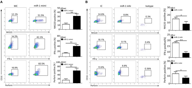
Peste des petits ruminants virus (PPRV) has emerged as a significant threat to the productivity of small ruminants worldwide. PPRV is lymphotropic in nature and induces in the hosts a transient but severe immunosuppression, especially innate immunity. However, it remains largely unknown how NK cells respond and are regulated at the earliest time points after an acute viral PPRV infection in goats. In this study, we revealed that multiple immune responses of goat peripheral NK cells were compromised during PPRV infection, including the cytolytic effector molecule expression and cytokine production. Importantly, we demonstrated that PPRV infection stimulated the expression of TWEAK, a negative regulator of cytotoxic function of NK cells, which may be involved in the suppression of cytotoxicity as well as cytokine production in infected goat NK cells. Furthermore, we found that PPRV infection induced TWEAK expression in goat NK cells involving post-transcription by suppressing miR-1, a novel negative miRNA directly targeting the TWEAK gene. Moreover, replication of virus is required for inhibition of miR-1 expression during PPRV infection, and the non-structural V protein of PPRV plays an important role in miR-1 mediated TWEAK upregulation. Additionally, we revealed that the regulation of NK cell immune responses by TWEAK is mediated by MyD88, SOCS1, and STAT3. Taken together, our results demonstrated that TWEAK may play a key role in regulating goat peripheral NK cell cytotoxicity and cytokine expression levels during PPRV infection.

Keywords: MicroRNA-1; NK cells; PPRV; TWEAK; goat.


Figures

Figure 1
Figure 1
The kinetics of virus replication in PPRV-infected goat NK cells. (A) Representative FACS plots showing cell surface expression levels of CD16+CD14 cells by PBMCs freshly obtained from goat. (B) Representative FACS plots showing freshly sorted goat CD16+CD14 cell labeled with anti-NKp46 mAb, anti-CD3 mAb, or isotype control mAb. (C) Freshly isolated goat CD16+CD14 cells were infected with PPRV at an MOI of 1 for the indicated times; the virus titers in the supernatants were measured by TCID50 assay. (D) The protein levels of PPRV-N expression in PPRV-infected CD16+CD14 NK cells at an MOI of 1 for the indicated times were measured by Western blot. The protein levels of PPRV-N were normalized to the levels of GAPDH. (E) The morphology of mock- and PPRV-infected NK cells at 48 h post-infection. Results are expressed as means ± standard error of mean (SEM). P-values were calculated using Student's t-test. An asterisk indicates a comparison with the indicated control. *P < 0.05; **P < 0.01. n.s., not significant. Scale bar = 50 μm.
Figure 2
Figure 2
NK cell expression of cytolytic effector molecules and cytokines after PPRV infection. Goat CD16+CD14 NK cells were infected with PPRV at an MOI of 1. On Day 1 post-infection, CD16+CD14 NK cells were stained for cell surface expression of NKG2D, and intracellular perforin, IFN-γ, and SLAM. (A) Representative FACS plots for NKG2D, perforin, IFN-γ, and SLAM by CD16+CD14-gated NK cells are shown. The numbers in the plots indicate the percentages of CD16+CD14 NK cells that stained for NKp46+, perforin+, IFN-γ+, and SLAM+. The protein expression of NKG2D, perforin, IFN-γ, and TNF-α (B) as well as TWEAK (C) in PPRV-infected PBMCs were measured by Western blot. The protein levels of PPRV-N were normalized to the levels of GAPDH. Results are expressed as means ± standard error of mean (SEM). P-values were calculated using Student's t-test. An asterisk indicates a comparison with the indicated control. *P < 0.05; **P < 0.01.
Figure 3
Figure 3
TWEAK suppresses NK cell cytolytic effector molecules and cytokines expression. (A,B) Goat CD16+CD14 NK cells were transfected with lentivirus control or different MOI of lentivirus TWEAK (A), or with siRNA control or different concentrations of si-TWEAK (B). Forty-eight hours later, the cells were stimulated with rh IL-12, and 24 h later, the protein levels of TWEAK were determined by Western blot. (C,D) Goat CD16+CD14 NK cells were transfected with different MOI of lentivirus TWEAK (C), different concentrations of si-TWEAK (D), or respective negative control. Forty-eight hours later, the cells were stimulated with rh IL-12, and 24 h later, the protein levels of NKG2D, perforin, IFN-γ, and TNF-α in isolated peripheral NK cells were determined by Western blot. (E–G) Goat CD16+CD14 NK cells were transfected with different MOI of lentivirus TWEAK (E), different concentrations of si-TWEAK (F), or respective negative control. Forty-eight hours later, the cells were stimulated with rh IL-12 followed by PPRV infection at an MOI of 1, and 24 h later, the expression levels of PPRV N protein and the virus titers in the supernatants of infected cells were evaluated by Western blot (E,F) and TCID50 assay (G). *P < 0.05; **P < 0.01. n.s., not significant.
Figure 4
Figure 4
miR-1 targets TWEAK in PPRV-infected goat PBMCs. (A) Heatmap of miRNAs predicted targets TWEAK in PPRV-infected goat PBMCs vs. mock-infected cells. (B) Predicted interaction between screened miRNAs and the putative binding sites in the TWEAK 3′UTRs. (C) Dual-luciferase assay of HEK293T cells transfected with luciferase constructs containing the putative chi-miR-204-3p, or the two putative miR-1-binding sites (binding site 1 or binding site 2) or a synthetic control miRNA with a scrambled sequence (miR-C). (D) Dual-luciferase assay of HEK293T cells transfected with luciferase constructs containing the wild-type 3′UTRs (3′WT UTRs) or mutated 3′UTRs (with deletion of the miR-1-responsive element) from TWEAK, plus miRNA. (E–H) Goat CD16+CD14 NK cells were infected with PPRV at an MOI of 1 for the indicated times (E,F), or at different MOIs for 24 h (G,H), and the expression of miR-1 (E,G) and TWEAK mRNA expression (F,H) were measured by qRT-PCR. *P < 0.05; **P < 0.01.
Figure 5
Figure 5
miR-1 suppresses TWEAK expression and virus replication during PPRV infection. (A–C) Goat CD16+CD14 NK cells were transfected with control miRNA (MC) or miR-1 mimics, or control inhibitor (IC) or miR-1 inhibitor, as indicated at a final concentration of 50 nM. Forty-eight hours later, the cells were infected with PPRV at an MOI of 1, and 24 h later, miR-1 (A) and TWEAK mRNA (B) expression was measured by qRT-PCR and the protein levels of TWEAK were determined by Western blot (C). (D,E) Goat CD16+CD14 NK cells were transfected with miR-1 mimics, miR-1 inhibitor, or respective negative control. Forty-eight hours later, the cells were infected with PPRV at an MOI of 1, and 24 h later, the expression levels of PPRV N protein and the virus titers in the supernatants of infected cells were evaluated by Western blot (D) and TCID50 assay (E). The levels of miR-1 were normalized to the levels of 5S. The levels of TWEAK and PPRV-N were normalized to the levels of GAPDH. Results are expressed as means ± standard error of mean (SEM). P-values were calculated using Student's t-test. An asterisk indicates a comparison with the indicated control. *P < 0.05; **P < 0.01.
Figure 6
Figure 6
miR-1 suppresses peripheral NK cell cytolytic effector molecules and cytokine expression during PPRV infection. (A,B) Goat CD16+CD14 NK cells were transfected with control miRNA (MC) or miR-1 mimics (A), or control inhibitor (IC) or miR-1 inhibitor (B), as indicated at a final concentration of 50 nM. Forty-eight hours later, the cells were infected with PPRV at an MOI of 1, and 24 h later, cells were stained for cell surface expression of NKG2D, IFN-γ, and intracellular perforin. Representative FACS plots for NKG2D, IFN-γ, and perforin by CD16+CD14 NK cells are shown. The numbers in the plots indicate the percentages of CD16+CD14 NK cells that stained for NKG2D+, IFN-γ+, and perforin+. *P < 0.05; **P < 0.01.
Figure 7
Figure 7
Replication of PPRV is required for inhibition of miR-1 expression. (A,B) Goat PBMCs were mock-infected or infected with PPRV (MOI = 1) or UV-inactivated PPRV (MOI = 1) for 72 hpi. At the end of the infection, cytopathic effect (CPE) (A) was detected and virus titers were measured by using the TCID50 method (B). (C–E) Goat CD16+CD14 NK cells were mock infected, infected with PPRV or with UV-inactivated (UV-PPRV) at an MOI of 1 for 24 h, and the levels of miR-1 (C) and TWEAK mRNA (D) were detected by qRT-PCR, while TWEAK protein expression was measured by Western blot (E). Histograms of the right panel showed densitometric analysis of indicated protein normalized to GAPDH to correct for protein loading. (F) Goat CD16+CD14 NK cells were transfected with pcDNA3.1-HA, pcDNA3.1-H-HA, pcDNA3.1-N-HA, and pCDNA3.1-V-HA plasmid for 48 h, and cell lysates from transfected cells were subjected to Western blot with antibody against HA. (G) Goat CD16+CD14 NK cells were transfected with pcDNA3.1-HA, pCDNA3.1-H-HA, pCDNA3.1-N-HA, and pCDNA3.1-V-HA plasmid for 48 h, and miR-1 expression in transfected and untransfected control cells was subjected to qRT-PCR analysis. (H,I) Goat CD16+CD14 NK cells were transfected with pcDNA3.1-HA, pCDNA3.1-H-HA, pCDNA3.1-N-HA, and pCDNA3.1-V-HA plasmid for 48 h, and the expression of TWEAK in transfected and untransfected control cells was subjected to qRT-PCR (H) and Western blot analysis (I). The levels of miR-1 were normalized to the level of 5S. GAPDH was used as a loading control in Western blot analysis. Results are expressed as means ± standard error of mean (SEM). P-values were calculated using Student's t-test. An asterisk indicates a comparison with the indicated control. *P < 0.05; **P < 0.01. n.s., not significant.
Figure 8
Figure 8
Regulation of NK cell function by miR-1 is mediated by TWEAK signaling. (A) Goat CD16+CD14 NK cells were transfected with control miRNA (MC) or miR-1 mimics, or miR-1 mimics and incubated with recombinant TWEAK at the final concentration of 50 nM. Forty-eight hours later, the cells were infected with PPRV at an MOI of 1, and 24 h later, the protein expression of NKG2D, perforin, IFN-γ, and TNF-α was measured by Western blot. Histograms of the right panel showed densitometric analysis of indicated protein normalized to GAPDH to correct for protein loading. (B) Goat CD16+CD14 NK cells were transfected with lentivirus TWEAK, si-TWEAK, or respective negative control. Forty-eight hours later, the cells were infected with PPRV at an MOI of 1, and 24 h later, the protein levels of SOCS-1, MyD88, STAT3, pSTAT3, and NF-κB p65 in NK cells were determined by Western blot and normalized to the levels of GAPDH. Results are expressed as means ± standard error of mean (SEM). P-values were calculated using Student's t-test. An asterisk indicates a comparison with the indicated control. *P < 0.05; **P < 0.01. n.s., not significant.
Figure 9
Figure 9
Proposed model for the inhibition of miR-1 expression upon PPRV infection and the role of miR-1 in the regulation of the NK cell immune responses. During PPRV infection, TWEAK expression in goat NK cells is stimulated through suppressing miR-1, which in turn suppresses NK cell cytotoxicity and cytokine expression in a SOCS1 and NF-κB-dependent manner. The non-structural V protein of PPRV plays an important role in miR-1-mediated TWEAK upregulation.

References

    1. Banyard AC, Parida S, Batten C, Oura C, Kwiatek O, Libeau G. Global distribution of peste des petits ruminants virus and prospects for improved diagnosis and control. J Gen Virol. (2010) 91:2885–97. 10.1099/vir.0.025841-0 - DOI - PubMed
    1. Baron MD, Diallo A, Lancelot R, Libeau G. Peste des petits ruminants virus. Adv Virus Res. (2016) 95:1–42. 10.1016/bs.aivir.2016.02.001 - DOI - PubMed
    1. Kumar N, Maherchandani S, Kashyap SK, Singh SV, Sharma S, Chaubey KK, et al. . Peste des petits ruminants virus infection of small ruminants: a comprehensive review. Viruses. (2014) 6:2287–327. 10.3390/v6062287 - DOI - PMC - PubMed
    1. Jagtap SP, Rajak KK, Garg UK, Sen A, Bhanuprakash V, Sudhakar SB, et al. . Effect of immunosuppression on pathogenesis of peste des petits ruminants (PPR) virus infection in goats. Microb Pathog. (2012) 52:217–26. 10.1016/j.micpath.2012.01.003 - DOI - PubMed
    1. Heaney J, Barrett T, Cosby SL. Inhibition of in vitro leukocyte proliferation by morbilliviruses. J Virol. (2002) 76:3579–84. 10.1128/JVI.76.7.3579-3584.2002 - DOI - PMC - PubMed

Publication types