Review
. 2019 Jul 19;1(4):312-328.
doi: 10.1016/j.jhepr.2019.07.002. eCollection 2019 Oct.

From NASH to diabetes and from diabetes to NASH: Mechanisms and treatment options

Affiliations
Review

From NASH to diabetes and from diabetes to NASH: Mechanisms and treatment options

Amalia Gastaldelli et al. JHEP Rep. .

Abstract

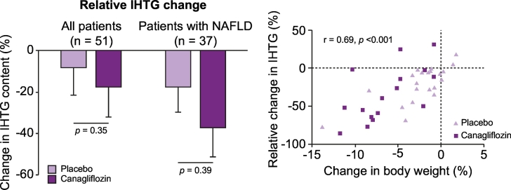
The worldwide prevalence of non-alcoholic fatty liver disease (NAFLD) is estimated to have reached 25% or more in adults. NAFLD is prevalent in obese individuals, but may also affect non-obese insulin-resistant individuals. NAFLD is associated with a 2- to 3-fold increased risk of developing type 2 diabetes (T2D), which may be higher in patients with more severe liver disease - fibrosis increases this risk. In NAFLD, not only the close association with obesity, but also the impairment of many metabolic pathways, including decreased hepatic insulin sensitivity and insulin secretion, increase the risk of developing T2D and related comorbidities. Conversely, patients with diabetes have a higher prevalence of steatohepatitis, liver fibrosis and end-stage liver disease. Genetics and mechanisms involving dysfunctional adipose tissue, lipotoxicity and glucotoxicity appear to play a role. In this review, we discuss the altered pathophysiological mechanisms that underlie the development of T2D in NAFLD and vice versa. Although there is no approved therapy for the treatment of NASH, we discuss pharmacological agents currently available to treat T2D that could potentially be useful for the management of NASH.


Figures

Fig. 1
Fig. 1
Natural history of T2D in the context of NAFLD. T2D results from an imbalance between insulin sensitivity and insulin secretion. In the progression from normal to impaired glucose tolerance to T2D, insulin secretion increases to overcome insulin resistance. At the same time there is a decrease in insulin clearance (mainly hepatic), especially in individuals with NAFLD, that determines higher peripheral insulin concentrations. However, despite high concentrations, insulin secreted is insufficient (beta-cell dysfunction) and therefore there is an increase in both fasting and postprandial glucose concentrations. Subjects become T2D only when the beta cells are unable to increase insulin secretion (beta-cell failure) and overcome peripheral IR. Redrawn from ref ,,. IR, insulin resistance; NAFLD, non-alcoholic fatty liver disease; NGT, normal glucose tolerance; T2D, type 2 diabetes
Fig. 2
Fig. 2
Insulin resistance causes excess substrate (amino acids, glycerol, FFAs) to be transported to the liver. Excess substrate (amino acids, glycerol, FFAs) stimulate GNG, glucose fluxes, de novo glycerol synthesis, TG synthesis and DNL. Moreover, the synthesis of lipotoxic compounds like DAGs and ceramides is enhanced. Modified from . DAGs, diacylglycerols; DNL, de novo lipogenesis; FFAs, free fatty acids; GNG, gluconeogenesis; TG, triglyceride; VLDL, very low-density lipoprotein.
Fig. 3
Fig. 3
Relationship between adipose tissue insulin resistance and dysfunction and insulin resistance in liver and muscle. Dysfunctional adipose tissue displays resistance to the antilipolytic effect of insulin with increased lipolysis and release of FFAs and glycerol that in turn are responsible for triglyceride accumulation and lipotoxicity in the liver, muscle and pancreas, impairing insulin secretion. Moreover, dysfunctional adipose tissue releases adipokines that activate pro-inflammatory pathways in these organs. FFAs, free fatty acids.
Fig. 4
Fig. 4
Relationship between lipo- and glucotoxicity, insulin resistance and beta-cell function. Both lipotoxicity and glucotoxicity contribute to insulin resistance, ectopic fat accumulation and beta-cell dysfunction and vice versa. In NAFLD, lipotoxicity and insulin resistance have been recognised as pathophysiological mechanisms responsible of development and progression to a more severe form of this disease. T2D is a chronic condition of glucotoxicity, although also lipotoxicity is often present and are responsible not only of insulin resistance but also of impaired insulin secretion. DAG, diacylglycerol; DNL, de novo lipogenesis; NAFLD, non-alcoholic fatty liver disease; ROS, reactive oxygen species; T2D, type 2 diabetes.
Fig. 5
Fig. 5
Effect of SGLT2 inhibitor on IHTG and body weight. Left panel: Effect of the SGLT2 inhibitor canagliflozin vs. placebo on relative IHTG content among all patients (left) or only patients with a diagnosis of NAFLD (right) at baseline. Right panel: Relationship between changes in body weight after canagliflozin or placebo treatment and change in IHTG. Reproduced with permission from . IHTG, intrahepatic triglyceride; NAFLD, non-alcoholic fatty liver disease.

References

    1. Younossi ZM, Koenig AB, Abdelatif D, Fazel Y, Henry L, Wymer M. Global epidemiology of nonalcoholic fatty liver disease-Meta-analytic assessment of prevalence, incidence, and outcomes. Hepatology. 2016;64:73–84. - PubMed
    1. EASL, Marchesini G, Day CP, Dufour J-F, Canbay A, Nobili V. EASL-EASD-EASO Clinical Practice Guidelines for the management of non-alcoholic fatty liver disease. J Hepatol. 2016;64:1388–1402. - PubMed
    1. Anstee QM, Targher G, Day CP. Progression of NAFLD to diabetes mellitus, cardiovascular disease or cirrhosis. Nat Rev Gastroenterol Hepatol. 2013;10:330–344. - PubMed
    1. Mantovani A, Byrne CD, Bonora E, Targher G. Nonalcoholic Fatty Liver Disease and Risk of Incident Type 2 Diabetes: A Meta-analysis. Diabetes Care. 2018;41:372–382. - PubMed
    1. Yki-Jarvinen H. Non-alcoholic fatty liver disease as a cause and a consequence of metabolic syndrome. Lancet Diabetes Endocrinol. 2014;2:901–910. - PubMed