. 2020 May;161(5):949-959.
doi: 10.1097/j.pain.0000000000001781.

Recovery from nerve injury induced behavioral hypersensitivity in rats parallels resolution of abnormal primary sensory afferent signaling

Affiliations

Recovery from nerve injury induced behavioral hypersensitivity in rats parallels resolution of abnormal primary sensory afferent signaling

M Danilo Boada et al. Pain. 2020 May.

Abstract

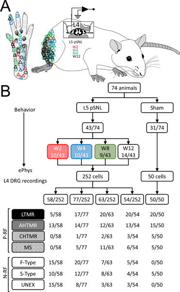
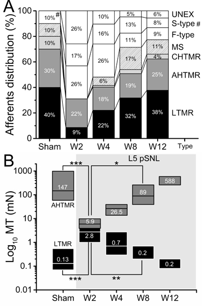
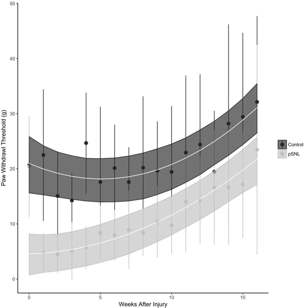
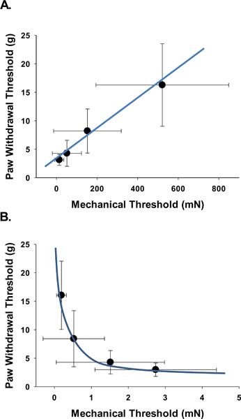
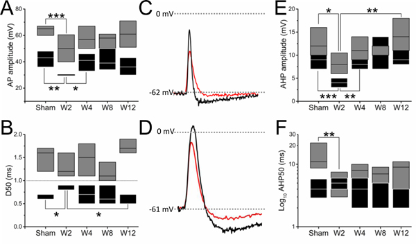
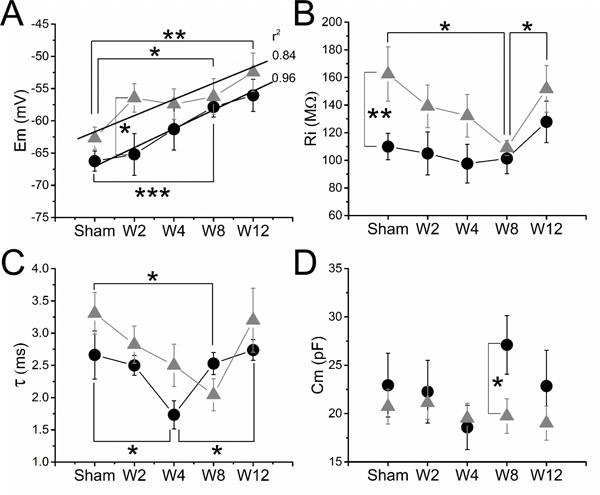
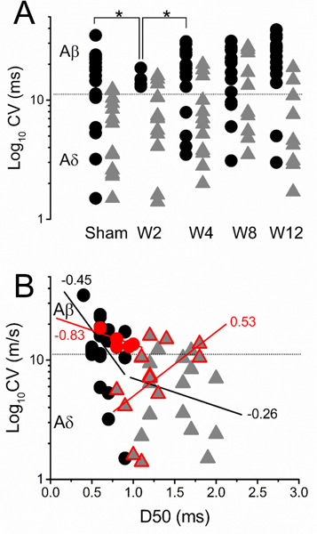
Pain and hypersensitivity months after peripheral injury reflect abnormal input from peripheral afferents likely in conjunction with central sensitization. We hypothesize that peripheral changes occur in defined sensory afferents and resolve as behavioral response to injury resolves. Male Sprague-Dawley rats underwent sham or partial L5 spinal nerve ligation, and paw withdrawal threshold (PWT) was sequentially measured during recovery. At 2, 4, 8, and 12 weeks after injury, randomized animals underwent electrophysiologic assessment of L4 fast-conducting high- and low-threshold mechanoreceptors, and individual neuronal mechanical thresholds (MTs) were contrasted with PWTs in the same animals. Paw withdrawal thresholds decreased after injury and resolved over time (P < 0.001). Similarly, MTs of fast-conducting high-threshold mechanoreceptors decreased after injury and resolved over time (P < 0.001). By contrast, MTs of low-threshold mechanoreceptors increased after injury and resolved over time (P < 0.001). Distributions of recordings from each afferent subtype were perturbed after injury, and this too resolved over time. After resolution of behavioral changes, several electrical abnormalities persisted in both neuronal subtypes. These data extend previous findings that mechanically sensitive nociceptors are sensitized, whereas tactile, largely Aβ afferents are desensitized after nerve injury by showing that the time course of resolution of these changes mirrors that of behavioral hypersensitivity in a surgical injury including neural damage. These data support a role of abnormal peripheral input, from both nociceptor and tactile afferents, during recovery from peripheral injury and underscore the potential importance of both classes of afferents as potential targets for pain treatment.


Conflict of interest statement

The other have no conflicts of interest to report.

Figures

Figure 1.
Figure 1.
A. Schematic diagram of the in vivo ePhys rat L4 preparation (lateral view) after pSNL in L5. Diagram of lateral flank illustrates the areas where the neuronal receptive field (RF) of the studied afferents were found in hairy skin and glabrous skin (Circles: tactile; Triangles: nociceptors) over time (red: week 2; blue: week 4; green: week 8 and black: week 12). B. Flowchart of animals studied and classification of the neurons included in the study per week (W): with mechanical RF (P-RF): low threshold mechanoreceptors (LTMR, black), high threshold mechanoreceptor with A fibers (AHTMR, dark gray), high threshold mechanoreceptor with C fibers (CHTMR, light gray) muscular spindle (MS) and without RF (N-RF): fast AP dynamic mechanically unresponsive (F-type), slow AP dynamics mechanically unresponsive (S-type), unresponsive (mechanically and electrically) (UNEX).
Figure 2.
Figure 2.
A. Effect of L5-pSNL in the L4 percent distribution of mechanically sensitive (LTMR, AHTMR, CHTMR and MS) and insensitive (F-type, S-type and UNEX) afferents over time (weeks: W) after L5-pSNL (#: S-type). There is a significant difference in distribution of N-RF and P-RF cells (p<0.001). The distribution is significantly different at 2 weeks after pSNL compared to control and remains different through 8 weeks (p=0.02), but is no different from control at 12 weeks after pSNL (p=0.09). B. Effects of the L5-pSNL in the mechanical sensitivity of L4 afferents over time (W). Numbers indicate medians (on top), with boxes representing the 25 and 75 percentiles. LTMR: low-threshold mechanoreceptor and AHTMR: A-fiber high-threshold mechanoreceptor. (*=p<0.05; **=p<0.01; ***=p<0.001).
Figure 3.
Figure 3.
Trajectory of L5-pSNL on paw withdrawal threshold (PWT) over time. The trajectory of the PWT recovery is nonlinear with a significant difference for the change trajectories between the sham and pSNL groups (p < 0.0001). Both sham and pSNL groups have a PWT that is not different at baseline. After the pSNL there is an immediate decrease in PWT (*, p<0.001). The difference in PWT persists through 13 weeks after the injury.
Figure 4.
Figure 4.
Correlation of single neuronal mechanical thresholds (MT) and behavior paw withdrawal thresholds (PWT). There is a negative correlation between LTMR MT and PWT with LTMR MT desensitizing (increasing thresholds) with sensitization (decreased PWT) (Spearman non-parametric correlation, r = −0.32, p = 0.012). At the same time there is a positive correlation between the AHTMR MT and the PWT with the AHTMR sensitizing (decreasing thresholds) with sensitization (decreasing PWT) (Spearman non-parametric correlation, r = 0.35, p = 0.011). The correlations for AHTMR MT and LTMR MT with PWT follow a non-linear pattern when compared to the AHTMR MT to PWT (blue lines).
Figure 5.
Figure 5.
Effect of L5-pSNL in the L4 mechanosensitive afferent active electrical signature of evoked action potentials over time in weeks (W): spike ((A) amplitude [AP amplitude] and (B) duration [AP duration; D50]) and AHP: ((E) amplitude and (F) duration [AHP50]). Representative traces of these effects are presented (black sham and red W2) in tactile (C) and nociceptive (D) afferents that demonstrate the changes after injury. Boxes represents 25 and 75 percentiles (black: LTMR; gray: AHTMR). (*=p<0.05; **=p<0.01; ***=p<0.001).
Figure 6.
Figure 6.
Effect of L5-pSNL in the L4 mechanosensitive afferents passive electrical properties over time in weeks (W): (A) membrane potential (Em), (B) input resistance (Ri), (C) time constant (τ) and (D) membrane capacitance (Cm). Data are medians ± SE. Regression lines and r2 values are shown for Em in LTMRs (black circles) and AHTMR (gray triangles) (*=p<0.05; **=p<0.01; ***=p<0.001).
Figure 7.
Figure 7.
A. Effect of L5-pSNL in the L4 mechanosensitive afferents CV (black circles: LTMR and gray triangles: AHTMR) over time and B. its effects on the relationship between CV and the spike duration (D50) (linear regression and r2 values) two weeks after injury (red circles: LTMR and red triangles: AHTMR). *=p<0.05.

References

    1. Arcourt A, Gorham L, Dhandapani R, Prato V, Taberner FJ, Wende H, Gangadharan V, Birchmeier C, Heppenstall PA, Lechner SG. Touch Receptor-Derived Sensory Information Alleviates Acute Pain Signaling and Fine-Tunes Nociceptive Reflex Coordination. Neuron 2017;93(1):179–193. - PubMed
    1. Bennett DL, Clark AJ, Huang J, Waxman SG, Dib-Hajj SD. The Role of Voltage-Gated Sodium Channels in Pain Signaling. Physiol Rev 2019;99(2):1079–1151. - PubMed
    1. Boada MD, Eisenach JC, Ririe DG. Mechanical sensibility of nociceptive and non-nociceptive fast-conducting afferents is modulated by skin temperature. J Neurophysiol 2016;115(1):546–553. - PMC - PubMed
    1. Boada MD, Gutierrez S, Aschenbrenner CA, Houle TT, Hayashida K, Ririe DG, Eisenach JC. Nerve injury induces a new profile of tactile and mechanical nociceptor input from undamaged peripheral afferents. J Neurophysiol 2015;113(1):100–109. - PMC - PubMed
    1. Boada MD, Gutierrez S, Giffear K, Eisenach JC, Ririe DG. Skin incision-induced receptive field responses of mechanosensitive peripheral neurons are developmentally regulated in the rat. J Neurophysiol 2012;108(4):1122–1129. - PMC - PubMed

Publication types