. 2020 Mar 2;217(4):1.
doi: 10.1084/jem.20191290.

Tumor-derived soluble CD155 inhibits DNAM-1-mediated antitumor activity of natural killer cells

Affiliations

Tumor-derived soluble CD155 inhibits DNAM-1-mediated antitumor activity of natural killer cells

Genki Okumura et al. J Exp Med. .

Abstract

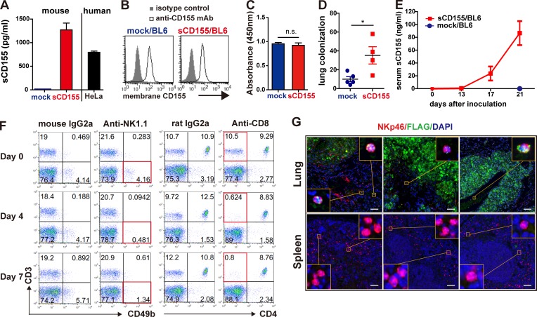
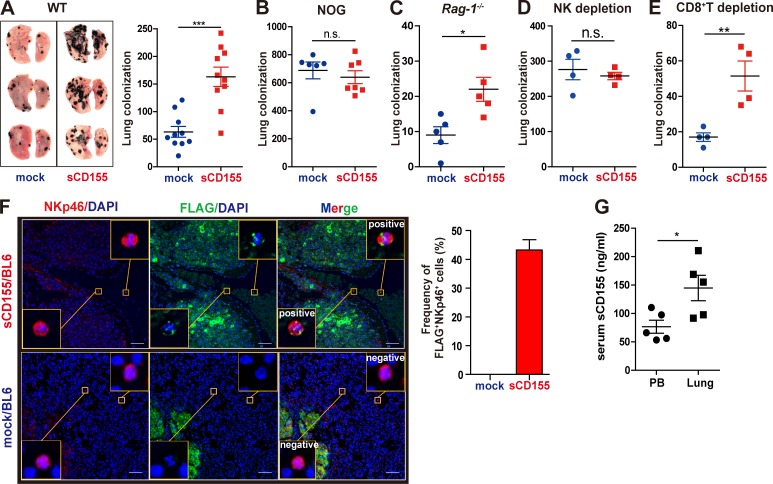
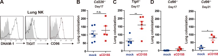
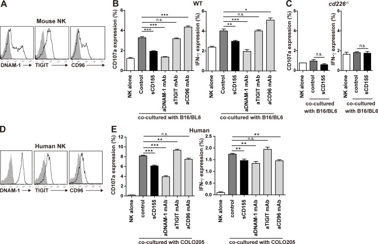
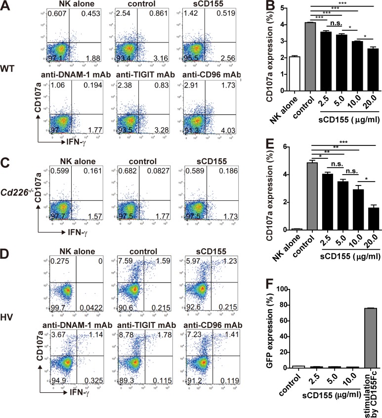
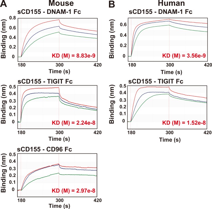
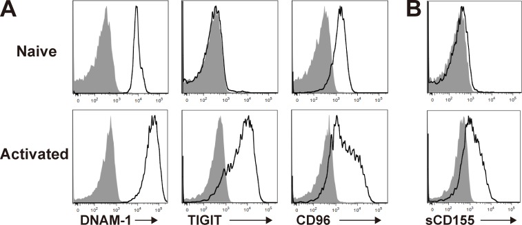
CD155 is a ligand for DNAM-1, TIGIT, and CD96 and is involved in tumor immune responses. Unlike mouse cells, human cells express both membranous CD155 and soluble CD155 (sCD155) encoded by splicing isoforms of CD155. However, the role of sCD155 in tumor immunity remains unclear. Here, we show that, after intravenous injection with sCD155-producing B16/BL6 melanoma, the numbers of tumor colonies in wild-type (WT), TIGIT knock-out (KO), or CD96 KO mice, but not DNAM-1 KO mice, were greater than after injection with parental B16/BL6 melanoma. NK cell depletion canceled the difference in the numbers of tumor colonies in WT mice. In vitro assays showed that sCD155 interfered with DNAM-1-mediated NK cell degranulation. In addition, DNAM-1 had greater affinity than TIGIT and CD96 for sCD155, suggesting that sCD155 bound preferentially to DNAM-1. Together, these results demonstrate that sCD155 inhibits DNAM-1-mediated cytotoxic activity of NK cells, thus promoting the lung colonization of B16/BL6 melanoma.


Conflict of interest statement

Disclosures: The authors declare no competing interests exist.

Figures

Figure S1.
Figure S1.
Characterization of generated sCD155/BL6 and mock/BL6, and serum levels of sCD155. (A) Concentrations of mouse and human sCD155 in the culture supernatant of sCD155/BL6 (n = 3), mock/BL6 (n = 3), and HeLa (n = 3) were analyzed 24 h after the start of the culture by CBA assay and ELISA, respectively. (B) Expression of membrane-bound CD155 on sCD155/BL6 and mock/BL6 was analyzed by using flow cytometry. (C) sCD155/BL6 (n = 3) and mock/BL6 (n = 3) were cultured (1.0 × 10⁵ cells/well) in 96-well flat plates for 24 h, and then BrdU reagent was added to the cultures. BrdU incorporation was measured after culture for 12 h. (D) C57BL/6 WT mice were intravenously injected with different clones of sCD155/BL6 (n = 4) and mock/BL6 (n = 5) from those used in Fig. 1. Colony numbers in the lung were counted on day 17. (E) C57BL/6 WT mice were intravenously injected with sCD155/BL6 (n = 5) or mock/BL6 (n = 5) used in Fig. 1 and Fig. 2, and analyzed for serum levels of sCD155 on days 0, 13, 17, and 21. (F) C57BL/6 WT mice were treated with mouse IgG2a, anti-NK1.1 antibody, rat IgG2a, or anti-CD8 antibody. Peripheral blood mononuclear cells on days 0, 4, and 7 were stained with antibodies against CD3, CD49b, and/or CD4. (G) C57BL/6 WT mice were intravenously injected with sCD155/BL6 or mock/BL6. Paraffin sections of lungs with colonized tumor and spleen on day 17 were stained as described in Fig. 1 F. Scale bars, 50 µm. Error bars indicate SD. Results were analyzed by using Student’s t test. For all analyses: *, P < 0.05; n.s., not significant.
Figure 1.
Figure 1.
sCD155 suppresses NK cell function against lung colonization of B16/BL6 melanoma. (A–C) C57BL/6 WT (n = 10 in each group), NOG (n = 7 and 6 for sCD155/BL6 and mock/BL6, respectively), or Rag-1−/− (n = 5 in each group) mice were intravenously inoculated with sCD155/BL6 or mock/BL6. Lung metastases were quantified by counting metastatic foci on the lung surface on day 17 (A and C) and day 12 (B). Representative images of lungs with metastases are shown (A). Results of three (A) or two (B and C) independent experiments were pooled for statistical analyses. (D and E) NK cells and CD8+ T cells were depleted from C57BL/6 WT mice by intraperitoneal administration of anti-NK1.1 mAb or anti-CD8 mAb. These mice (n = 4 in each group) were intravenously inoculated with sCD155/BL6 or mock/BL6 on day 0. Lung metastases were quantified by counting metastatic foci on the lung surface on day 17. Data are representative of two independent experiments. (F) C57BL/6 mice were intravenously inoculated with sCD155/BL6 or mock/BL6. Paraffin sections of lung with metastases were stained for FLAG (sCD155-FLAG; green) and NKp46 (red) by using the Opal multiplex immunofluorescence staining with DAPI (blue). Note that mock/BL6 also express Flag. Slides were then observed in a Mantra Quantitative Pathology Workstation. Representative FLAG-positive and -negative images are shown in the yellow squares. Scale bars, 50 µm. For the bar graph, the frequencies (%) of FLAG+ NKp46+ cells among NKp46+ cells in three lungs each for the sCD155/BL6 and mock/BL6 treatments were quantified. (G) Serum levels of sCD155 in peripheral blood (PB) and pulmonary vein of mice were analyzed on day 17 after injection of sCD155/BL6 (n = 5 in each group). Results were analyzed by using Student’s t test. Error bars indicate SD. For all analyses: *, P < 0.05; **, P < 0.01; ***, P < 0.001; n.s., not significant.
Figure 2.
Figure 2.
sCD155 interferes with DNAM-1–mediated immunity against lung metastasis of B16/BL6 melanoma. (A) C57BL/6 mice were intravenously inoculated with B16/BL6. On day 17, lung metastatic foci were resected under a stereo microscope. The resected tissues were mashed and passed through a mesh to prepare a single-cell suspension. Expression of DNAM-1, TIGIT, and CD96 on tumor-infiltrating NK cells was analyzed by using flow cytometry. (B–D) C57BL/6 Cd226−/− (n = 6 and 5 for sCD155/BL6 and mock/BL6, respectively), Tigit−/− (n = 6 and 5 for sCD155/BL6 and mock/BL6, respectively), and Cd96−/− (n = 3 in each group) mice were intravenously inoculated with sCD155/BL6 (sCD155) or mock/BL6 (mock). WT control that were performed in parallel are shown in Fig 1 A. Lung metastases were quantified by counting metastatic foci on the lung surface on day 17 (B–D, left). In the case of Cd96−/− mice, lung metastases were also counted on day 21 (D, right). Results of two independent experiments were pooled for statistical analysis (B and C). Data are representative of two independent experiments (D). Error bars indicate SD. Results were analyzed by using Student’s t test. For all analyses: *, P < 0.05; **, P < 0.01; n.s., not significant.
Figure 3.
Figure 3.
sCD155 suppresses DNAM-1–mediated cytotoxic activity of NK cells. (A) Mouse NK cells were isolated from the spleens of C57BL/6 WT mice and cultured with IL-2 for 5 d. Expression of DNAM-1, TIGIT, and CD96 on IL-2–activated mouse NK cells was analyzed by using flow cytometry. The shaded histogram indicates isotype control, and the open histogram indicates each specific antibody. (B and C) IL-2–activated mouse WT or Cd226−/− NK cells were co-cultured with B16/BL6 (E:T = 1:1) in the presence of sCD155 (10 μg/ml), anti-mouse DNAM-1 mAb, anti-mouse TIGIT mAb, anti-mouse CD96 mAb, or isotype control. CD107a and IFN-γ expression was analyzed. (D) Human DNAM-1, TIGIT, and CD96 expression on peripheral blood NK cells isolated from a healthy donor was analyzed by using flow cytometry. Representative histograms are shown. The shaded histogram indicates isotype control, and the open histogram indicates each specific antibody. (E) Human NK cells were co-cultured with COLO 205 (E:T = 1:1) in the presence of sCD155 (10 μg/ml), anti-human DNAM-1 mAb, anti-human TIGIT mAb, anti-human CD96 mAb, or isotype control. CD107a and IFN-γ expression was analyzed. Error bars indicate SD. Results were analyzed by using Student’s t test. For all analyses: *, P < 0.05; **, P < 0.01; ***, P < 0.001; n.s., not significant.
Figure S2.
Figure S2.
sCD155 suppressed mouse and human NK cell activation in a DNAM-1–dependent manner. (A and C) IL-2–activated mouse NK cells derived from WT (A) and Cd226−/− (C) mice were co-cultured with B16/BL6 (E:T = 1:1) in the presence of sCD155; mAbs against mouse DNAM-1, mouse TIGIT, or mouse CD96; or isotype control and analyzed for CD107a and IFN-γ expression. (D) Human NK cells isolated from PBMC in a healthy volunteer (HV) were co-cultured with COLO 205 (E:T = 1:1) in the presence of sCD155; mAbs against human DNAM-1, human TIGIT, or human CD96; or isotype control and analyzed for CD107a and IFN-γ expression. (B and E) IL-2–activated mouse NK cells (B) and human NK cells (E) were co-cultured with B16/BL6 (E:T = 1:1) and COLO 205 (E:T = 1:1) in the presence of mouse and human sCD155, respectively, or BSA (control) and analyzed for CD107a expression. (F) Mouse DNAM-1–GFP reporter cells were cultured in the presence of sCD155 or BSA (control) or stimulated with plate-coated mouse CD155-Fc fusion protein and analyzed for GFP expression by flow cytometry. Data are representative of two experiments (A, C, and D). Error bars indicate SD. Results were analyzed by using Student’s t test. For all analyses: *, P < 0.05; **, P < 0.01; ***, P < 0.001; n.s., not significant.
Figure 4.
Figure 4.
DNAM-1 is the molecule with the greatest affinity for sCD155. (A) Mouse His-tagged sCD155 was coated onto a Ni-NTA biosensor and reacted with the analytes indicated. Mouse DNAM-1–Fc fusion protein (Fc) was used at 125 nM (red), 62.5 nM (blue), or 31.25 nM (green). Mouse TIGIT-Fc was used at 307 nM (red), 153.5 nM (blue), or 76.75 nM (green). Mouse CD96-Fc was used at 762 nM (red), 381 nM (blue), or 190.5 nM (green). Antibody affinity was measured in the global analysis mode of the BLItz system. Quantified affinities (KD) are shown in red font. (B) Human His-tagged sCD155 was coated onto a Ni-NTA biosensor and reacted with the analytes indicated. Human DNAM-1–Fc was used at 500 nM (red), 250 nM (blue), or 125 nM (green). Human TIGIT-Fc was used at 615 nM (red), 307.5 nM (blue), or 153.75 nM (green). Antibody affinity was measured in the global analysis mode of the BLItz system. Quantified affinities (KD) are shown in red font.
Figure S3.
Figure S3.
sCD155 binds to activated CD8+ T cells. (A) DNAM-1, TIGIT, and CD96 expression on spleen CD8+ T cells stimulated with or without plate-coated anti-CD3 mAb and soluble anti-CD28 mAb in the presence of IL-2 was analyzed by using flow cytometry. The shaded histogram indicates isotype control, and the open histogram indicates each specific antibody. (B) sCD155 binding to CD8+ T cells. The naive (upper) or activated (bottom) CD8+ T cells were incubated with recombinant mouse CD155-His and then were stained with PE-conjugated anti-His mAb. The shaded histogram indicates isotype control, and the open histogram indicates anti-His mAb staining.

Similar articles

Cited by

References

    1. Baury B., Masson D., McDermott B.M. Jr., Jarry A., Blottière H.M., Blanchardie P., Laboisse C.L., Lustenberger P., Racaniello V.R., and Denis M.G.. 2003. Identification of secreted CD155 isoforms. Biochem. Biophys. Res. Commun. 309:175–182. 10.1016/S0006-291X(03)01560-2 - DOI - PubMed
    1. Bevelacqua V., Bevelacqua Y., Candido S., Skarmoutsou E., Amoroso A., Guarneri C., Strazzanti A., Gangemi P., Mazzarino M.C., D’Amico F., et al. . 2012. Nectin like-5 overexpression correlates with the malignant phenotype in cutaneous melanoma. Oncotarget. 3:882–892. 10.18632/oncotarget.594 - DOI - PMC - PubMed
    1. Blake S.J., Stannard K., Liu J., Allen S., Yong M.C., Mittal D., Aguilera A.R., Miles J.J., Lutzky V.P., de Andrade L.F., et al. . 2016. Suppression of metastases using a new lymphocyte checkpoint target for cancer immunotherapy. Cancer Discov. 6:446–459. 10.1158/2159-8290.CD-15-0944 - DOI - PubMed
    1. Bottino C., Castriconi R., Pende D., Rivera P., Nanni M., Carnemolla B., Cantoni C., Grassi J., Marcenaro S., Reymond N., et al. . 2003. Identification of PVR (CD155) and Nectin-2 (CD112) as cell surface ligands for the human DNAM-1 (CD226) activating molecule. J. Exp. Med. 198:557–567. 10.1084/jem.20030788 - DOI - PMC - PubMed
    1. Cuff S., Dolton G., Matthews R.J., and Gallimore A.. 2010. Antigen specificity determines the pro- or antitumoral nature of CD8+ T cells. J. Immunol. 184:607–614. 10.4049/jimmunol.0804089 - DOI - PubMed

Publication types

Substances