. 2020 Apr;153(2):216-229.
doi: 10.1111/jnc.14981. Epub 2020 Mar 2.

Complex sex and estrous cycle differences in spontaneous transient adenosine

Affiliations

Complex sex and estrous cycle differences in spontaneous transient adenosine

Jason R Borgus et al. J Neurochem. 2020 Apr.

Abstract

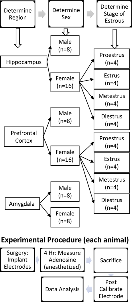
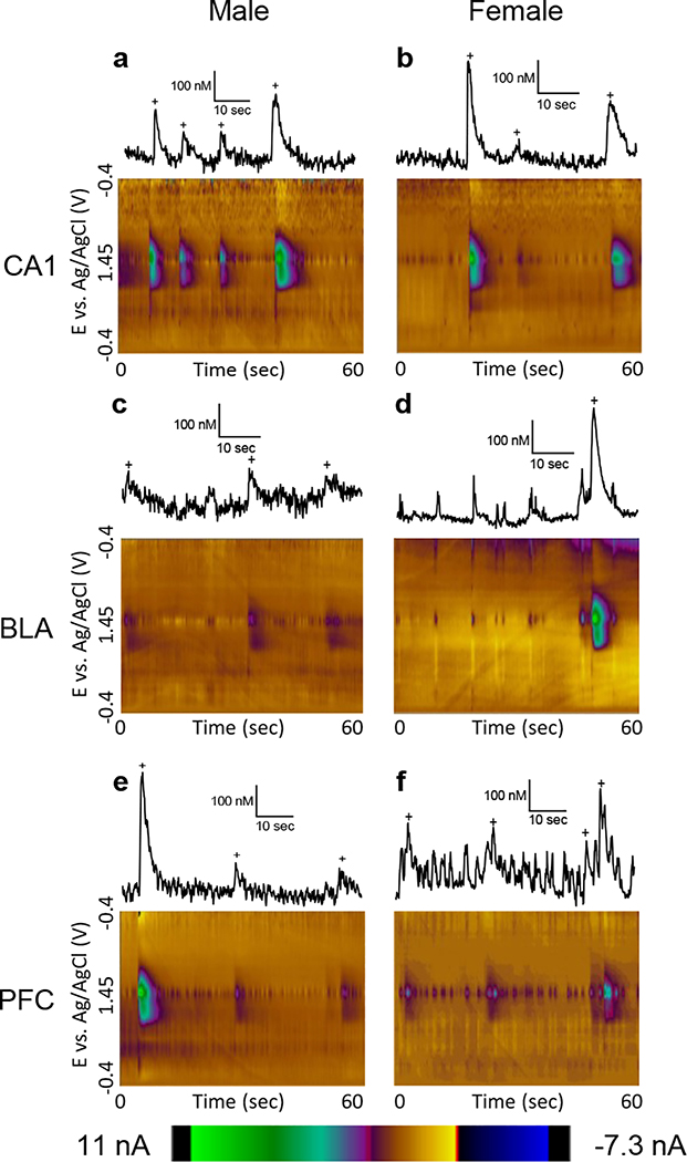
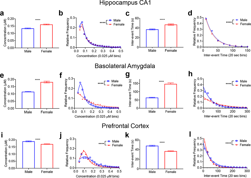
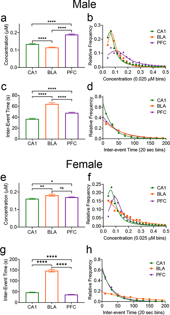
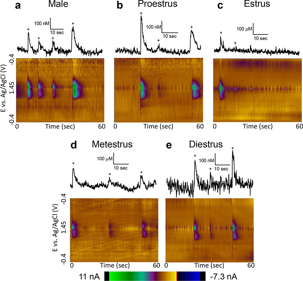
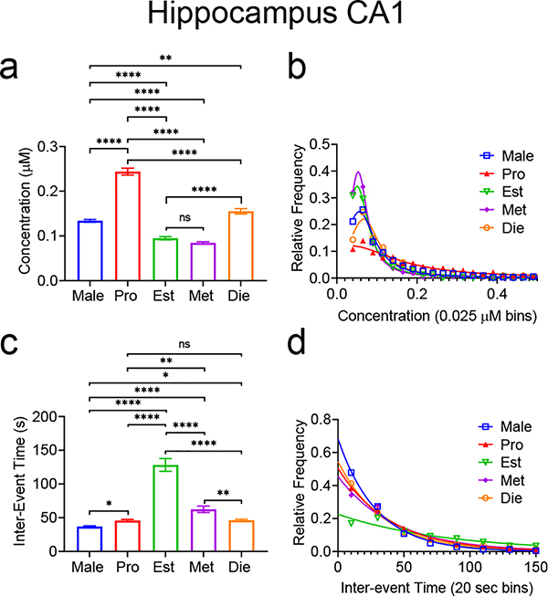
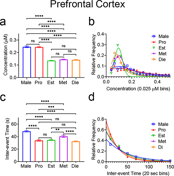
Adenosine is a ubiquitous neuromodulator that plays a role in sleep, vasodilation, and immune response and manipulating the adenosine system could be therapeutic for Parkinson's disease or ischemic stroke. Spontaneous transient adenosine release provides rapid neuromodulation; however, little is known about the effect of sex as a biological variable on adenosine signaling and this is vital information for designing therapeutics. Here, we investigate sex differences in spontaneous, transient adenosine release using fast-scan cyclic voltammetry to measure adenosine in vivo in the hippocampus CA1, basolateral amygdala, and prefrontal cortex. The frequency and concentration of transient adenosine release were compared by sex and brain region, and in females, the stage of estrous. Females had larger concentration transients in the hippocampus (0.161 ± 0.003 µM) and the amygdala (0.182 ± 0.006 µM) than males (hippocampus: 0.134 ± 0.003, amygdala: 0.115 ± 0.002 µM), but the males had a higher frequency of events. In the prefrontal cortex, the trends were reversed. Males had higher concentrations (0.189 ± 0.003 µM) than females (0.170 ± 0.002 µM), but females had higher frequencies. Examining stages of the estrous cycle, in the hippocampus, adenosine transients are higher concentration during proestrus and diestrus. In the cortex, adenosine transients were higher in concentration during proestrus, but were lower during all other stages. Thus, sex and estrous cycle differences in spontaneous adenosine are complex, and not completely consistent from region to region. Understanding these complex differences in spontaneous adenosine between the sexes and during different stages of estrous is important for designing effective treatments manipulating adenosine as a neuromodulator.

Keywords: FSCV; adenosine; estrous cycle; fast scan cyclic voltammetry; in vivo; sex differences.


Conflict of interest statement

conflict of interest disclosure

The authors declare no competing financial interests.

Figures

Figure 1.
Figure 1.. Flow chart of experimental procedure and groups for animal experiments.
The experimenter decided the region that would be studied and the sex of the animal was recorded. If the animal was female, stage of estrous was determined by vaginal swab after the animal had been anesthetized. Surgery was performed under urethane anesthesia and the electrodes implanted. Spontaneous adenosine was measured for 4 hr. under anesthesia in the selected brain region. The animal was sacrificed by decapitation and a post experiment calibration of the electrode performed. The data were analyzed by software to find the adenosine transients and then statistical analysis performed.
Figure 2.
Figure 2.. Example data from male and female rats from each brain region.
Concentration traces (top) and 3D color plots (bottom) compare adenosine release in the CA1 region of the hippocampus in (a) male) and (b) female rats, the basolateral amygdala in (c) male and (d) female rats, and the prefrontal cortex in (e) male and (f) female rats. Adenosine transients are marked with plus (+) signs in the concentration traces, which are all scaled the same to highlight the varying concentrations.
Figure 3.
Figure 3.. Comparison of spontaneous adenosine between males and females in the hippocampus, basolateral amygdala, and prefrontal cortex.
Mean transient concentrations (a) in females were significantly larger in the hippocampus (Welch’s t-test, n=3080 M/4639 F transients, p<0.0001, n is same for panels a-d) and (b) the distribution of transient concentrations was wider in the females (K-S test, p<0.0001). (c) Mean inter-event time was significantly longer in females in the hippocampus (Welch’s t-test, p<0.0001) (d) and the distribution of inter-event times was significantly wider in females (K-S test, p<0.0001). In the basolateral amygdala, the same trends are observed where (e) mean concentration (Welch’s t-test, n=1934 M/1057 F transients, p<0.0001, n is same for panels e-h) is significantly larger in females and the (f) frequency distribution is wider in females (K-S test, p<0.0001). The BLA (g) inter-event time is also significant longer in females (Welch’s t-test, p<0.0001) and (h) the distribution of inter-event times is wider in females (K-S test, p<0.0001). In the prefrontal cortex, the trend reverses. (i) Mean transient concentrations were significantly larger in males (Welch’s t-test, n=2393 M/6233 F transients, p<0.0001, n is same for panels i to l) and (j) the distribution was wider for males (K-S test, p<0.0001), while (k) inter-event time (Welch’s t-test, p<0.0001) was significantly longer in males with (l) the distribution was wider for males (K-S test, p<0.0001). All error bars are SEM. **** p<0.0001
Figure 4.
Figure 4.. Comparison of spontaneous adenosine transients in the hippocampus, basolateral amygdala, and prefrontal cortex in male (top, a-d) and female (bottom, e-h) rats.
(a) In males, there was an overall effect of region on the mean transient concentrations and all pairs of columns were significantly different from each other (One-way ANOVA, p<0.0001 Tukey’s multiple comparisons test, significance marked, n=3080 CA1/2393 PFC/1934 BLA transients, n is the same for all statistics, panels a-d). (b) There was a main effect of region on distribution of concentrations, and comparisons between individual regions were significant except between the CA1 and the BLA (Kruskal-Wallace test, p<0.0001, CA1-PFC & PFC-BLA p<0.0001). (c) There was a significant effect of region on inter-event time in males, with significant differences between all columns (One-way ANOVA, p<0.0001, Tukey’s multiple comparisons test, p<0.0001). (d) There was a significant main effect of frequency distribution in males, with all regions being significantly different (Kruskal-Wallace test, p<0.0001). (e) In females, there is a significant main effect of region on transient concentration and the concentration differed between the CA1-BLA and CA1-PFC, but not the PFC-BLA (One-way ANOVA, p=0.0009, with Tukey’s multiple comparisons test, n=4639 CA1/6258 PFC/1057 BLA transients, CA1-PFC p=0.0321, CA1-BLA p=0.0076, n is same for all female statistics, panels e-h). (f) In females, there is a main effect of region on concentration distribution and the CA1 is significantly different from both the PFC and the BLA, but the PFC and the BLA were not different (Kruskal-Wallace test, p<0.0001, CA1-PFC & CA1-BLA p<0.0001). (g) There is a significant main effect of region on inter-event time in females and comparison between all regions are significantly different (One-way ANOVA, p<0.0001, Tukey’s multiple comparisons test, p<0.0001). (h) Overall, there was a main effect of region on inter-event time distribution. However, the frequency distributions (Kruskal Wallace test, p<0.0001, CA1-BLA/PFC-BLA p<0.0001, CA1-PFC) were not significantly different between the CA1 and the PFC, but were significantly different between all other regions and overall. All error bars are SEM. **** p<0.0001, *** p<0.001, ** p<0.01, * p<0.05
Figure 5.
Figure 5.. Example male data and female data by estrous cycle in the hippocampus.
Concentration traces (top) and 3D color plots (bottom) for adenosine transients in the CA1 region of the hippocampus in (a) male and female rats during (b) proestrus, (c) estrus, (d) metestrus, and (e) diestrus highlighting the differences in transient concentration and frequency between estrous stages. Adenosine transients are marked with plus (+) signs on the concentration traces, which are all scaled the same to highlight the variety of concentrations.
Figure 6.
Figure 6.. Spontaneous adenosine transient comparison in the hippocampus between male rats and female rats in various stages of estrous.
(a) Overall, there was a main effect of stages of estrous/sex on mean transient concentration (One-way ANOVA with Tukey’s multiple comparisons test, n=3080 Male/1237 Pro/558 Est/858 Met/1232 Die transients, p<0.0001, n is the same for all statistics in this figure). Differences between groups are marked (Tukey’s multiple comparison). (b) There is a main effect of stage of estrous/sex on the concentration distribution (Kruskal-Wallace, p<0.0001). Pairwise comparisons of different stages were also significantly different, except between estrus and metestrus, where there was no significance (Kruskal-Wallace test, Male-Pro/Male-Est/Male-Met/Male-Die/Pro-Est/Pro-Met/Pro-Die/Est-Die/Met-Die p<0.0001). (c) There was a significant main effect of estrous cycle/sex on mean inter-event time (One-way ANOVA, p<0.0001), with the mean inter-event time of the stages being significantly different, except between proestrus and diestrus (Tukey’s multiple comparisons test, p<0.0001, Pro-Met p=0.0010, Met-Die p=0.0014, Male-Pro p=0.0425, Male-Die p=0.0313). (d) There was a significant main effect of estrous stage/sex on inter-event time distribution (Kruskal-Wallace, p<0.0001) and pairwise comparisons were significantly different except between proestrus and metestrus, proestrus and diestrus, and metestrus and diestrus (Male-Pro/MaleEst/Male-Met/Male-Die/Pro-Est/Est-Male, Est-Die p<0.0001). All error bars are SEM. **** p<0.0001, *** p<0.001, ** p<0.01, * p<0.05
Figure 7.
Figure 7.. Spontaneous adenosine transient comparison in the prefrontal cortex between male rats and female rats in various stages of estrous.
(a) There is a main effect of stage of estrous and sex on mean transient concentrations (One-way ANOVA, p<0.0001, n=2367 Male/1699 Pro/1668 Est/1765 Met/1776 Die transients, n is same for all statistics in this figure). Pairwise comparisons are marked (Tukey’s multiple comparisons test). (b) Overall, there was a main effect of stage of estrous/sex on transient concentration distribution (Kruskal-Wallace, p<0.0001). Pairwise comparisons of distributions were also significantly different, except between estrus and metestrus, and estrus and diestrus where there was no significant difference (Kruskal-Wallace test, Male-Est/Male-Met/Male-Die/ProEst/Pro-Met/Pro-Die p<0.0001, Male-Pro p=0.0001, Male-Die p=0.0453). (c) There was an overall main effect of stage of estrous/sex on inter-event time (One-way ANOVA, p<0.0001). Individual comparisons are marked (Tukey’s multiple comparison test). (d) There was a significant main effect of estrous stage/sex on the distribution of inter-event times (Kruskal-Wallace, p<0.0001). Pairwise comparisons of distributions were significantly different except between proestrus and diestrus, and estrus and metestrus (Kruskal-Wallace test, Male-Pro/Male-Est/Male-Met/Male-Die/Pro-Est/Pro-Met p<0.0001, Met-Die p=0.0001, Est-Die p=0.0101). All error is in SEM. **** p<0.0001, *** p<0.001, ** p<0.01, * p<0.05

Similar articles

Cited by

References

    1. Abel KM, Drake R, Goldstein JM (2010) Sex differences in schizophrenia. Int. Rev. Psychiatry 22, 417–428. - PubMed
    1. Akhondzadeh S, Shasavand E, Jamilian HR, Shabestari O, Kamalipour A (2000) Dipyridamole in the treatment of schizophrenia: Adenosine-dopamine receptor interactions. J. Clin. Pharm. Ther 25, 131–137. - PubMed
    1. Aronica SM, Kraus WL, Katzenellenbogen BS (1994) Estrogen action via the cAMP signaling pathway: Stimulation of adenylate cyclase and cAMP-regulated gene transcription (estrogens/antlesrgens/breast cancer/uterus). Proc. Nati. Acad. Sci. USA 91, 8517–8521. - PMC - PubMed
    1. Barker JM, Galea LAM, Eb D- (2009) General and Comparative Endocrinology Sex and regional differences in estradiol content in the prefrontal cortex, amygdala and hippocampus of adult male and female rats. Gen. Comp. Endocrinol 164, 77–84. - PubMed
    1. Borman RP, Wang Y, Nguyen MD, Ganesana M, Lee ST, Venton BJ (2017) Automated Algorithm for Detection of Transient Adenosine Release. ACS Chem. Neurosci 8, 386–393. - PMC - PubMed

Publication types