. 2020 Feb 10;15(2):e0229092.
doi: 10.1371/journal.pone.0229092. eCollection 2020.

Development of a sheep model of atrioventricular block for the application of novel therapies

Affiliations

Development of a sheep model of atrioventricular block for the application of novel therapies

Melad Farraha et al. PLoS One. .

Abstract

Introduction: Sheep have been adopted as a pre-clinical large animal for scientific research as they are good models of cardiac anatomy and physiology, and allow for investigation of pathophysiological processes which occur in the large mammalian heart. There is, however, no defined model of atrioventricular block in sheep to allow for pre-clinical assessment of new cardiac treatment options. We therefore aimed to develop an adult sheep model of atrioventricular block with the focus on future novel applications.

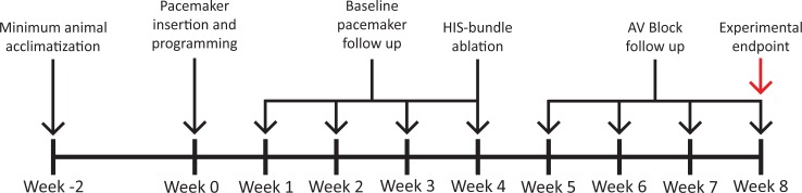
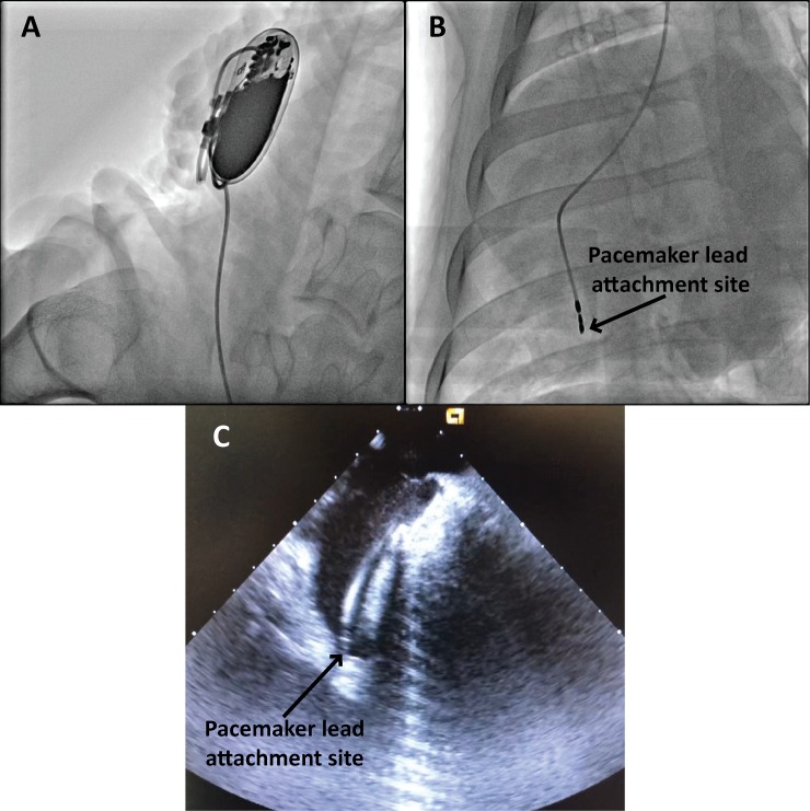
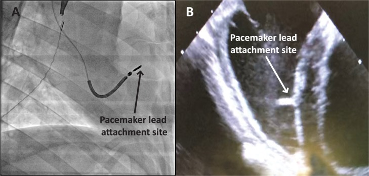
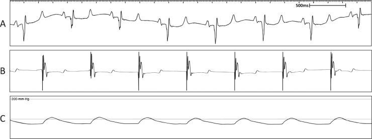
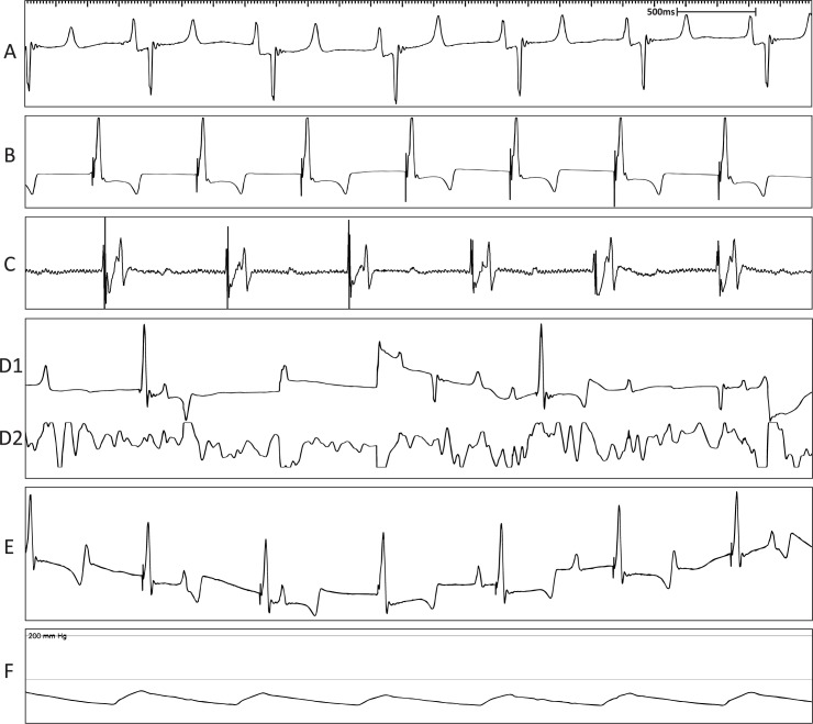
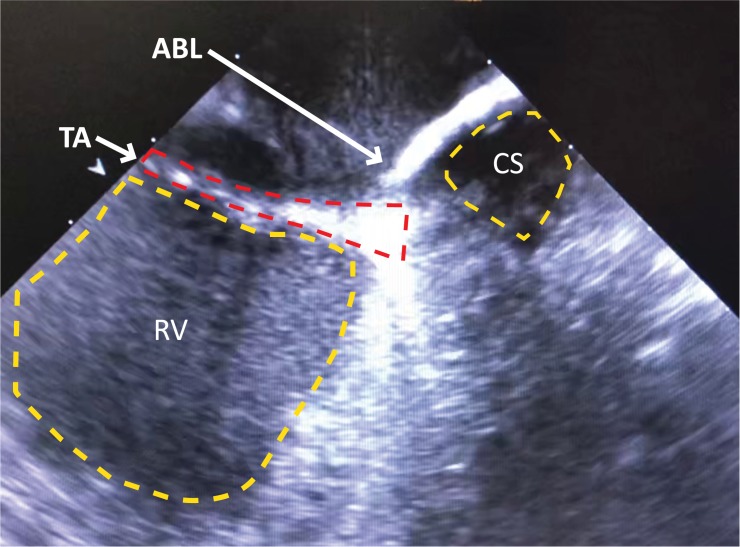
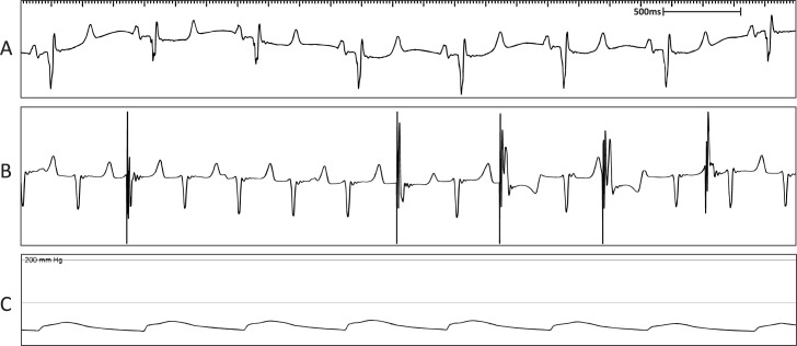
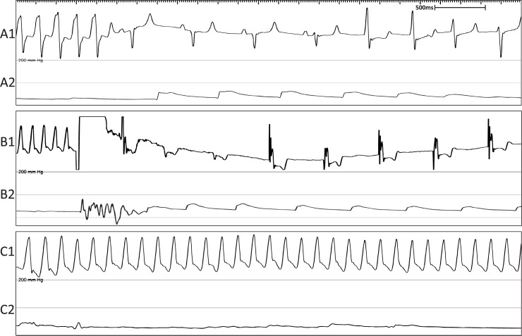
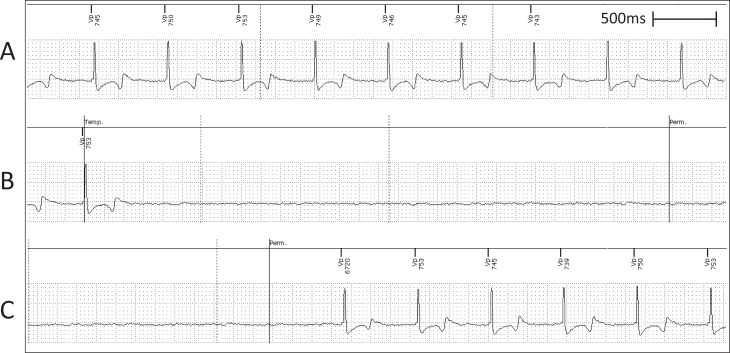
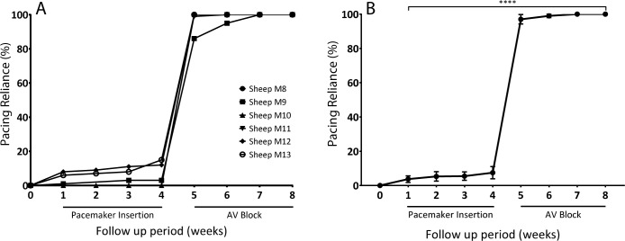
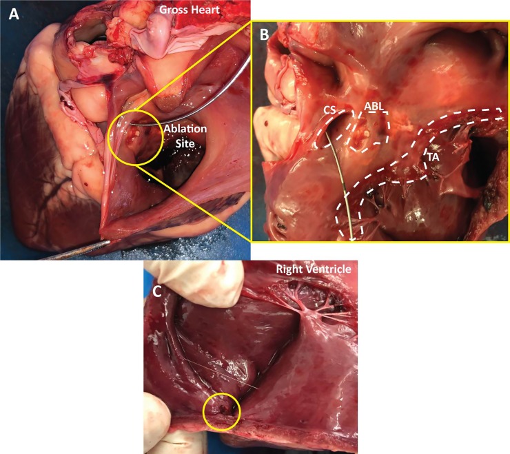
Methods and results: We utilized six sheep to undergo two procedures each. The first procedure involved implantation of a single chamber pacemaker into the right ventricular apex, for baseline assessment over four weeks. The second procedure involved creating atrioventricular block by radiofrequency ablation of the His bundle, before holding for a further four weeks. Interrogation of pacemakers and electrocardiograms determined the persistence of atrioventricular block during the follow up period. Pacemakers were inserted, and atrioventricular block created in 6 animals using a conventional approach. One animal died following ablation of the His bundle, due to procedural complications. Four unablated sheep were assessed for baseline data over four weeks and showed 5.53 ± 1.28% pacing reliance. Five sheep were assessed over four weeks following His bundle ablation and showed continuous (98.89 ± 0.81%) ventricular pacing attributable to persistent atrioventricular block, with no major complications.

Conclusion: We have successfully developed, characterized and validated a large animal model of atrioventricular block that is stable and technically feasible in adult sheep. This model will allow for the advancement of novel therapies, including the development of cell and gene-based therapies.


Conflict of interest statement

The authors have declared that no competing interests exist.

Figures

Fig 1
Fig 1. Experimental timeline depicting the stages at which the sheep were subjected to pacemaker insertion, His bundle ablation and monitoring for data collection.
Fig 2
Fig 2. Representative fluoroscopy and ICE confirm anatomical location of the ventricular leads in the RV apex.
(A) Fluoroscopic image demonstrating the anatomical location of the pacemaker device in the subcutaneous pocket. (B) Fluoroscopic image showing ventricular lead in the RV apex. (C) ICE image confirming position of the same ventricular lead in the RV apex.
Fig 3
Fig 3. Representative fluoroscopy and ICE confirm attachment site of ventricular leads at the RV septal wall.
(A) Fluoroscopic image showing the ventricular lead attached at the RV septal wall. (B) ICE recording confirming the ventricular lead attached at the RV septal wall.
Fig 4
Fig 4. Representative surface ECG recordings during the pacemaker implantation to confirm lead placement.
(A) Surface ECG depicting normal sinus rhythm under anaesthesia. (B) Surface ECG depicting paced rhythm during pacemaker diagnostics. (C) Mean arterial pressure trace indicating maintenance of blood pressure throughout the procedure.
Fig 5
Fig 5. Representative surface ECG recordings during the ablation procedure confirming AVB model creation.
(A) Surface ECG depicting normal sinus rhythm under anaesthesia. (B) Surface ECG depicting pacing rhythm during pacemaker diagnostics. (C) Bipolar electrogram from the ablation catheter proximal pair showing His electrogram before ablation. (D1) Surface ECG during ablation. (D2) Bipolar electrogram from the ablation catheter proximal pair during the ablation. (E) Surface ECG showing AVB evident by AV dissociation. (F) Mean arterial blood pressure trace indicating maintenance of blood pressure throughout the procedure.
Fig 6
Fig 6. ICE recording confirms site tricuspid annulus (TA) and coronary sinus (CS) to assist with location of His bundle.
Representative ICE recording confirming the location of the ablation catheter tip (ACT) at the site of the TA relative to the ostium of the CS and the right ventricle (RV).
Fig 7
Fig 7. Representative surface ECG recordings following creation of AVB in sheep M9 showing a fast, fascicular rhythm.
(A) Surface ECG depicting normal sinus rhythm. (B) Surface ECG showing AVB evident by AV dissociation with intermittent pacing due to the presence of a fast, fascicular rhythm competing with the pacemaker programmed at 80 bpm. (C) Mean arterial blood pressure trace indicating maintenance of blood pressure during the procedure.
Fig 8
Fig 8. Representative surface ECG recordings showing VT and VF experienced during the ablation procedure.
(A1) Surface ECG showing non-sustained VT. (A2) Mean arterial blood pressure trace indicating return of normal blood pressure following termination of VT. (B1) Surface ECG showing cardioverted VF. (B2) Mean arterial blood pressure trace indicating return of normal blood pressure following cardioversion of VF. (C1) Surface ECG showing VF causing death. (C2) Mean arterial blood pressure trace showing no blood pressure during VF.
Fig 9
Fig 9. Lack of escape rhythm in the post-ablation follow up period.
(A) Representative intracardiac ventricular lead recording depicting ventricular paced rhythm. (B) Representative lead recording showing lack of sinus rhythm when sensing feature activated by the pacemaker. Sensing period between the Temp. and Perm. lines. (C) Representative lead recording depicting return of ventricular pacing rhythm when sensing feature deactivated at Perm. line.
Fig 10
Fig 10. Diagnostic pacing data collected from pacemakers during baseline and post-ablation periods.
(A) Pacing reliance was collected for 4 weeks following pacemaker implantation. His bundle ablation was performed at the week 4 timepoint, after which pacing reliance was collected for a further 4 weeks (B) Mean pacing reliance trends presented over the total examined period (% pacing reliance). Data is presented as mean ± SEM. N = 5. Statistically significant if p<0.05.
Fig 11
Fig 11. Gross anatomy of an explanted sheep heart showing landmarks, ablation lesions and lead attachment site.
(A) This view depicts the gross heart anatomy with the view looking into the right atrium. The ablation site is shown within the circle. A wire was placed in the coronary sinus for orientation purposes. (B) Zoomed in perpendicular view of the coronary sinus (CS), tricuspid annulus (TA) and the ablation lesions (ABL). (C) View showing the lead attachment site within the circle at the apex of right ventricle.

References

    1. Protze SI, Liu J, Nussinovitch U, Ohana L, Backx PH, Gepstein L, et al. Sinoatrial node cardiomyocytes derived from human pluripotent cells function as a biological pacemaker. Nature biotechnology. 2017;35(1):56–68. 10.1038/nbt.3745 - DOI - PubMed
    1. Saito Y, Nakamura K, Ito H. Cell-based Biological Pacemakers: Progress and Problems. Acta medica Okayama. 2018;72(1):1–7. 10.18926/AMO/55656 - DOI - PubMed
    1. Kapoor N, Liang W, Marbán E, Cho HC. Direct conversion of quiescent cardiomyocytes to pacemaker cells by expression of Tbx18. Nature Biotechnology. 2012;31:54–62. 10.1038/nbt.2465 - DOI - PMC - PubMed
    1. Chauveau S, Anyukhovsky EP, Ben-Ari M, Naor S, Jiang YP, Danilo P Jr., et al. Induced Pluripotent Stem Cell-Derived Cardiomyocytes Provide in vivo Biological Pacemaker Function. Circulation Arrhythmia and electrophysiology. 2017;10(5):1–10. - PMC - PubMed
    1. Plotnikov AN, Sosunov EA, Qu J, Shlapakova IN, Anyukhovsky EP, Liu L, et al. Biological pacemaker implanted in canine left bundle branch provides ventricular escape rhythms that have physiologically acceptable rates. Circulation. 2004;109(4):506–12. 10.1161/01.CIR.0000114527.10764.CC - DOI - PubMed

Publication types