. 2020 Apr;99(4):463-471.
doi: 10.1177/0022034520904018. Epub 2020 Feb 10.

Genome-wide Identification of Foxf2 Target Genes in Palate Development

Affiliations

Genome-wide Identification of Foxf2 Target Genes in Palate Development

J Xu et al. J Dent Res. 2020 Apr.

Abstract

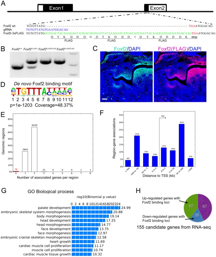
Cleft palate is among the most common structural birth defects in humans. Previous studies have shown that mutations in FOXF2 are associated with cleft palate in humans and mice and that Foxf2 acts in a Shh-Foxf-Fgf18-Shh molecular network controlling palatal shelf growth. In this study, we combined RNA-seq and ChIP-seq approaches to identify direct transcriptional target genes mediating Foxf2 function in palate development in mice. Of 155 genes that exhibited Foxf2-dependent expression in the developing palatal mesenchyme, 88 contained or were located next to Foxf2-binding sites. Through in situ hybridization analyses, we demonstrate that expression of many of these target genes, including multiple genes encoding transcription factors and several encoding extracellular matrix-modifying proteins, were specifically upregulated in the posterior region of palatal shelves in Foxf2-/- mouse embryos. Foxf2 occupancy at many of these putative target loci, including Fgf18, in the developing palatal tissues was verified by ChIP-polymerase chain reaction analyses. One of the Foxf2 target genes, Chst2, encodes a carbohydrate sulfotransferase integral to glycosaminoglycan sulfation. Correlating with ectopic Chst2 expression, Foxf2-/- embryos a exhibited region-specific increase in sulfated keratan sulfate and a concomitant reduction in chondroitin sulfate accumulation in the posterior palatal mesenchyme. However, expression of the core protein of versican, a major chondroitin sulfate proteoglycan important in palatal shelf morphogenesis, was increased, whereas expression of collagen I was reduced in the corresponding region of the palatal mesenchyme. These results indicate that, in addition to regulating palatal shelf growth through the Fgf18-Shh signaling network, Foxf2 controls palatal shelf morphogenesis through regulating expression of multiple transcription factors as well as through directly controlling the synthesis and processing of extracellular matrix components in the palatal mesenchyme. Our ChIP-seq and RNA-seq data sets provide an excellent resource for comprehensive understanding of the molecular network controlling palate development.

Keywords: ChIP-seq; RNA-seq; cleft palate; craniofacial; extracellular matrix; palatogenesis.


Conflict of interest statement

The authors declare no potential conflicts of interest with respect to the authorship and/or publication of this article.

Figures

Figure 1.
Figure 1.
Generation of Foxf2FLAG/+ mice and identification of Foxf2 direct target genes through combined analyses of ChIP-seq and RNA-seq data. (A) Schematic diagram of the Foxf2 gene structure and strategy for CRISPR/Cas9-mediated insertion of the 3xFLAG epitope tag at the C-terminus of the endogenous Foxf2 protein. The gRNA target sequence is shown in blue font. The sequence of the midportion of the oligonucleotide donor template for homology-directed repair contains the 3xFLAG tag-coding sequence (shown in green font). The endogenous Foxf2 stop codon is shown in red font. (B) Foxf2FLAG/+ and Foxf2FLAG/FLAG mice were identified by polymerase chain reaction genotyping. (C) Immunofluorescent detection of the FLAG epitope (red) and the Foxf2 protein (green) on frontal sections of E13.5 (embryonic day 13.5) Foxf2FLAG/+ embryonic head. Scale bar in C, 100 μm. (D) The most enriched Foxf2-binding motif identified from Foxf2-FLAG ChIP-seq peaks. (E, F) Region-gene association analysis of Foxf2-FLAG ChIP-seq data with the GREAT online tool (http://great.stanford.edu). The “basal plus extension” parameter was used to associate the ChIP-seq peaks to genes within 1,000 kb. The majority of peaks were associated with 1 or 2 genes (E) and located in 50 to 500 kb from the transcription start sites (TSS). (G) Gene ontology analysis of FLAG-Foxf2 ChIP-seq peaks performed with GREAT. The top GO biological process terms are shown. The plot indicates the –log10 of binomial P values of each GO term. (H) Among 155 genes that showed differential expression in the E13.5 palatal mesenchyme in Foxf2-/- and littermates, 88 genes (62 upregulated and 26 downregulated in Foxf2-/- palate) were associated with FLAG-Foxf2 binding peaks.
Figure 2.
Figure 2.
Analysis of Foxf2-dependent expression of direct target genes in the developing palatal shelves by whole-mount in situ hybridization assays. (A–R, A′–R′) Comparison of patterns of expression of Fgf18, Chst2, Foxd1, Foxq1, Exoc2, Adamts9, Smoc2, Spon1, Tbx15, Corin, Lmcd1, Pcdh19, Creb5, Jazf1, Lrrc32, Ddah1, Sim2, and Dlx5 mRNAs in E13.5 (embryonic day 13.5) Foxf2-/- mutant and wild-type (Wt) control embryos. Note that Fgf18, Chst2, and Foxd1 mRNAs are upregulated in specific anterior (arrows) and posterior (arrowheads) subdomains of Foxf2-/- mutant palatal shelves. Foxq1 and Exoc2 are increased along the anterior-posterior axis of Foxf2-/- mutant palatal shelves. Adamts9, Smoc2, Spon1, Tbx15, Corin, Lmcd1, Pcdh19, Creb5, Jazf1, and Lrrc32 mRNAs were upregulated in specific posterior (arrowheads) subdomains in the Foxf2-/- mutant palatal shelves. Scale bar, 200 μm.
Figure 3.
Figure 3.
Validation of Foxf2 occupancy at specific genomic sites identified by ChIP-seq and associated with extracellular matrix–related and transcription factor genes. (A–I) Genome browser views of genomic regions containing Fgf18, Chst2, Adamts9, Smoc2, Spon1, Jazf1, Creb5, Tbx15, and Sim2 genes. Red asterisks, triangles, and circles indicate 3 distinct Foxf2 binding peaks associated with each locus. The binding of Foxf2 at each peak-associated genomic sequence was confirmed by ChIP–polymerase chain reaction.
Figure 4.
Figure 4.
Foxf2-/- embryos exhibited increased accumulation of highly sulfated keratan sulfate (KS) and concomitant decrease in chondroitin sulfate in the posterior palatal mesenchyme region where ectopic Chst2 expression was also detected. (A, B) Frontal sections show expression of Chst2 mRNAs (detected in blue) in the posterior regions of the developing palatal shelves in embryonic day 13.5 (E13.5) wild-type (Wt) and Foxf2-/- mutant embryos. (C–F) Immunofluorescent detection of highly sulfated KS (red) on sections from posterior regions of the palatal shelves in E13.5 Wt and Foxf2-/- mutant embryos. Panels E and F show enlarged views of the boxed areas in panels C and D, respectively. Arrows indicate ectopic 5D4 antibody staining signals in the specific domain in posterior palatal mesenchyme. (G–J) Immunofluorescent detection of highly sulfated KS (red) and Foxf2 (green) on sections from posterior regions of the palatal shelves in E13.5 Wt and Foxf2-/- mutant embryos. Panels I and J show enlarged views of the boxed areas in panels G and H, respectively. Arrows indicate ectopic 5D4 antibody staining signals in the specific domain in posterior palatal mesenchyme. (K–N) Immunofluorescent detection of chondroitin sulfate (green) on sections from posterior regions of the palatal shelves in E13.5 Wt and Foxf2-/- mutant embryos. Panels M and N show enlarged views of the boxed areas in panels K and L, respectively. Arrows indicate decreased chondroitin sulfate staining signals in the specific domain in posterior palatal mesenchyme. (O, P) Fluorescent detection of hyaluronan (red) on sections from posterior regions of the palatal shelves in E13.5 Wt and Foxf2-/- mutant embryos. Sections were counterstained with DAPI. Scale bar, 50 μm.
Figure 5.
Figure 5.
Comparison of patterns of expression of versican, Runx2, and collagen I in palatal mesenchyme in E13.5 (embryonic day 13.5) wild-type (Wt) and Foxf2-/- mutant embryos. (A, B) Frontal sections show patterns of expression of Vcan mRNAs (detected in blue) in the posterior regions of the developing palatal shelves in Wt and Foxf2-/- mutant embryos. (C, D) Immunofluorescent detection of Vcan core protein (red) on sections from posterior regions of the palatal shelves in Wt and Foxf2-/- mutant embryos. Arrows indicate expanded Vcan mRNAs and Vcan protein in the specific domain in the posterior palatal mesenchyme in Foxf2-/- embryos. (E, F) Immunofluorescent detection of Vcan core proteins (red) and Foxf2 (green) on sections from posterior regions of the palatal shelves in Wt and Foxf2-/- mutant embryos. (G, H) Immunofluorescent detection of Vcan core protein (red) and chondroitin sulfate (green) on sections from posterior regions of the palatal shelves in Wt and Foxf2-/- mutant embryos. (I–L) Immunofluorescent detection of highly sulfated keratan sulfate (red) and Runx2 (green) on sections from posterior regions of the palatal shelves in Wt and Foxf2-/- mutant embryos. Panels K and L show the enlarged view of the boxed areas in panels I and K, respectively. Arrows indicate ectopic 5D4 antibody staining signals in the specific domain in posterior palatal mesenchyme. (M, N) Frontal sections show expression of Col1a1 mRNAs (detected in blue) in the posterior regions of the developing palatal shelves in Wt and Foxf2-/- mutant embryos. (O, P) Immunofluorescent detection of collagen I (red) on sections from posterior regions of the palatal shelves in Wt and Foxf2-/- mutant embryos. Scale bar, 50 μm.

Similar articles

Cited by

References

    1. Bolte C, Ren X, Tomley T, Ustiyan V, Pradhan A, Hoggatt A, Kalin TV, Herring BP, Kalinichenko VV. 2015. Forkhead box f2 regulation of platelet-derived growth factor and myocardin/serum response factor signaling is essential for intestinal development. J Biol Chem. 290(12):7563–7575. - PMC - PubMed
    1. Brinkley LL, Vickerman MM. 1982. The effects of chlorcyclizine-induced alterations of glycosaminoglycans on mouse palatal shelf elevation in vivo and in vitro. J Embryol Exp Morphol. 69:193–213. - PubMed
    1. Bush JO, Jiang R. 2012. Palatogenesis: morphogenetic and molecular mechanisms of secondary palate development. Development. 139(2):231–243. - PMC - PubMed
    1. Caterson B, Melrose J. 2018. Keratan sulfate, a complex glycosaminoglycan with unique functional capability. Glycobiology. 28(4):182–206. - PMC - PubMed
    1. Chai Y, Maxson RE., Jr. 2006. Recent advances in craniofacial morphogenesis. Dev Dyn. 235(9):2353–2375. - PubMed

Publication types

Substances