Review
. 2020 Feb 6;21(3):1071.
doi: 10.3390/ijms21031071.

Dual Role of Chondrocytes in Rheumatoid Arthritis: The Chicken and the Egg

Affiliations
Review

Dual Role of Chondrocytes in Rheumatoid Arthritis: The Chicken and the Egg

Chia-Chun Tseng et al. Int J Mol Sci. .

Abstract

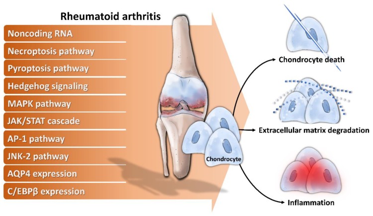
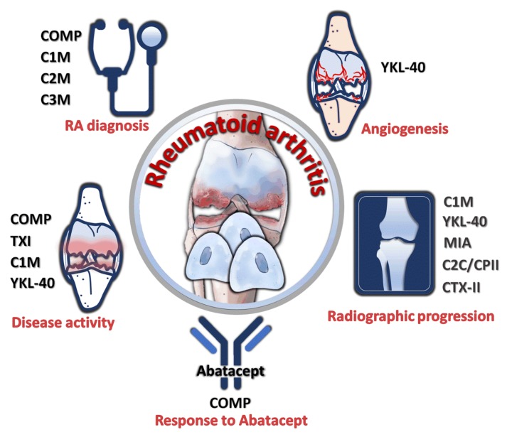
Rheumatoid arthritis (RA) is one of the inflammatory joint diseases that display features of articular cartilage destruction. The underlying disturbance results from immune dysregulation that directly and indirectly influence chondrocyte physiology. In the last years, significant evidence inferred from studies in vitro and in the animal model offered a more holistic vision of chondrocytes in RA. Chondrocytes, despite being one of injured cells in RA, also undergo molecular alterations to actively participate in inflammation and matrix destruction in the human rheumatoid joint. This review covers current knowledge about the specific cellular and biochemical mechanisms that account for the chondrocyte signatures of RA and its potential applications for diagnosis and prognosis in RA.

Keywords: cartilage; chondrocyte; rheumatoid arthritis.


Conflict of interest statement

The authors declare no conflicts of interest.

Figures

Figure 1
Figure 1
Mechanisms of chondrocytes dysfunction in rheumatoid arthritis. Noncoding RNA and activation of necroptosis pathway, pyroptosis pathway, hedgehog signaling, mitogen-activated protein kinase (MAPK) pathway, Janus kinase/Signal transducer and activator of transcription protein (JAK/STAT) cascade, AP-1 pathway, c-Jun N-terminal kinase 2 (JNK-2) pathway, combined with enhanced aquaporin-4 (AQP4) expression and CCAAT/enhancer binding protein β (C/EBPβ) contribute to increased chondrocyte death, ECM degradation and inflammation.
Figure 2
Figure 2
Inhibitors of chondrocytes dysfunction and corresponding targeting pathways. Resveratrol interferes with lymphotoxin α signaling, necrostatin-1 inhibits necroptosis pathway, hyaluronan blocks MAPK pathway, and paclitaxel suppresses AP-1 activity which contribute to chondrocyte dysfunction in rheumatoid arthritis.
Figure 3
Figure 3
Utility of chondrocyte products as diagnostic and prognostic markers. Cartilage oligomeric matrix protein (COMP), collagen type I (TXI), C1M, C2M, C3M, YKL-40, MIA, C2C/CPII ratio, and type II collagen (CTX-II) were reported to have diagnostic prognostic significance for disease activity, treatment response, radiographic progression, and angiogenesis.

References

    1. Karouzakis E., Neidhart M., Gay R.E., Gay S. Molecular and cellular basis of rheumatoid joint destruction. Immunol. Lett. 2006;106:8–13. doi: 10.1016/j.imlet.2006.04.011. - DOI - PubMed
    1. Li F., Tang Y., Song B., Yu M., Li Q., Zhang C., Hou J., Yang R. Nomenclature clarification: Synovial fibroblasts and synovial mesenchymal stem cells. Stem Cell Res. Ther. 2019;10:260. doi: 10.1186/s13287-019-1359-x. - DOI - PMC - PubMed
    1. Li Y., Liu Y., Wang C., Xia W.R., Zheng J.Y., Yang J., Liu B., Liu J.Q., Liu L.F. Succinate induces synovial angiogenesis in rheumatoid arthritis through metabolic remodeling and HIF-1α/VEGF axis. Free Radic. Biol. Med. 2018;126:1–14. doi: 10.1016/j.freeradbiomed.2018.07.009. - DOI - PubMed
    1. Tanaka S. RANKL is a therapeutic target of bone destruction in rheumatoid arthritis. F1000Research. 2019;8:533. doi: 10.12688/f1000research.17296.1. - DOI - PMC - PubMed
    1. Cai X., Zhang L., Wei W. Regulatory B cells in inflammatory diseases and tumor. Int. Immunopharmacol. 2019;67:281–286. doi: 10.1016/j.intimp.2018.12.007. - DOI - PubMed