. 2020 Feb 6;9(2):448.
doi: 10.3390/jcm9020448.

Total, Bioavailable, and Free Vitamin D Levels and Their Prognostic Value in Pulmonary Arterial Hypertension

Affiliations

Total, Bioavailable, and Free Vitamin D Levels and Their Prognostic Value in Pulmonary Arterial Hypertension

Maria Callejo et al. J Clin Med. .

Abstract

Introduction: Epidemiological studies suggest a relationship between vitamin D deficiency and cardiovascular and respiratory diseases. However, whether total, bioavailable, and/or free vitamin D levels have a prognostic role in pulmonary arterial hypertension (PAH) is unknown. We aimed to determine total, bioavailable, and free 25-hydroxy-vitamin D (25(OH)vitD) plasma levels and their prognostic value in PAH patients. Methods: In total, 67 samples of plasma from Spanish patients with idiopathic, heritable, or drug-induced PAH were obtained from the Spanish PH Biobank and compared to a cohort of 100 healthy subjects. Clinical parameters were obtained from the Spanish Registry of PAH (REHAP). Results: Seventy percent of PAH patients had severe vitamin D deficiency (total 25(OH)vitD < 10 ng/mL) and secondary hyperparathyroidism. PAH patients with total 25(OH)vitD plasma above the median of this cohort (7.17 ng/mL) had better functional class and higher 6-min walking distance and TAPSE (tricuspid annular plane systolic excursion). The main outcome measure of survival was significantly increased in these patients (age-adjusted hazard ratio: 5.40 (95% confidence interval: 2.88 to 10.12)). Vitamin D-binding protein (DBP) and albumin plasma levels were downregulated in PAH. Bioavailable 25(OH)vitD was decreased in PAH patients compared to the control cohort. Lower levels of bioavailable 25(OH)vitD (<0.91 ng/mL) were associated with more advanced functional class, lower exercise capacity, and higher risk of mortality. Free 25(OH)vitD did not change in PAH; however, lower free 25(OH)vitD (<1.53 pg/mL) values were also associated with high risk of mortality. Conclusions: Vitamin D deficiency is highly prevalent in PAH, and low levels of total 25(OH)vitD were associated with poor prognosis.

Keywords: prognosis; pulmonary arterial hypertension; survival; vitamin D.


Conflict of interest statement

The authors declare no conflicts of interest regarding the present study.

Figures

Figure 1
Figure 1
PAH patients present decreased total 25-hydroxy-vitamin D (25(OH)vitD) and increased intact parathyroid hormone (iPTH). (A) Total 25(OH)vitD and (B) iPTH plasma levels from controls and PAH patients. Colors indicate the ranges of levels as follows: for panels A and C, red encodes severe deficiency (<10 ng/mL), yellow encodes moderate deficiency (10–20 ng/mL), and green encodes sufficiency (>20 ng/mL); for panels B and D, green encodes normal levels (10–55 pg/mL) and red encodes hyperparathyroidism (>55 pg/mL). (C) Percentage of patients according to total 25(OH)vitD levels. (D) Percentage of patients according to iPTH range. Results in panels A and B are presented as scatter plots and medians; *** indicates p < 0.001 vs. controls, Mann–Whitney test. In panels C and D, *** denotes p < 0.001 vs. controls, chi-square test for trend.
Figure 2
Figure 2
VitD-binding protein (DBP), albumin, and bioavailable but not free 25(OH)vitD are decreased in PAH patients. (A) Plasma DBP concentration, (B) albumin, (C) calculated bioavailable 25(OH)vitD, and (D) calculated free 25(OH)vitD from controls and PAH patients. Data are represented as scatter plots and medians. * and *** indicate p < 0.05 and p < 0.001 vs. controls, respectively, Mann–Whitney test.
Figure 3
Figure 3
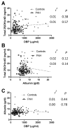
VitD-binding protein (DBP), albumin and total 25(OH)vitD plasma concentrations are independent variables. Correlations between (A) DBP and total 25(OH)vitD, (B) albumin and total 25(OH)vitD, and (C) DBP and albumin in controls and in PAH patients. The calculated Pearson r2 and p are shown in each panel.
Figure 4
Figure 4
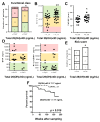
PAH patients with lower total 25(OH)vitD levels present worse prognosis. PAH patients were categorized according to total 25(OH)vitD levels (above vs. below the median (7.17 ng/mL) in the cohort). (A) NYHA functional class; (B) 6-min walking distance test (6MWD); (C) TAPSE; (D) BNP (left) and NT-proBNP (right). Colors in panels A, B, and D identify the ranges of these biomarkers considered of low (green), intermediate (yellow), or high (red) risk according to ERS/ESC guidelines for PAH. (E) Non-invasive risk score showing the number of patients with zero, one, two, or three low risk factors. In panels A and E, * denotes p < 0.05, chi-square test for trend. In panels B and D, data are scatter plots and medians; * p < 0.05 and ** p < 0.01 Mann–Whitney test. (F) Kaplan–Meier analysis of survival in PAH patients with total 25(OH)vitD levels above vs. below the median (7.17 ng/mL); p < 0.05, log-rank test.
Figure 5
Figure 5
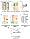
Bioavailable 25(OH)vitD levels in PAH patients and prognosis. PAH patients were categorized according to bioavailable 25(OH)vitD levels (above vs. below the median (0.91 ng/mL) in the cohort). (A) NYHA functional class; (B) 6-min walking distance test (6MWD); (C) TAPSE; (D) BNP (left) and NT-proBNP (right). Color codes are as in Figure 4. (E) Non-invasive risk score showing the number of patients with zero, one, two, or three low risk factors. In panels A and E, * denotes p < 0.05, chi-square test for trend. In panels B and D, data are scatter plots and medians, ** p < 0.01 Mann–Whitney test. (F) Kaplan–Meier analysis of survival in PAH patients with bioavailable 25(OH)vitD levels above vs. below the median (0.91 ng/mL); p = 0.09, log-rank test.
Figure 6
Figure 6
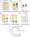
Free 25(OH)vitD levels in PAH patients and prognosis. PAH patients were categorized according to free 25(OH)vitD levels (above vs. below the median (1.53 pg/mL) in the cohort). (A) NYHA functional class; (B) 6 min walking distance test (6MWD); (C) TAPSE; (D) BNP (left) and NT-proBNP (right). Color codes are as in Figure 4. (E) Non-invasive risk score showing the number of patients with zero, one, two, or three low risk factors. In panels A and E, * denotes p < 0.05, chi-square test for trend. In panels B and D, data are scatter plots and medians; ** p < 0.01 Mann–Whitney test. (F) Kaplan–Meier analysis of survival in PAH patients with bioavailable 25(OH)vitD levels above vs. below the median (0.91 ng/mL); p = 0.10, log-rank test.

References

    1. Simonneau G., Montani D., Celermajer D.S., Denton C.P., Gatzoulis M.A., Krowka M., Williams P.G., Souza R. Haemodynamic definitions and updated clinical classification of pulmonary hypertension. Eur. Respir. J. 2019:53. doi: 10.1183/13993003.01913-2018. - DOI - PMC - PubMed
    1. Thenappan T., Ormiston M.L., Ryan J.J., Archer S.L. Pulmonary arterial hypertension: Pathogenesis and clinical management. BMJ. 2018;360:j5492. doi: 10.1136/bmj.j5492. - DOI - PMC - PubMed
    1. Demerouti E., Karyofyllis P., Manginas A., Anthi A., Karatasakis G., Athanassopoulos G., Voudris V. Improving Survival in Patients with Pulmonary Arterial Hypertension: Focus on Intravenous Epoprostenol. Am. J. Cardiovasc. Drugs. 2019;19:99–105. doi: 10.1007/s40256-018-00319-z. - DOI - PubMed
    1. Galie N., Humbert M., Vachiery J.L., Gibbs S., Lang I., Torbicki A., Simonneau G., Peacock A., Vonk Noordegraaf A., Beghetti M., et al. 2015 ESC/ERS Guidelines for the diagnosis and treatment of pulmonary hypertension: The Joint Task Force for the Diagnosis and Treatment of Pulmonary Hypertension of the European Society of Cardiology (ESC) and the European Respiratory Society (ERS): Endorsed by: Association for European Paediatric and Congenital Cardiology (AEPC), International Society for Heart and Lung Transplantation (ISHLT) Eur. Respir. J. 2015;46:903–975. doi: 10.1183/13993003.01032-2015. - DOI - PubMed
    1. Norman P.E., Powell J.T. Vitamin D and cardiovascular disease. Circ. Res. 2014;114:379–393. doi: 10.1161/CIRCRESAHA.113.301241. - DOI - PubMed