

Differences in the Inflammatory Response of White Adipose Tissue and Adipose-Derived Stem Cells
- PMID: 32041245
- PMCID: PMC7037886
- DOI: 10.3390/ijms21031086
Differences in the Inflammatory Response of White Adipose Tissue and Adipose-Derived Stem Cells
Abstract
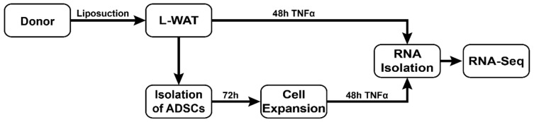
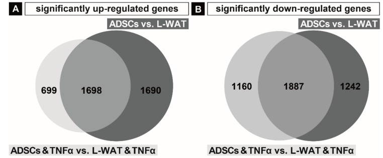
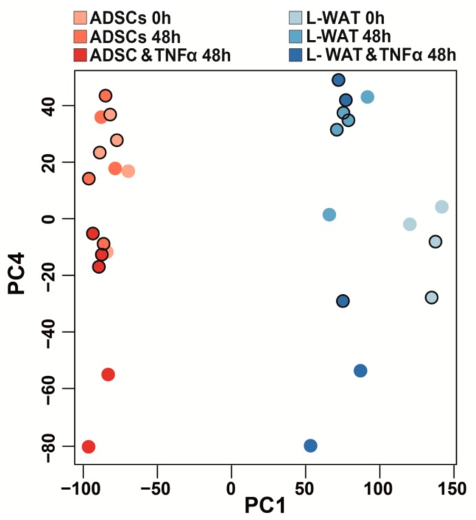
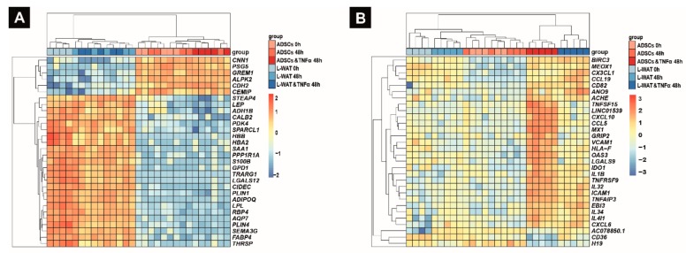
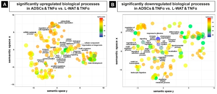
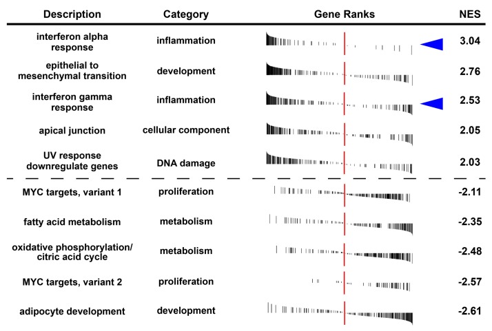
The application of liposuctioned white adipose tissue (L-WAT) and adipose-derived stem cells (ADSCs) as a novel immunomodulatory treatment option is the currently subject of various clinical trials. Because it is crucial to understand the underlying therapeutic mechanisms, the latest studies focused on the immunomodulatory functions of L-WAT or ADSCs. However, studies that examine the specific transcriptional adaptation of these treatment options to an extrinsic inflammatory stimulus in an unbiased manner are scarce. The aim of this study was to compare the gene expression profile of L-WAT and ADSCs, when subjected to tumor necrosis factor alpha (TNFα), and to identify key factors that might be therapeutically relevant when using L-WAT or ADSCs as an immuno-modulator. Fat tissue was harvested by liposuction from five human donors. ADSCs were isolated from the same donors and shortly subjected to expansion culture. L-WAT and ADSCs were treated with human recombinant TNFα, to trigger a strong inflammatory response. Subsequently, an mRNA deep nextgeneration sequencing was performed to evaluate the different inflammatory responses of L-WAT and ADSCs. We found significant gene expression changes in both experimental groups after TNFα incubation. However, ADSCs showed a more homogenous gene expression profile by predominantly expressing genes involved in immunomodulatory processes such as CCL19, CCL5, TNFSF15 and IL1b when compared to L-WAT, which reacted rather heterogeneously. As RNA sequencing between L-WAT and ADSCS treated with TNFα revealed that L-WAT responded very heterogeneously to TNFα treatment, we therefore conclude that ADSCs are more reliable and predictable when used therapeutically. Our study furthermore yields insight into potential biological processes regarding immune system response, inflammatory response, and cell activation. Our results can help to better understand the different immunomodulatory effects of L-WAT and ADSCs.
Keywords: TNFalpha; adiposederived stem cells; immunomodulation; inflammation; white fat tissue.
Conflict of interest statement
The authors have declared that no competing interests exist.
Figures
Similar articles
-
CCL5 promotes macrophage recruitment and survival in human adipose tissue.Arterioscler Thromb Vasc Biol. 2010 Jan;30(1):39-45. doi: 10.1161/ATVBAHA.109.197442. Epub 2009 Nov 5. Arterioscler Thromb Vasc Biol. 2010. PMID: 19893003
-
Exposure to an organometal compound stimulates adipokine and cytokine expression in white adipose tissue.Cytokine. 2011 Mar;53(3):355-62. doi: 10.1016/j.cyto.2010.11.015. Epub 2010 Dec 30. Cytokine. 2011. PMID: 21194965 Free PMC article.
-
Evaluation of CD49f as a novel surface marker to identify functional adipose-derived mesenchymal stem cell subset.Cell Prolif. 2021 May;54(5):e13017. doi: 10.1111/cpr.13017. Epub 2021 Mar 11. Cell Prolif. 2021. PMID: 33704842 Free PMC article.
-
Single-Cell Profiles and Clinically Useful Properties of Human Mesenchymal Stem Cells of Adipose and Bone Marrow Origin.Am J Sports Med. 2019 Jun;47(7):1722-1733. doi: 10.1177/0363546519848678. Epub 2019 May 17. Am J Sports Med. 2019. PMID: 31100005 Review.
-
Omega-3 fatty acids and adipose tissue biology.Mol Aspects Med. 2018 Dec;64:147-160. doi: 10.1016/j.mam.2018.01.004. Epub 2018 Jan 17. Mol Aspects Med. 2018. PMID: 29329795 Review.
Cited by
-
Transcriptome organization of white blood cells through gene co-expression network analysis in a large RNA-seq dataset.Front Immunol. 2024 Apr 2;15:1350111. doi: 10.3389/fimmu.2024.1350111. eCollection 2024. Front Immunol. 2024. PMID: 38629067 Free PMC article.
-
Adipose tissue in bone regeneration - stem cell source and beyond.World J Stem Cells. 2022 Jun 26;14(6):372-392. doi: 10.4252/wjsc.v14.i6.372. World J Stem Cells. 2022. PMID: 35949397 Free PMC article. Review.
-
Recent Advances of Adipose-Tissue-Derived Mesenchymal Stem Cell-Based Therapy for Retinal Diseases.J Clin Med. 2023 Nov 9;12(22):7015. doi: 10.3390/jcm12227015. J Clin Med. 2023. PMID: 38002628 Free PMC article. Review.
-
Adipose tissue aging is regulated by an altered immune system.Front Immunol. 2023 Feb 17;14:1125395. doi: 10.3389/fimmu.2023.1125395. eCollection 2023. Front Immunol. 2023. PMID: 36875140 Free PMC article. Review.
-
Synovial Fluid Derived from Human Knee Osteoarthritis Increases the Viability of Human Adipose-Derived Stem Cells through Upregulation of FOSL1.Cells. 2023 Jan 15;12(2):330. doi: 10.3390/cells12020330. Cells. 2023. PMID: 36672268 Free PMC article.
References
-
- Cavaillon J.M. Pro-versus anti-inflammatory cytokines: Myth or reality. Cell. Mol. Biol. 2001;47:695–702. - PubMed
-
- Schuerwegh A.J., Dombrecht E.J., Stevens W.J., Van Offel J.F., Bridts C.H., De Clerck L.S. Influence of pro-inflammatory (IL-1α, IL-6, TNF-α, IFN-γ) and anti-inflammatory (IL-4) cytokines on chondrocyte function. Osteoarthr. Cartil. 2003;11:681–687. doi: 10.1016/S1063-4584(03)00156-0. - DOI - PubMed
MeSH terms
Substances
Grants and funding
LinkOut - more resources
Full Text Sources
Research Materials
