. 2020 Feb 6;12(2):425.
doi: 10.3390/nu12020425.

Whey Peptides Stimulate Differentiation and Lipid Metabolism in Adipocytes and Ameliorate Lipotoxicity-Induced Insulin Resistance in Muscle Cells

Affiliations

Whey Peptides Stimulate Differentiation and Lipid Metabolism in Adipocytes and Ameliorate Lipotoxicity-Induced Insulin Resistance in Muscle Cells

Kenneth D'Souza et al. Nutrients. .

Abstract

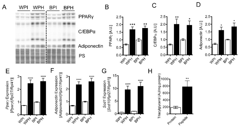
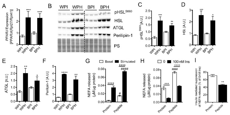
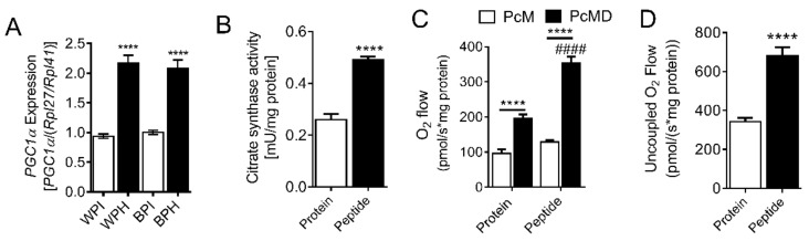
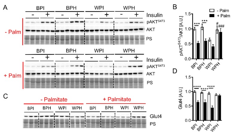
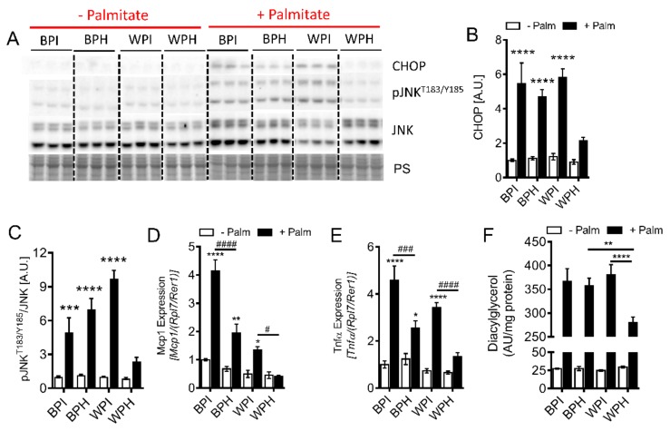
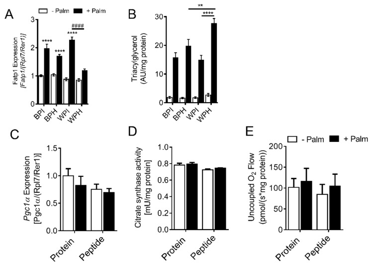
Deregulation of lipid metabolism and insulin function in muscle and adipose tissue are hallmarks of systemic insulin resistance, which can progress to type 2 diabetes. While previous studies suggested that milk proteins influence systemic glucose homeostasis and insulin function, it remains unclear whether bioactive peptides generated from whey alter lipid metabolism and its accumulation in muscle and adipose tissue. Therefore, we incubated murine 3T3-L1 preadipocytes and C2C12 myotubes with a whey peptide mixture produced through pepsin-pancreatin digestion, mimicking peptides generated in the gut from whey protein hydrolysis, and examined its effect on indicators of lipid metabolism and insulin sensitivity. Whey peptides, particularly those derived from bovine serum albumin (BSA), promoted 3T3-L1 adipocyte differentiation and triacylglycerol (TG) accumulation in accordance with peroxisome proliferator-activated receptor γ (PPARγ) upregulation. Whey/BSA peptides also increased lipolysis and mitochondrial fat oxidation in adipocytes, which was associated with the upregulation of peroxisome proliferator-activated receptor δ (PPARδ). In C2C12 myotubes, whey but not BSA peptides ameliorated palmitate-induced insulin resistance, which was associated with reduced inflammation and diacylglycerol accumulation, and increased sequestration of fatty acids in the TG pool. Taken together, our study suggests that whey peptides generated via pepsin-pancreatin digestion profoundly alter lipid metabolism and accumulation in adipocytes and skeletal myotubes.

Keywords: PPAR; adipocytes; differentiation; insulin resistance; lipolysis; lipotoxicity; metabolism; mitochondria; myocytes; whey peptides.


Conflict of interest statement

The authors declare no conflict of interest.

Figures

Figure 1
Figure 1
Effect of whey peptides on 3T3-L1 adipocyte differentiation. 3T3-L1 preadipocytes were differentiated in the presence of 2.5 mg/mL WPI, WPH, BPI or BPH. Immunoblotting and densitometric analysis of (A,B) PPARγ, (A,C) C/EBPα, and (A,D) adiponectin (n = 6). mRNA levels of PPARγ (E) and PPARγ target genes, (F) adiponectin and (G) stearoyl-CoA desaturase (Scd1) (n = 8) were determined. TG levels (H) were measured using a targeted lipidomics approach in adipocytes incubated with BSA protein isolate or whey peptides (n = 8). (BH): * p < 0.05, ** p < 0.01, *** p < 0.001, **** p < 0.0001 vs. protein isolate controls. BPI, BSA protein isolate; BPH, BSA peptide hydrolysate; WPI, whey protein isolate; WPH, whey peptide hydrolysate; PPARγ, peroxisome proliferator-activated receptor γ; C/EBPα, CCAAT/enhancer-binding protein α; PS, protein stain; Scd1, stearoyl-Coenzyme A desaturase 1; A.U., arbitrary units.
Figure 2
Figure 2
Influence of whey peptides on lipolysis in 3T3-L1 adipocytes. 3T3-L1 preadipocytes were differentiated in presence of 2.5 mg/mL WPI, WPH, BPI, or BPH. (A) PPARδ mRNA levels were determined (n = 8). Immunoblotting and densitometric analysis were performed to assess protein levels of (B,C) pHSLS660, (B,D) HSL, (B,E) ATGL, and (B,F) perilipin-1 (n = 6). (G) Basal and isoproterenol-stimulated lipolysis and (H,I) insulin-mediated suppression of lipolysis was determined in adipocytes incubated with BSA protein isolate or whey peptides (n = 6). A, CI: * p < 0.05, ** p < 0.01, *** p < 0.001, **** p < 0.0001 vs. protein isolate controls or as indicated. # p < 0.05, #### p < 0.0001 in peptide vs. protein-treated adipocytes. HSL, hormone-sensitive lipase; ATGL, adipose triglyceride lipase; PS, protein stain; NEFA, non-esterified fatty acids; A.U., arbitrary units.
Figure 3
Figure 3
Effect of whey peptides on mitochondrial function in adipocytes. 3T3-L1 preadipocytes were differentiated in the presence of 2.5 mg/mL WPI, WPH, BPI, or BPH. (A) Pgc1α mRNA levels and (B) citrate synthase activity (n = 7–8). (C) Fatty acid-linked mitochondrial respiration and (D) uncoupled respiration was assessed in permeabilized adipocytes incubated with BSA protein isolate or whey peptides (n = 7). (AD): **** p < 0.0001 vs. protein controls or as indicated. #### p < 0.0001 for peptide vs. protein-treated adipocytes. Pgc1α, PPARγ coactivator 1-α; Pc, palmitoylcarnitine; M, malate; D, ADP.
Figure 4
Figure 4
Effect of whey peptides on insulin signaling in C2C12 myotubes. C2C12 myotubes were incubated in the presence or absence of 0.4 mM palmitate and co-incubated with 2.5 mg/mL BPI, BPH, WPI, or WPH. Immunoblotting and densiometric analysis were performed to assess (A,B) AKT phosphorylation at S473 and (C,D) Glut4 protein levels (n = 6). (B,D): *** p < 0.001, **** p < 0.0001 as indicated. # p < 0.05, ### p < 0.001 for peptide vs. protein treated myotubes. A.U., arbitrary units.
Figure 5
Figure 5
Influence of whey peptides on palmitate-induced ER stress, inflammation, and DG accumulation in C2C12 myotubes. C2C12 myotubes were incubated in the presence or absence of 0.4 mM palmitate and co-incubated with 2.5 mg/mL BPI, BPH, WPI or WPH. Immunoblotting and densiometric analysis were performed to assess (A,B) protein levels of CHOP and (A,C) phosphorylation JNK at T183/Y185 (n = 6). mRNA levels of inflammatory markers, (D) Mcp1 and (E) Tnfα (n = 8) were determined. Levels of (F) DGs were measured using a targeted lipidomic approach (n = 6–8). (BE): * p < 0.05, ** p < 0.01, *** p < 0.001, **** p < 0.0001 vs. no palmitate controls or as indicated. # p < 0.05, ### p < 0.001, #### p < 0.0001 as indicated. CHOP, C/EBP homologous protein; JNK, c-Jun N-terminal kinase; Mcp1, monocyte chemoattractant protein 1; Tnfα, tumor necrosis factor α; Palm, palmitate; A.U., arbitrary units.
Figure 6
Figure 6
Effect of whey peptides on fatty acid transporter levels, TG accumulation, and mitochondrial abundance. C2C12 myotubes were incubated in the presence or absence of 0.4 mM palmitate and co-incubated with 2.5 mg/mL BPI, BPH, WPI, or WPH. (A) mRNA levels of Fatp1 (n = 8) were assessed. (B) TG levels were measured using a targeted lipidomics approach (n = 8). (C) Pgc1α mRNA levels (n = 8), (D) citrate synthase activity (n = 3), and (E) uncoupled respiration (n = 7) were determined in myotubes treated with BSA protein isolate or whey peptides. ** p < 0.01, **** p < 0.0001 vs. no palmitate controls or as indicated, #### p < 0.0001 as indicated. Fatp1, fatty acid transport protein 1; Pgc1α, peroxisome proliferator-activated receptor γ coactivator 1-α; A.U., arbitrary units.

Similar articles

Cited by

References

    1. DeFronzo R.A., Tripathy D. Skeletal muscle insulin resistance is the primary defect in type 2 diabetes. Diabetes Care. 2009;32(Suppl. 2):S157–S163. doi: 10.2337/dc09-S302. - DOI - PMC - PubMed
    1. Baron A.D., Brechtel G., Wallace P., Edelman S.V. Rates and tissue sites of non-insulin- and insulin-mediated glucose uptake in humans. (6 Pt 1)Am. J. Physiol. 1988;255:E769–E774. doi: 10.1152/ajpendo.1988.255.6.E769. - DOI - PubMed
    1. Rutkowski J.M., Stern J.H., Scherer P.E. The cell biology of fat expansion. J. Cell. Biol. 2015;208:501–512. doi: 10.1083/jcb.201409063. - DOI - PMC - PubMed
    1. Griffin M.E., Marcucci M.J., Cline G.W., Bell K., Barucci N., Lee D., Goodyear L.J., Kraegen E.W., White M.F., Shulman G.I. Free fatty acid-induced insulin resistance is associated with activation of protein kinase C theta and alterations in the insulin signaling cascade. Diabetes. 1999;48:1270–1274. doi: 10.2337/diabetes.48.6.1270. - DOI - PubMed
    1. Kim J.Y., van de Wall E., Laplante M., Azzara A., Trujillo M.E., Hofmann S.M., Schraw T., Durand J.L., Li H., Li G., et al. Obesity-associated improvements in metabolic profile through expansion of adipose tissue. J. Clin. Invest. 2007;117:2621–2637. doi: 10.1172/JCI31021. - DOI - PMC - PubMed

LinkOut - more resources