. 2020 Feb 6;8(1):72.
doi: 10.3390/vaccines8010072.

Statin Drugs Plus Th1 Cytokines Potentiate Apoptosis and Ras Delocalization in Human Breast Cancer Lines and Combine with Dendritic Cell-Based Immunotherapy to Suppress Tumor Growth in a Mouse Model of HER-2pos Disease

Affiliations

Statin Drugs Plus Th1 Cytokines Potentiate Apoptosis and Ras Delocalization in Human Breast Cancer Lines and Combine with Dendritic Cell-Based Immunotherapy to Suppress Tumor Growth in a Mouse Model of HER-2pos Disease

Crystal M Oechsle et al. Vaccines (Basel). .

Abstract

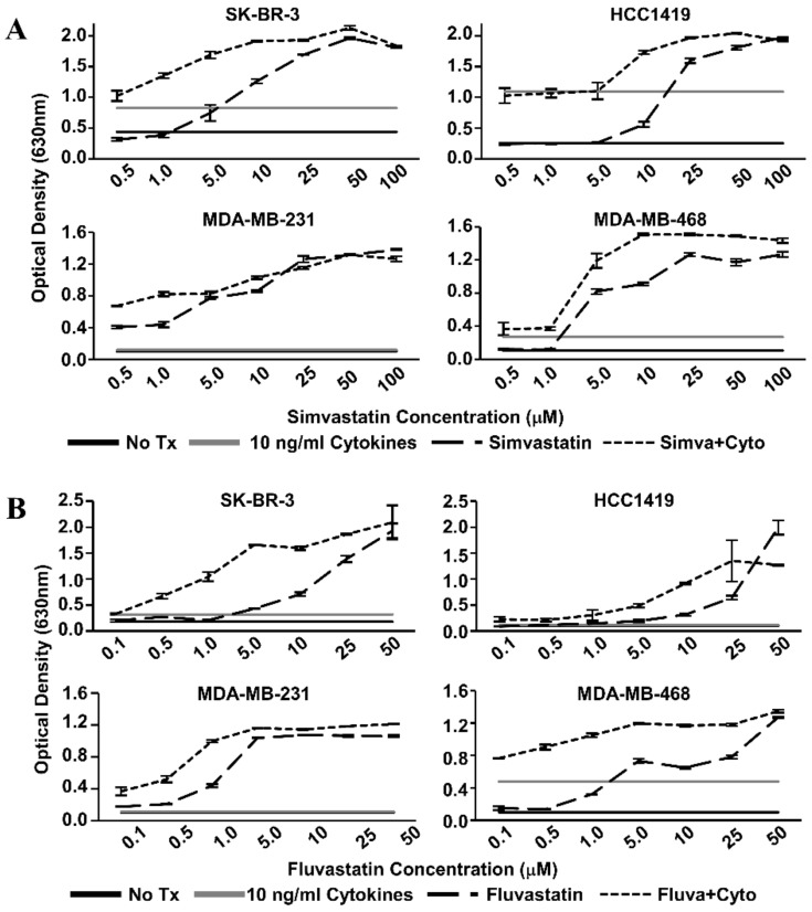
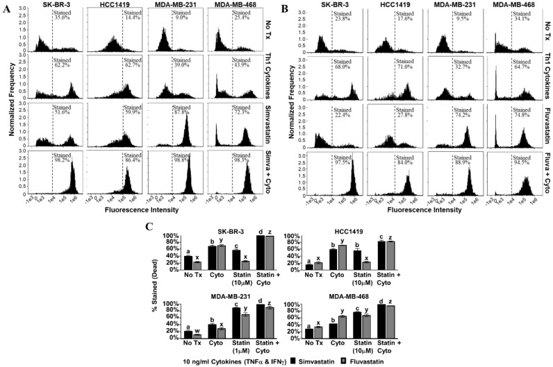
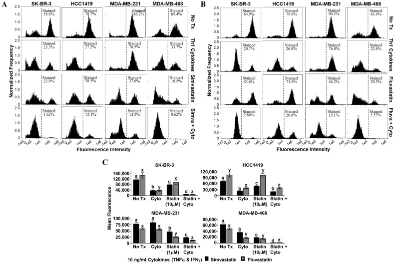
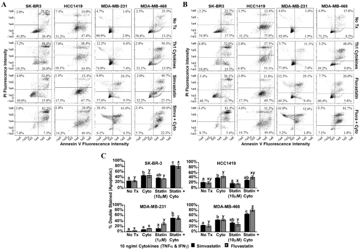
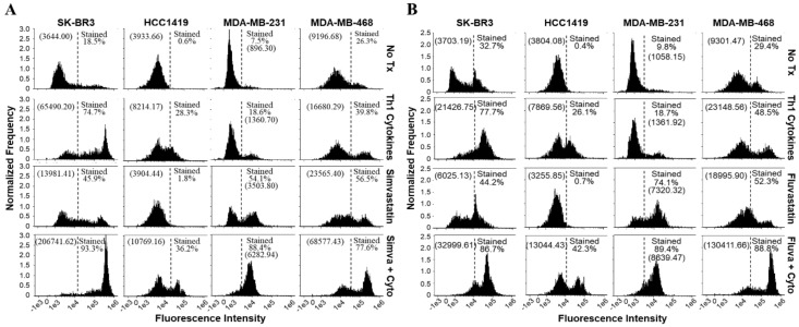
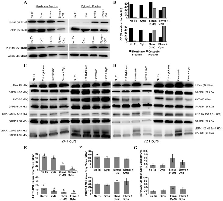
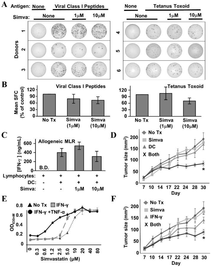
A dendritic cell-based, Type 1 Helper T cell (Th1)-polarizing anti-Human Epidermal Growth Factor Receptor-2 (HER-2) vaccine supplied in the neoadjuvant setting eliminates disease in up to 30% of recipients with HER-2-positive (HER-2pos) ductal carcinoma in situ (DCIS). We hypothesized that drugs with low toxicity profiles that target signaling pathways critical for oncogenesis may work in conjunction with vaccine-induced immune effector mechanisms to improve efficacy while minimizing side effects. In this study, a panel of four phenotypically diverse human breast cancer lines were exposed in vitro to the combination of Th1 cytokines Interferon-gamma (IFN-γ) and Tumor Necrosis Factor-alpha (TNF-α) and lipophilic statins. This combination was shown to potentiate multiple markers of apoptotic cell death. The combination of statin drugs and Th1 cytokines minimized membrane K-Ras localization while maximizing levels in the cytoplasm, suggesting a possible means by which cytokines and statin drugs might cooperate to maximize cell death. A combined therapy was also tested in vivo through an orthotopic murine model using the neu-transgenic TUBO mammary carcinoma line. We showed that the combination of HER-2 peptide-pulsed dendritic cell (DC)-based immunotherapy and simvastatin, but not single agents, significantly suppressed tumor growth. Consistent with a Th1 cytokine-dependent mechanism, parenterally administered recombinant IFN-γ could substitute for DC-based immunotherapy, likewise inhibiting tumor growth when combined with simvastatin. These studies show that statin drugs can amplify a DC-induced effector mechanism to improve anti-tumor activity.

Keywords: Ras; apoptosis; breast cancer; cytokine; statin; vaccine.


Conflict of interest statement

The authors declare that cancer vaccine technologies developed by GKK and BJC (patents pending) are currently licensed by Immunorestoration LLC, Warminster Pennsylvania, USA. The funders had no role in the design of the study; in the collection, analyses, or interpretation of data; in the writing of the manuscript, or in the decision to publish the results.

Figures

Figure 1
Figure 1
Statin dose–response curves via Alamar Blue dye reduction assay. Human breast cancer cell lines (SK-BR-3, HCC1419, MDA-MB-231, and MDA-MB-468) were treated with increasing concentrations of (A) Simvastatin or (B) Fluvastatin in the presence (short dash) or absence (long dash) of recombinant Th1 cytokines (Tumor Necrosis Factor-alpha, TNF-α and Interferon-gamma, IFN-γ, 10 ng/mL each) for 72 h. Alamar Blue dye was added and, following color change, the optical density of the dye in the culture supernatants was determined. Optical Density (OD) values of untreated controls (black) and cytokine only treatment (gray) are represented as horizontal lines.
Figure 2
Figure 2
Combination of Th1 cytokines and statin drugs potentiates metabolic suppression in breast cancer lines. SK-BR-3, HCC1419, MDA-MB-231, and MDA-MB-468 human breast cancer cell lines were cultured with no additives (No Tx), treated with recombinant Th1 cytokines (“Cyto” TNF-α and IFN-γ, 10 ng/mL each), statin drugs (Simvastatin or Fluvastatin, 1 µM MDA-MB-231; 10 µM remaining cell lines), or the combination of Th1 cytokines and a statin drug (“Statin + Cyto”). After 72h incubation, Alamar Blue dye was added and, following color change, optical density of culture supernatants was determined. Results displayed are from one representative experiment of at least four trials +/− Standard Error of the Mean (SEM). Letter designations represent Tukey’s Honest Significant Difference (HSD) comparisons: treatments with the same letter designation are not statistically different; when letter designations differ between treatments, the p-value is less than 0.05.
Figure 3
Figure 3
Combination of Th1 cytokines and statin drugs maximize cell death in breast cancer lines. SK-BR-3, HCC1419, MDA-MB-231, and MDA-MB-468 human breast cancer cell lines were cultured with no additives (No Tx), treated with recombinant Th1 cytokines (TNF-α and IFN-γ, 10 ng/mL each), a statin drug (A) Simvastatin or (B) Fluvastatin (1 µM MDA-MB-231; 10 µM remaining cell lines), or the combination of Th1 cytokines and a statin drug (A) “Simva + Cyto” or (B) “Fluva + Cyto”. Flow cytometric results displayed in panels A and B are from one representative experiment. (C) Graphical interpretation of gated flow cytometric results comparing the percentage of stained events between groups: no additives (No Tx), treated with recombinant Th1 cytokines (TNFα and IFNγ, 10 ng/mL each), a statin drug (Simvastatin or Fluvastatin, 1 µM MDA-MB-231; 10 µM remaining cell lines), or the combination of Th1 cytokines and a statin drug (“Statin + Cyto”). Results displayed are from at least three trials +/− SEM. Letter designations represent Tukey’s HSD comparisons: treatments with the same letter designation are not statistically different; when letter designations differ between treatments, the p-value is less than 0.05.
Figure 4
Figure 4
Statin drugs plus Th1 cytokines maximize mitochondrial membrane depolarization as assessed by tetramethylrhodamine ethyl ester (TMRE) staining. SK-BR-3, HCC1419, MDA-MB-231, and MDA-MB-468 human breast cancer cell lines were cultured with no additives (No Tx), treated with recombinant Th1 cytokines (TNF-α and IFN-γ, 10 ng/mL each), a statin drug (A) Simvastatin or (B) Fluvastatin (1 µM MDA-MB-231; 10 µM remaining cell lines), or the combination of Th1 cytokines and a statin drug (A) “Simva + Cyto” or (B) “Fluva + Cyto” for approximately 48 h. Flow cytometric results displayed in panels A and B are from one representative experiment. (C) Statistical analysis of flow cytometry results comparing the mean channel fluorescent intensity between groups: no additives (No Tx), treated with recombinant Th1 cytokines (TNF-α and IFN-γ, 10 ng/mL each), a statin drug (Simvastatin or Fluvastatin, 1 µM MDA-MB-231; 10 µM remaining cell lines), or the combination of Th1 cytokines and a statin drug (“Statin + Cyto”). Results displayed are from at least five trials +/− SEM. Letter designations represent Tukey’s HSD comparisons: treatments with the same letter designation are not statistically different; when letter designations differ between treatments, the p-value is less than 0.05.
Figure 5
Figure 5
Statin drugs plus Th1 cytokines maximize apoptotic markers in breast cancer lines. SK-BR-3, HCC1419, MDA-MB-231, and MDA-MB-468 human breast cancer cell lines were cultured with no additives (No Tx), treated with recombinant Th1 cytokines (TNF-α and IFN-γ, 10 ng/mL each), a statin drug (A) Simvastatin or (B) Fluvastatin (1 µM MDA-MB-231; 10 µM remaining cell lines), or the combination of Th1 cytokines and a statin drug (A) “Simva + Cyto” or (B) “Fluva + Cyto”. Flow cytometric results displayed in panels A and B are of one representative experiment from 3 separate trials with each drug. (C) Statistical analysis of composite results for proportions of double-staining cell events between groups: no additives (No Tx), treated with recombinant Th1 cytokines (TNF-α and IFN-γ, 10 ng/mL each), a statin drug (Simvastatin or Fluvastatin, 1 µM MDA-MB-231; 10 µM remaining cell lines), or the combination of Th1 cytokines and a statin drug (“Statin + Cyto”). Results displayed are from three trials +/− SEM. Letter designations represent Tukey’s HSD comparisons: treatments with the same letter designation are not statistically different; when letter designations differ between treatments, the p-value is less than 0.05.
Figure 6
Figure 6
Cells treated with a statin drug and Th1 cytokines demonstrate enhanced activation of caspase 3/7. SK-BR-3, HCC1419, MDA-MB-231, and MDA-MB-468 human breast cancer cell lines were cultured with no additives (No Tx), treated with recombinant Th1 cytokines (TNF-α and IFN-γ, 10 ng/mL each), a statin drug (A) Simvastatin or (B) Fluvastatin (1 µM MDA-MB-231; 10 µM remaining cell lines), or the combination of Th1 cytokines and a statin drug (A) “Simva + Cyto” or (B) “Fluva + Cyto” and stained with CellEvent™ fluorescent caspase substrate. The experiment was repeated at least three times; the displayed flow cytometric results are from one representative experiment. The percentage of stained events, as well as the geometric mean channel fluorescence (in parentheses), is annotated on each histogram.
Figure 7
Figure 7
Combined treatment with Th1 cytokines and statin drugs potentiates K-Ras delocalization from membrane. MDA-MB-231 human breast cancer cells were cultured with no additives (No Tx), treated with recombinant Th1 cytokines (TNF-α and IFN-γ, 10 ng/mL each), a statin drug (Simvastatin or Fluvastatin, 1 µM), or the combination of Th1 cytokines and a statin drug (“Simva + Cyto” or “Fluva + Cyto”) for 24 h. Membrane proteins were separated from cytosolic proteins and the distribution of the K-Ras protein was analyzed by Western blot. (A) Representative Western blot images and (B) corresponding semi-quantitative densitometry analysis of K-Ras normalized to β-actin loading control. Representative Western blot images of whole-cell K-Ras, AKT, ERK ½, and phospho-ERK ½ after (C) 24 and (D) 72 h of treatment. Semi-quantitative densitometry analysis of the (E) GAPDH-normalized total AKT, (F) GAPDH-normalized total ERK-1/2, and (G) GAPDH-normalized phosphorylated ERK-1/2. All Western blot experiments were repeated at least three times. Displayed graphical results in panels (E), (F), and (G) are from at least three trials +/− SEM. Letter designations in panel (E) represent Tukey’s HSD comparisons: treatments with the same letter designation are not statistically different; when letter designations differ between treatments, the p-value is less than 0.05.
Figure 8
Figure 8
Simvastatin does not interfere with T cell function and enhances effects of immunotherapy in a mouse model of HER-2pos breast cancer. (A) Example IFN-γ EliSpot wells from individual healthy donor Peripheral Blood Mononuclear Cells (PBMCs) stimulated with a mixture of common viral peptide recall antigens (left panel) or tetanus toxoid (right panel) in the presence or absence of simvastatin (“Simva”; 1–10 µM). (B) Composite analysis of each of the 3 donors for viral recall peptides (left panel) and 3 donors for tetanus toxoid (right panel). Statistical analysis by one-way ANOVA indicated no significant difference between simvastatin-treated and untreated groups. (C) IFN-γ ELISA analysis of 72 h culture supernatants from allogeneic Mixed Leukocyte Reactions (MLRs) where activated dendritic cells (DC) and lymphocyte-rich elutriation fractions were co-cultured at 1:40 stimulator:responder ratios in the presence or absence of 1–10 µM simvastatin. Data displayed represents the mean IFN-γ production from seven unique allogeneic DC:lymphocyte pairings. Statistical analysis by one-way ANOVA indicated no significant difference between simvastatin-treated and untreated groups (B.D. = below detection). (D) TUBO-bearing Balb/c mice, 7 days after implantation, were either left untreated (“No Rx”) or treated with Simvastatin (“Simva”), peptide-pulsed DC-based therapy (“DC”), or given combined treatment with simvastatin plus peptide-pulsed DCs (“Both”). Tumor size was measured periodically with calipers during treatment. Tumor size denoted as average area +/− SEM. Statistical significance is indicated by the asterisk (*). (E) TUBO cells were cultured in vitro with IFN-γ plus TNF-α, IFN-γ only, or no treatment in the presence of increasing concentrations of Simvastatin for 72 h, then assessed for metabolic activity by the Alamar Blue dye assay. (F) TUBO-bearing Balb/c mice, 7 days after implantation, were either left untreated (“No Rx”) or treated with Simvastatin (“Simva”), Interferon-γ (“IFN-γ”) or given combined treatment with simvastatin plus IFN-γ (“Both”). Tumor size was measured periodically with calipers during treatment. Tumor size denoted as average area +/− SEM. Statistical significance is indicated by the asterisk (*).

Similar articles

Cited by

References

    1. Seshadri R., Firgaira F.A., Horsfall D.J., McCaul K., Setlur V., Kitchen P. Clinical significance of HER-2/neu oncogene amplification in primary breast cancer. The South Australian Breast Cancer Study Group. J. Clin. Oncol. 1993;11:1936–1942. doi: 10.1200/JCO.1993.11.10.1936. - DOI - PubMed
    1. Marks J.R., Humphrey P.A., Wu K., Berry D., Bandarenko N., Kerns B.J., Iglehart J.D. Overexpression of p53 and HER-2/neu proteins as prognostic markers in early stage breast cancer. Ann. Surg. 1994;219:332–341. doi: 10.1097/00000658-199404000-00002. - DOI - PMC - PubMed
    1. Czerniecki B.J., Koski G.K., Koldovsky U., Xu S., Cohen P.A., Mick R., Nisenbaum H., Pasha T., Xu M., Fox K.R., et al. Targeting HER-2/neu in early breast cancer development using dendritic cells with staged interleukin-12 burst secretion. Cancer Res. 2007;67:1842–1852. doi: 10.1158/0008-5472.CAN-06-4038. - DOI - PubMed
    1. Sharma A., Koldovsky U., Xu S., Mick R., Roses R., Fitzpatrick E., Weinstein S., Nisenbaum H., Levine B.L., Fox K., et al. HER-2 pulsed dendritic cell vaccine can eliminate HER-2 expression and impact ductal carcinoma in situ. Cancer. 2012;118:4354–4362. doi: 10.1002/cncr.26734. - DOI - PMC - PubMed
    1. Lowenfeld L., Mick R., Datta J., Xu S., Fitzpatrick E., Fisher C.S., Fox K.R., DeMichele A., Zhang P., Weinstein S., et al. Dendritic Cell Vaccination Enhances Immune Responses and Induces Regression of HER2pos DCIS Independent of Route: Results of Randomized Selection Design Trial. Clin. Cancer Res. 2016;23:2961–2971. doi: 10.1158/1078-0432.CCR-16-1924. - DOI - PubMed