

NANOG expression in human development and cancerogenesis
- PMID: 32041418
- PMCID: PMC7082888
- DOI: 10.1177/1535370220905560
NANOG expression in human development and cancerogenesis
Abstract
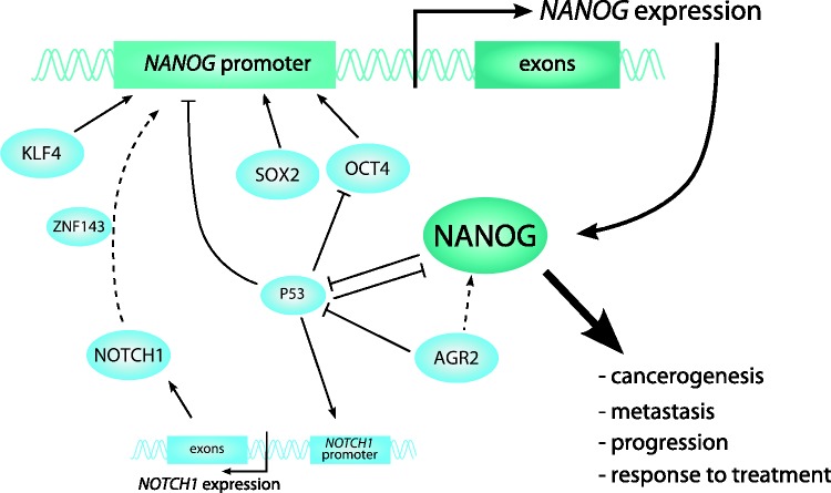
NANOG is an important stem cell transcription factor involved in human development and cancerogenesis. Its expression is complex and regulated on different levels. Moreover, NANOG protein might regulate hundreds of target genes at the same time. NANOG is crucial for preimplantation development phase and progressively decreases during embryonic stem cells differentiation, thus regulating embryonic and fetal development. Postnatally, NANOG is undetectable or expressed in very low amounts in the majority of human tissues. NANOG re-expression can be detected during cancerogenesis, already in precancerous lesions, with increasing levels of NANOG in high grade dysplasia. NANOG is believed to enable cancer cells to obtain stem-cell like properties, which are believed to be the source of expanding growth, tumor maintenance, metastasis formation, and tumor relapse. High NANOG expression in cancer is frequently associated with advanced stage, poor differentiation, worse overall survival, and resistance to treatment, and is therefore a promising prognostic and predictive marker. We summarize the current knowledge on the role of NANOG in cancerogenesis and development, including our own experience. We provide a critical overview of NANOG as a prognostic and diagnostic factor, including problems regarding its regulation and detection.
Impact statement: NANOG has emerged as a key stem cell transcription factor in normal development and cancerogenesis. It is generally regarded as a good prognostic and predictive factor in various human cancers. It is less known that it is expressed already at precancerous stages in various organs, suggesting that finally an ideal candidate diagnostic marker has been discovered, enabling to distinguish between true dysplasia and reactive atypia. NANOG regulation is complex, and new insights into our understanding of its regulation might provide important information for future development in a broad field of two entirely different processes, i.e. normal development and cancerogenesis, showing how a physiologic mechanism can be used and abused, transforming itself into a key mechanism of disease development and progression.
Keywords: Nanog; cancer; cancerogenesis; development; precancerosis; stem cells.
Figures
References
-
- Mitsui K, Tokuzawa Y, Itoh H, Segawa K, Murakami M, Takahashi K, Maruyama M, Maeda M, Yamanaka S. The homeoprotein nanog is required for maintenance of pluripotency in mouse epiblast and ES cells. Cell 2003; 113:631–42 - PubMed
-
- Chambers I, Colby D, Robertson M, Nichols J, Lee S, Tweedie S, Smith A. Functional expression cloning of nanog, a pluripotency sustaining factor in embryonic stem cells. Cell 2003; 113:643–55 - PubMed
-
- Yates B, Braschi B, Gray KA, Seal RL, Tweedie S, Bruford EA. Genenames.org: the HGNC and VGNC resources in 2017. Nucleic Acids Res 2017; 45:D619–d25 - PMC - PubMed
-
- Kerr CL, Hill CM, Blumenthal PD, Gearhart JD. Expression of pluripotent stem cell markers in the human fetal ovary. Hum Reprod 2008; 23:589–99 - PubMed
Publication types
MeSH terms
Substances
LinkOut - more resources
Full Text Sources
Research Materials