

Tissue and cancer-specific expression of DIEXF is epigenetically mediated by an Alu repeat
- PMID: 32041475
- PMCID: PMC7574385
- DOI: 10.1080/15592294.2020.1722398
Tissue and cancer-specific expression of DIEXF is epigenetically mediated by an Alu repeat
Abstract
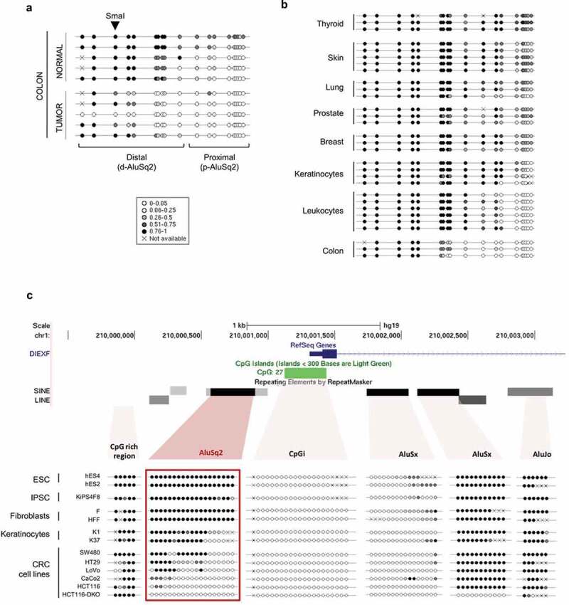
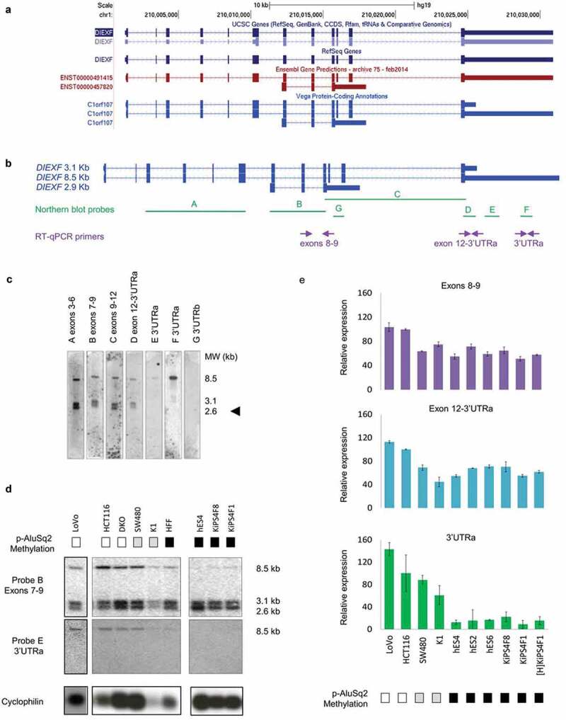
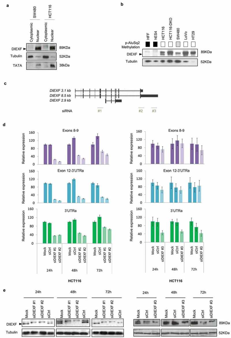
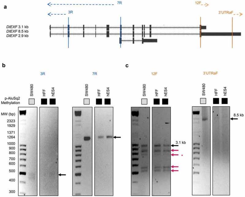
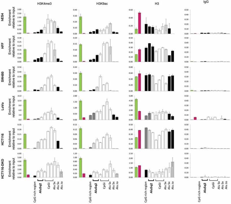
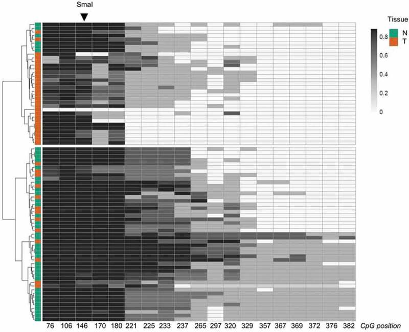
Alu repeats constitute a major fraction of human genome and for a small subset of them a role in gene regulation has been described. The number of studies focused on the functional characterization of particular Alu elements is very limited. Most Alu elements are DNA methylated and then assumed to lie in repressed chromatin domains. We hypothesize that Alu elements with low or variable DNA methylation are candidates for a functional role. In a genome-wide study in normal and cancer tissues, we pinpointed an Alu repeat (AluSq2) with differential methylation located upstream of the promoter region of the DIEXF gene. DIEXF encodes a highly conserved factor essential for the development of zebrafish digestive tract. To characterize the contribution of the Alu element to the regulation of DIEXF we analysed the epigenetic landscapes of the gene promoter and flanking regions in different cell types and cancers. Alternate epigenetic profiles (DNA methylation and histone modifications) of the AluSq2 element were associated with DIEXF transcript diversity as well as protein levels, while the epigenetic profile of the CpG island associated with the DIEXF promoter remained unchanged. These results suggest that AluSq2 might directly contribute to the regulation of DIEXF transcription and protein expression. Moreover, AluSq2 was DNA hypomethylated in different cancer types, pointing out its putative contribution to DIEXF alteration in cancer and its potential as tumoural biomarker.
Keywords: DIEXF (UTP25); Alu repeat; DNA methylation; alternative polyadenylation sites; alternative transcription start sites (TSSs); cancer; histone marks; multiple 3ʹUTRs.
Conflict of interest statement
MAP is cofounder and equity holder of Aniling, a biotech company with no interests in this paper. MAP lab has received research funding from Celgene. The rest of the authors declare no conflict of interest.
Figures
Similar articles
-
The epigenetic landscape of Alu repeats delineates the structural and functional genomic architecture of colon cancer cells.Genome Res. 2017 Jan;27(1):118-132. doi: 10.1101/gr.207522.116. Epub 2016 Dec 20. Genome Res. 2017. PMID: 27999094 Free PMC article.
-
Genome-wide tracking of unmethylated DNA Alu repeats in normal and cancer cells.Nucleic Acids Res. 2008 Feb;36(3):770-84. doi: 10.1093/nar/gkm1105. Epub 2007 Dec 15. Nucleic Acids Res. 2008. PMID: 18084025 Free PMC article.
-
Epigenomic analysis of Alu repeats in human ependymomas.Proc Natl Acad Sci U S A. 2010 Apr 13;107(15):6952-7. doi: 10.1073/pnas.0913836107. Epub 2010 Mar 29. Proc Natl Acad Sci U S A. 2010. PMID: 20351280 Free PMC article.
-
Epigenetic implication of gene-adjacent retroelements in Helicobacter pylori-infected adults.Epigenomics. 2012 Oct;4(5):527-35. doi: 10.2217/epi.12.51. Epigenomics. 2012. PMID: 23130834 Review.
-
The transcript repeat element: the human Alu sequence as a component of gene networks influencing cancer.Funct Integr Genomics. 2010 Aug;10(3):307-19. doi: 10.1007/s10142-010-0168-1. Funct Integr Genomics. 2010. PMID: 20393868 Review.
Cited by
-
Mycobacterium manresensis induces trained immunity in vitro.iScience. 2023 Jun 16;26(6):106873. doi: 10.1016/j.isci.2023.106873. Epub 2023 May 13. iScience. 2023. PMID: 37250788 Free PMC article.
-
Analysis of hepatic lentiviral vector transduction; implications for preclinical studies and clinical gene therapy protocols.bioRxiv [Preprint]. 2024 Aug 31:2024.08.20.608805. doi: 10.1101/2024.08.20.608805. bioRxiv. 2024. Update in: Viruses. 2025 Feb 17;17(2):276. doi: 10.3390/v17020276. PMID: 39229157 Free PMC article. Updated. Preprint.
-
Gene body methylation in cancer: molecular mechanisms and clinical applications.Clin Epigenetics. 2022 Nov 28;14(1):154. doi: 10.1186/s13148-022-01382-9. Clin Epigenetics. 2022. PMID: 36443876 Free PMC article. Review.
-
Analysis of Hepatic Lentiviral Vector Transduction: Implications for Preclinical Studies and Clinical Gene Therapy Protocols.Viruses. 2025 Feb 17;17(2):276. doi: 10.3390/v17020276. Viruses. 2025. PMID: 40007031 Free PMC article.
-
Epigenetic Research in Stem Cell Bioengineering-Anti-Cancer Therapy, Regenerative and Reconstructive Medicine in Human Clinical Trials.Cancers (Basel). 2020 Apr 21;12(4):1016. doi: 10.3390/cancers12041016. Cancers (Basel). 2020. PMID: 32326172 Free PMC article. Review.
References
-
- International Human Genome Sequencing Consortium . Initial sequencing and analysis of the human genome. Nature. 2001:409(6822):860–921. - PubMed
-
- Wicker T, Sabot F, Hua-Van A, et al. A unified classification system for eukaryotic transposable elements. Nat Rev Genet. 2007;8(12):973–982. - PubMed
-
- Chen LL, Yang L. ALUternative regulation for gene expression. Trends Cell Biol. 2017;27(7):480–490. - PubMed
Publication types
MeSH terms
Substances
LinkOut - more resources
Full Text Sources
Other Literature Sources
Medical
Molecular Biology Databases